Genetics Chapters 1-2
1/88
There's no tags or description
Looks like no tags are added yet.
Name | Mastery | Learn | Test | Matching | Spaced | Call with Kai |
|---|
No analytics yet
Send a link to your students to track their progress
89 Terms
Transmission Genetics
The basic principles of heredity
Focus is on the individual organism
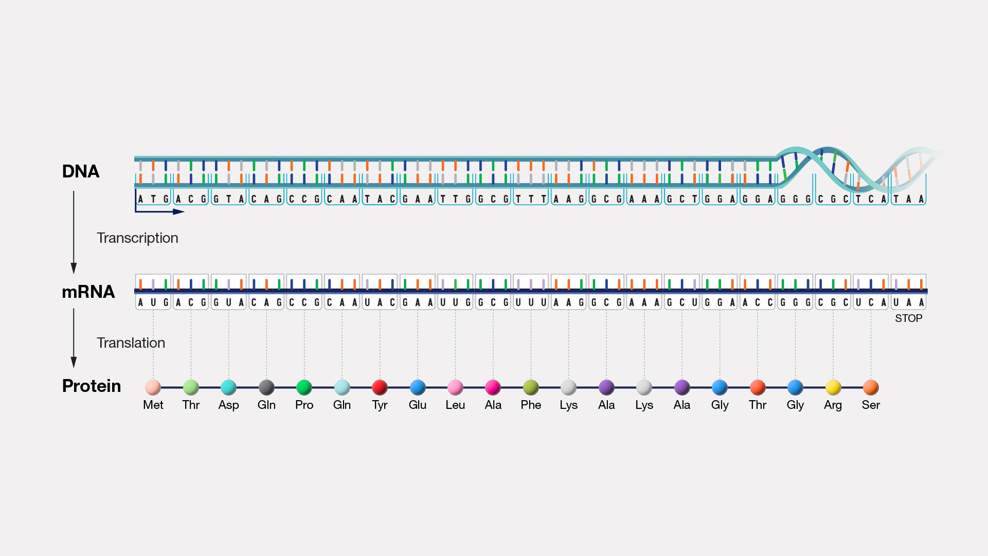
Molecular Genetics
The chemical nature of the gene
How genetic information is encoded, replicated, and expressed
Focus is the gene and its structure, organization, and function
Population genetics
The genetic composition of populations (group of organisms of the same species)
How genetic composition changes geographically and with the passage of time
Focus is the group of genes found in a population
Model Genetic Organisms
Organisms with characteristics that make them useful for genetic analysis
Characteristics of Model Organisms
Short generation time
Production of numerous progeny
Ability to carry out controlled genetic crosses
Ability to be reared in a laboratory environment
Availability of numerous genetic variants
Accumulated body of knowledge about their genetic systems
Why can’t humans be model organisms?
Humans take 20-30 years for one full generation, making experiments take decades to get results.
Humans have very few offspring per reproductive cycle (usually one at a time), so sample sizes would be too small for a reliable genetic study.
Controlled genetic crosses: We cannot ethically choose who breeds with whom. Controlled mating is impossible in humans.
Reared in a laboratory environment: Humans cannot be raised or kept in lab conditions for experimentation—this is unethical and impractical.
Availability of numerous genetic variants: While humans do have genetic diversity, we cannot create or selectively breed variants for study the way we can with model organisms.
Accumulated body of genetic knowledge: Human genetics is well-studied, but the knowledge is partly based on model organisms. We needed simpler organisms first to understand fundamental genetics.
Don’t follow the common characteristics
Name the 6 Common Model Organisms
Drosophila melanogaster (fruit fly)
Escherichia coli (Bacterium)
Caenorhabditis elegans (Nematode)
Arabidopsis thaliana (Thale-cress plant)
Mus musculus (House Mouse)
Saccharomyces cerevisiae (Baker’s yeast)
Pangenesis
Theory of Heredity
Each part of the body contains genetic information for that particular part
Specific particles (gemmules) carry information from parts of the body to the reproductive organs
Then passed to the embryo at the moment of conception
Inheritance of acquired characteristics
Theory of Heredity
Traits acquired in a person’s lifetime
Become incorporated into that person’s hereditary information
Are passed on to offspring
Preformationism
Theory of Heredity/Embryology
Inside the egg or sperm exists a full formed miniature adult (a homunculus)
Simply enlarges during development
Blending Inheritance
Theory of Heredity
Traits of offspring are a blend, or mixture, of parental traits
What are the defining characteristics of Prokaryotes?
Unicellular
No membrane-bound organelles
Made up of eubacteria and archaea
DNA isn’t highly ordered or in a packed arrangement like Eukaryotes
What are the defining characteristics of Eukaryotes?
Both unicellular and multicellular
Membrane-bound organelles
Genetic material is surrounded in a nuclear envelope to form a nucleus
DNA is closely associated with histones to form tightly packed chromosomes
Parts of a Prokaryote
Cell Wall, Plasma membrane, cytoplasm, ribosomes, nucleioid, flagella

Parts of a Eukaryote
Nucleus, plasma membrane, cytoplasm, ribosomes, mitochondria, endoplasmic reticulum, Golgi apparatus, chloroplast (plant), cell wall (plant), vacuole (plant)
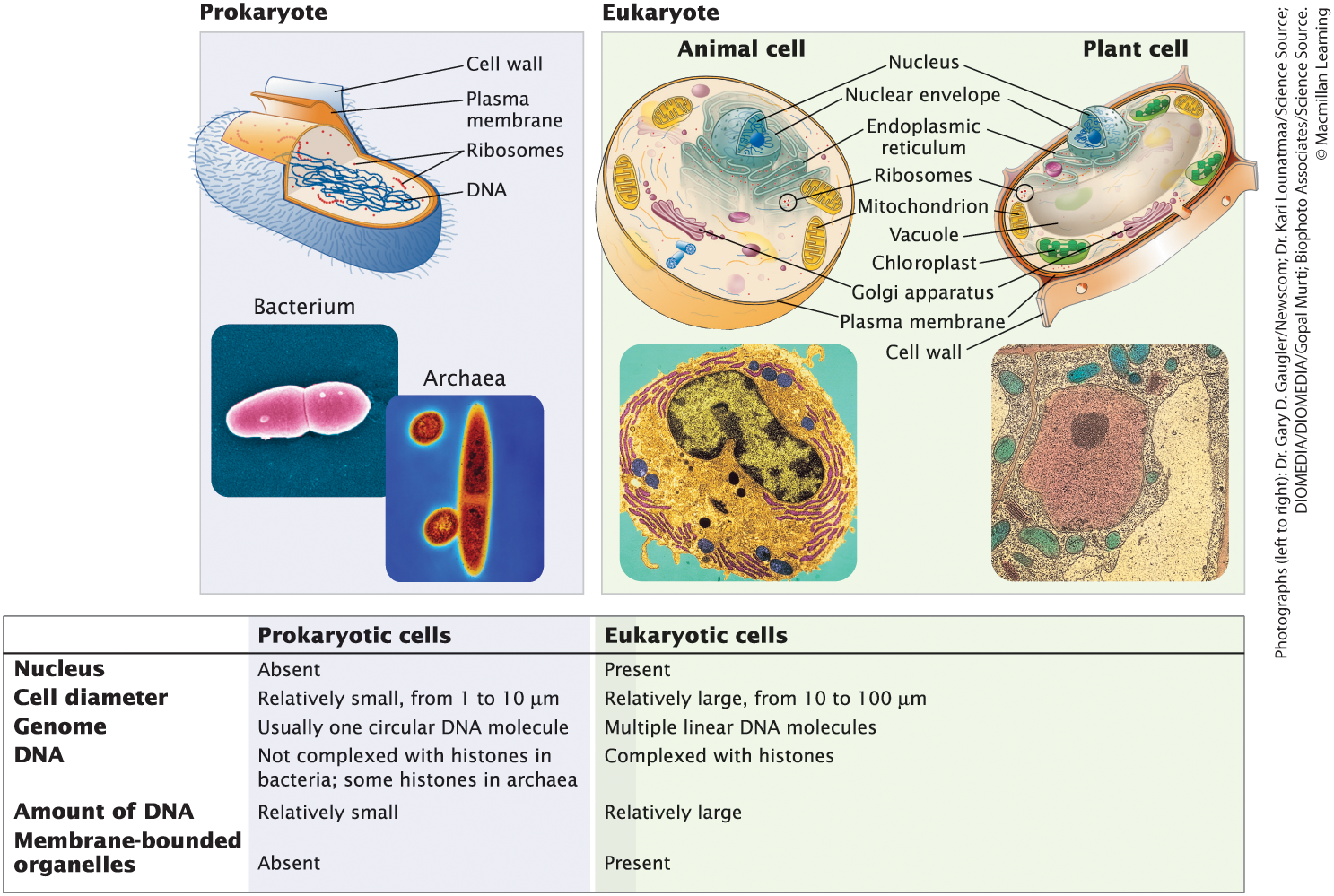
Nucleus - Prokaryote vs Eukaryote
Absent in Prokaryotes (Nucleioid region instead)
Present in Eukaryotes (Genetic material is surrounded in a nuclear envelope to form a nucleus)
Cell Diameter Range - Prokaryotes vs Eukaryotes
For prokaryotes, relatively small, from 1-10 μm
For eukaryotes, relatively large, from 10-100 μm
Genome structure in Prokaryotes vs Eukaryotes
Prokaryotes, one circular DNA molecule
Eukaryotes, multiple linear DNA molecules
DNA Structure, Arrangement, and Amount in Prokaryotes vs Eukaryotes
Prokaryotic, isn’t highly ordered or packed, relatively small amount of DNA
Eukaryotic, closely associated with histones to form tightly packed chromosomes, relatively large amount of DNA
Viruses
Neither prokaryotic nor Eukaryotic
Outer protein coat surrounding nucleic acid
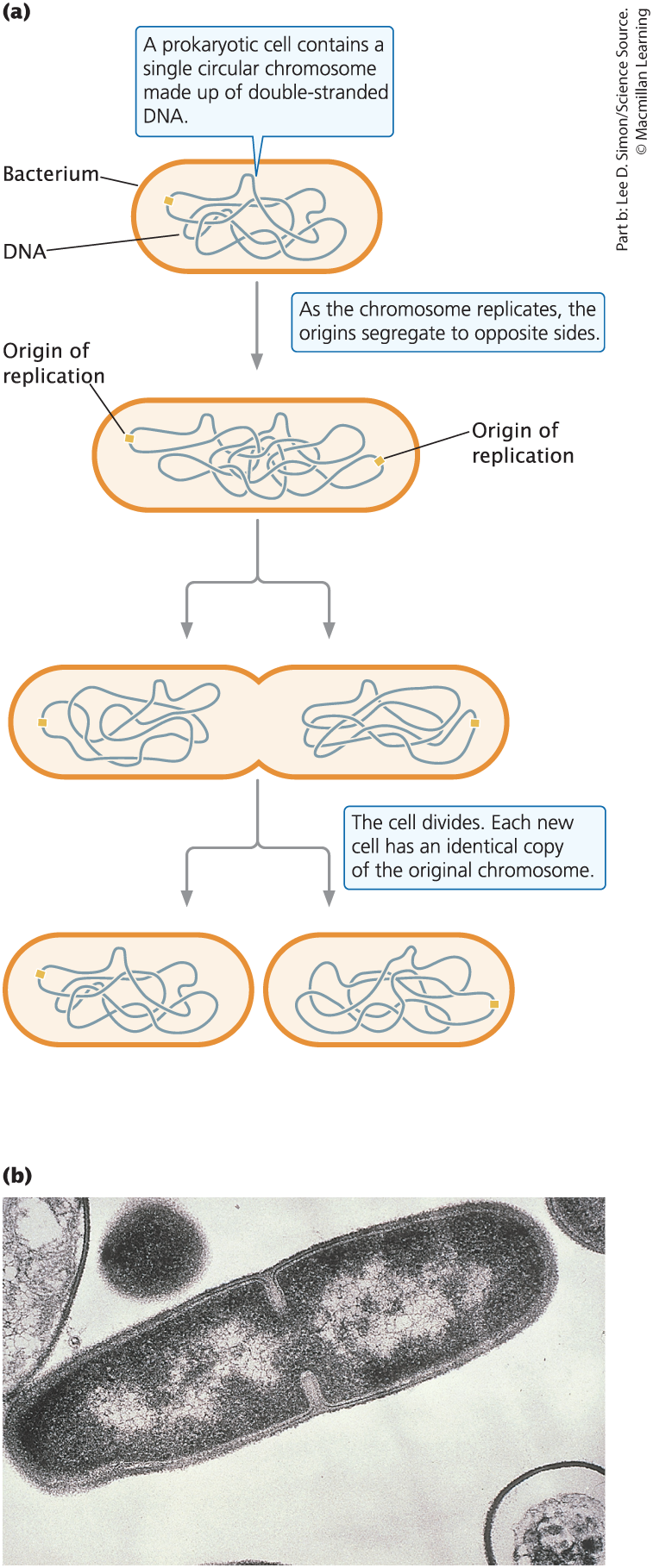
Prokaryotic Cell Reproduction
Occurs through binary fission, involving the simple division and separation of the replicated circular chromosome, high rate of replication
Structural Maintenance of Chromosomes
Proteins that encircle DNA and compact and organize into chromosomes
Eukaryotic Cell Reproduction
By mitosis for growth and repair, or by meiosis for sexual reproduction
Do all eukaryotic organisms have the same amount of chromosomes?
No. And there is no relation between complexity of organism and amount of chromosomes they have.
Homologous Chromosomes
pairs of chromosomes that have the same length, centromere position, and genes in the same order, with one chromosome inherited from each parent
Diploid and Haploid
Diploid: two sets of genetic information/chromosomes
Haploid: one set of genetic information/chromosomes
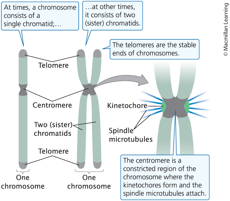
Centromere
middle point of chromosome, where sister chromatids are attached, contains a kinetochore (Attachment point for spindle microtubules)
What would be the result if a chromosome didn’t have a kinetochore or something was wrong with it?
Nondisjunction (failure of correct separation of homologous chromosomes)
Spindle microtubules wouldn’t attach to the chromosome
The chromosome wouldn’t be drawn into a newly formed nucleus
The resulting daughter cells would be missing a chromosome (have the incorrect amount)
Telomere
Tips of a linear chromosome (only in eukaryotes)
Origin of Replication
A specific DNA sequence where replication or synthesis starts. One in prokaryotes, a lot in eukaryotes
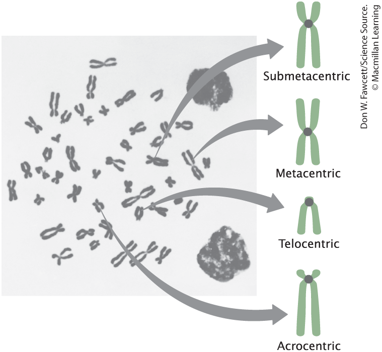
Chromosome Structure
Types of Chromosome Structures
Metacentric (Middle
Submetacentric (Almost middle)
Telocentric (end)
Afrocentric (almost end)
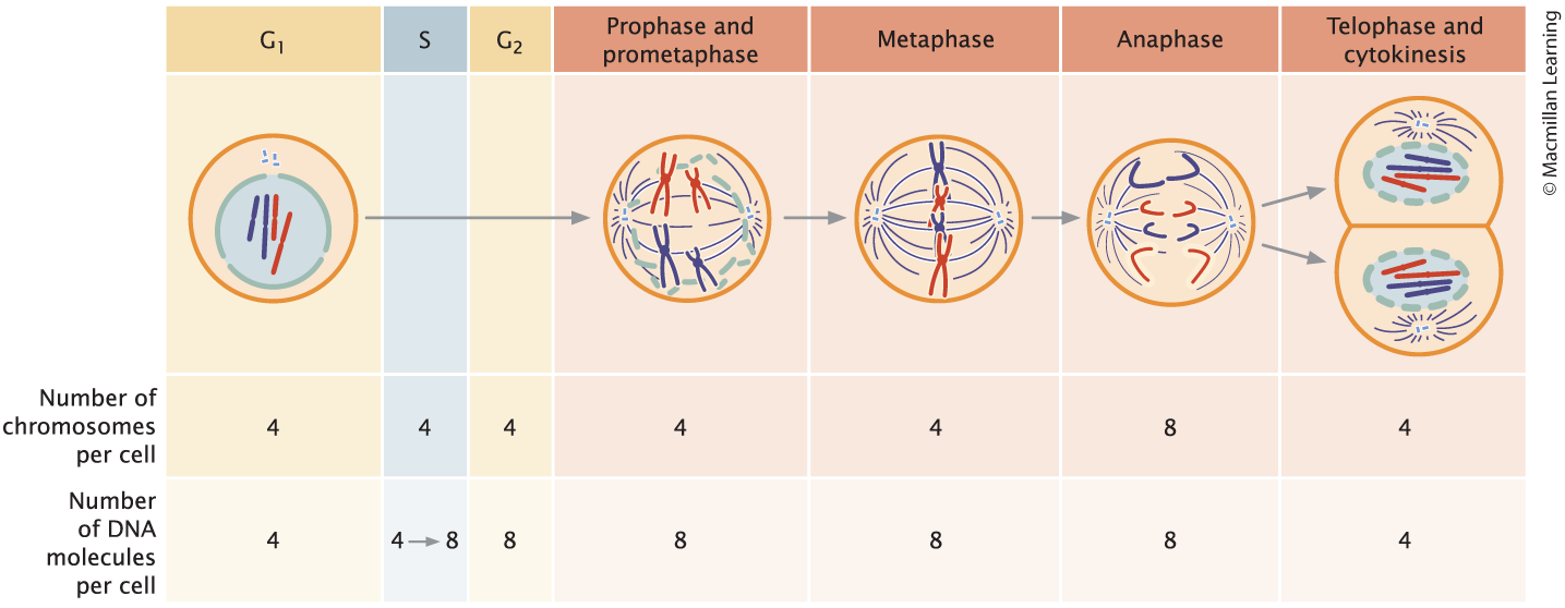
Cell Cycle
The life cycle of the cell. Consists of:
Interphase (cell growth, DNA synthesis before division): G1, S, G2
M Phase (cell division): Prophase, Prometaphase, Metaphase, Anaphase, and Telophase
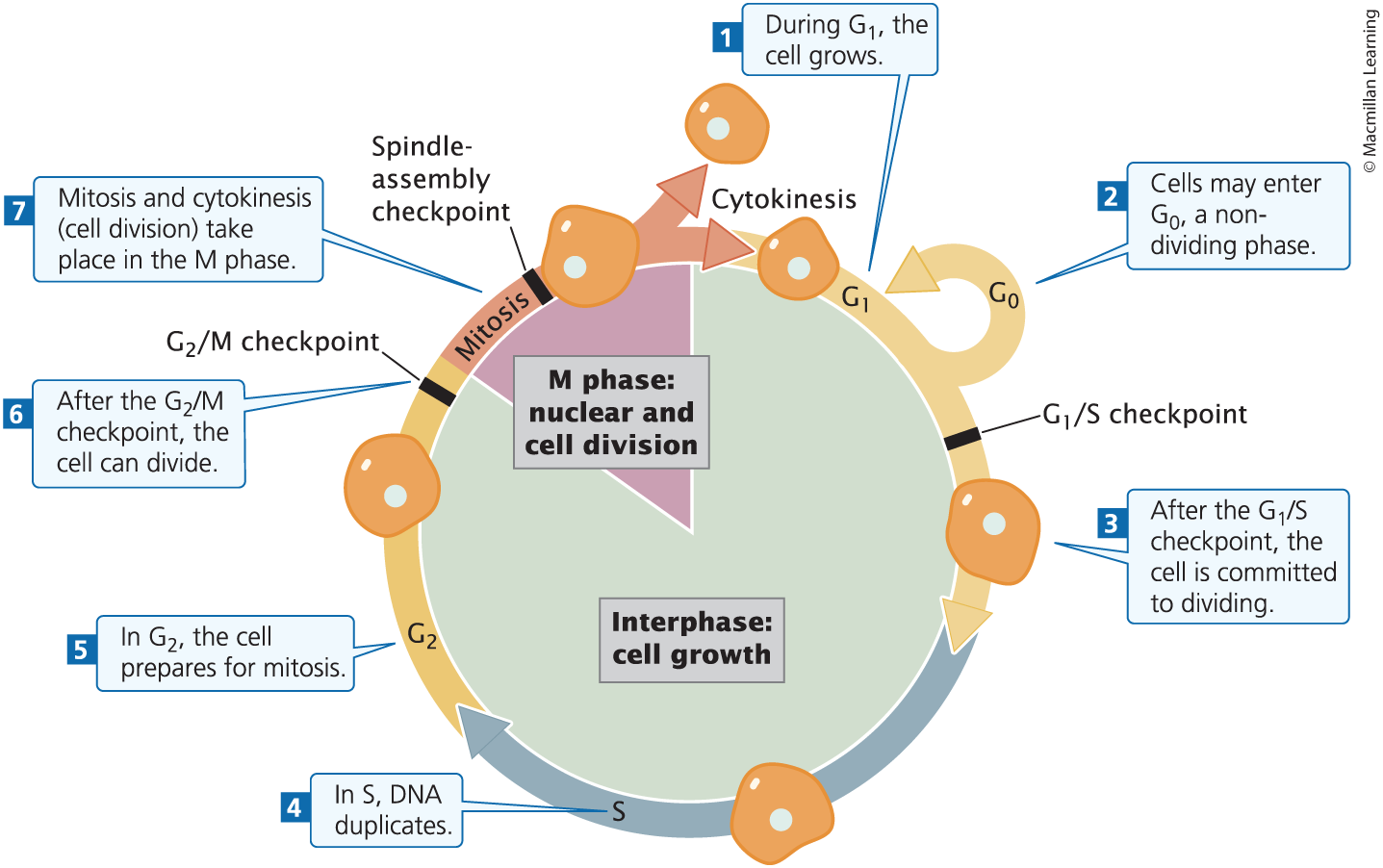
G0
A stable, nondividing period of variable length that some cells enter temporarily and others enter permanently during G1
Interphase
An extended period between cell divisions, including cell growth, DNA synthesis. It consists of G1, S, and G2 phases
G1
First phase of growth, where cell grows/increases in size, proteins necessary for DNA synthesis and cell division are synthesized; G1/S Checkpoint
G1/S Checkpoint
A regulated decision point in the cell cycle. Access whether cell is ready to undergo DNA synthesis
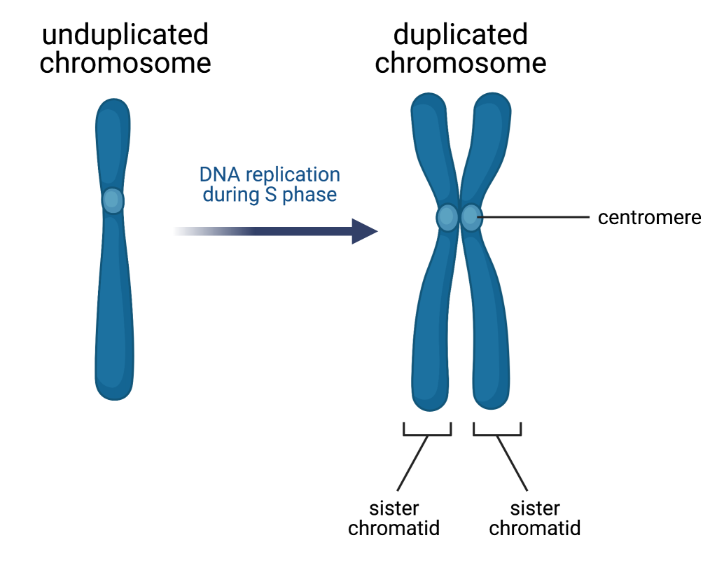
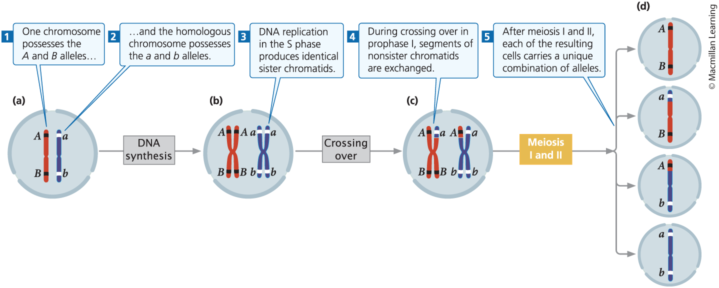
S Phase
DNA synthesis, the chromosome is replicated, resulting in two identical sister chromatids joined together
G2
Preparation for division, cell growths, and protein synthesis necessary for cell division; G2/M Checkpoint
G2/M Checkpoint
A regulated decision point in the cell cycle. only passed if DNA is completely replicated and undamaged
M Phase
Mitotic phase: includes mitosis and cytokinesis
Includes Prophase, Prometaphase, Metaphase, Anaphase, and Telophase
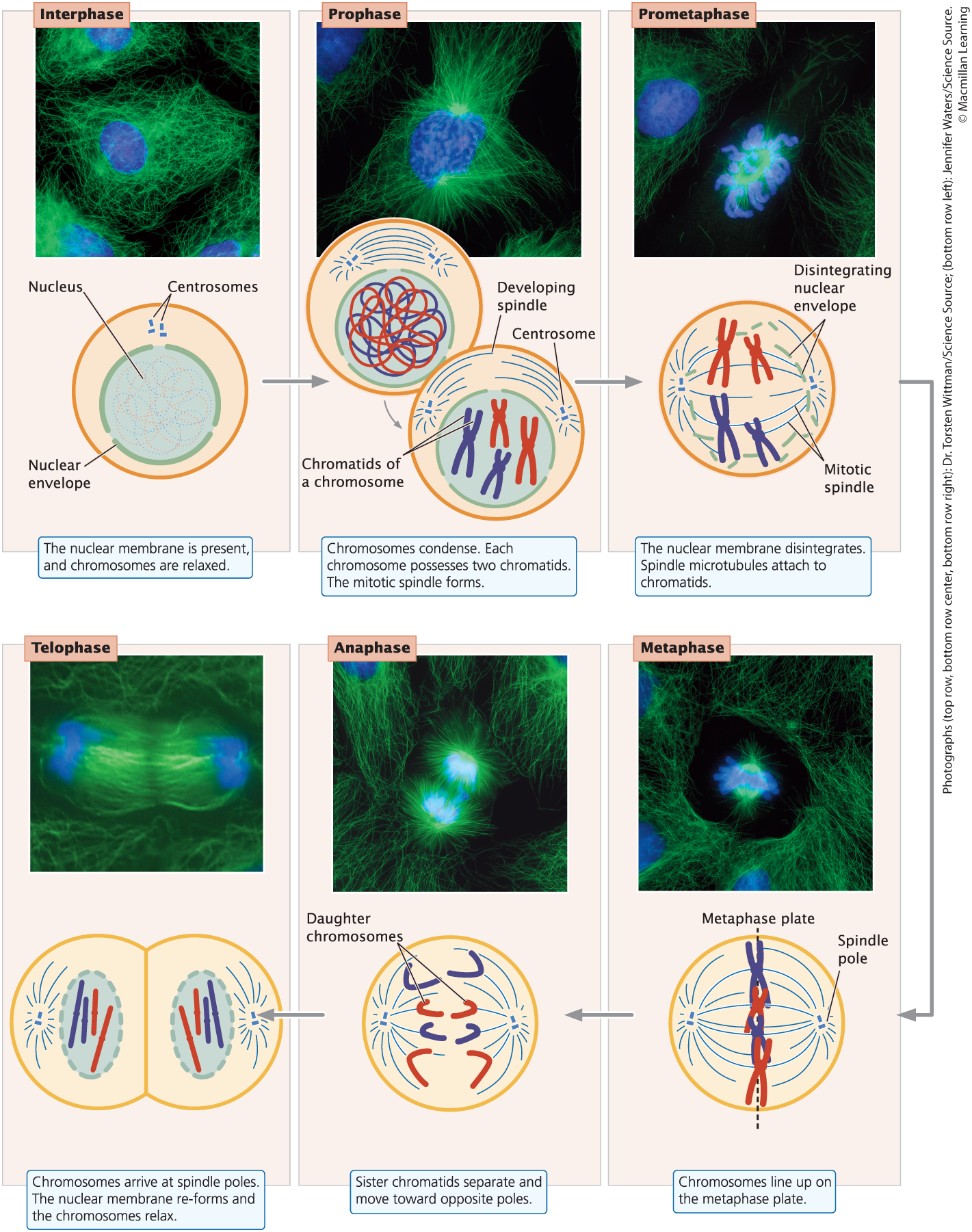
Prophase
Chromosomes condense and mitotic spindle forms
Prometaphase
Nuclear envelope disintegrates, and spindle microtubules anchor to kinetochores
Metaphase
Chromosomes align on the metaphase plate; spindle assembly checkpoint
Spindle-Assembly Checkpoint
Ensures that each chromosome is aligned on the metaphase plate and attached to spindle microtubules from opposite poles before anaphase
Anaphase
Early anaphase: sister chromatids separate, becoming individual chromosomes that migrate toward the poles.
Late anaphase: contractile ring forms (animal cells); cell plate begins to form (plant cells)
Telophase
Chromosomes arrive at spindle poles, the nuclear envelope reforms, and condensed chromosomes relax. Cleavage furrow forms and deepens (animal cells); cell plate becomes obvious and grows (plant cells)
Cytokinesis
Cytoplasm divides; animal cells split completely; cell plate/wall completes separating cells
Mitosis and Consequences of Cell Cycle
Process of cell division for growth and repair; separation of sister chromatids
Consequences:
Two genetically identical cells
Each cell with approximately have the cytoplasm as the parent
Cytokinesis
Separation of cytoplasm
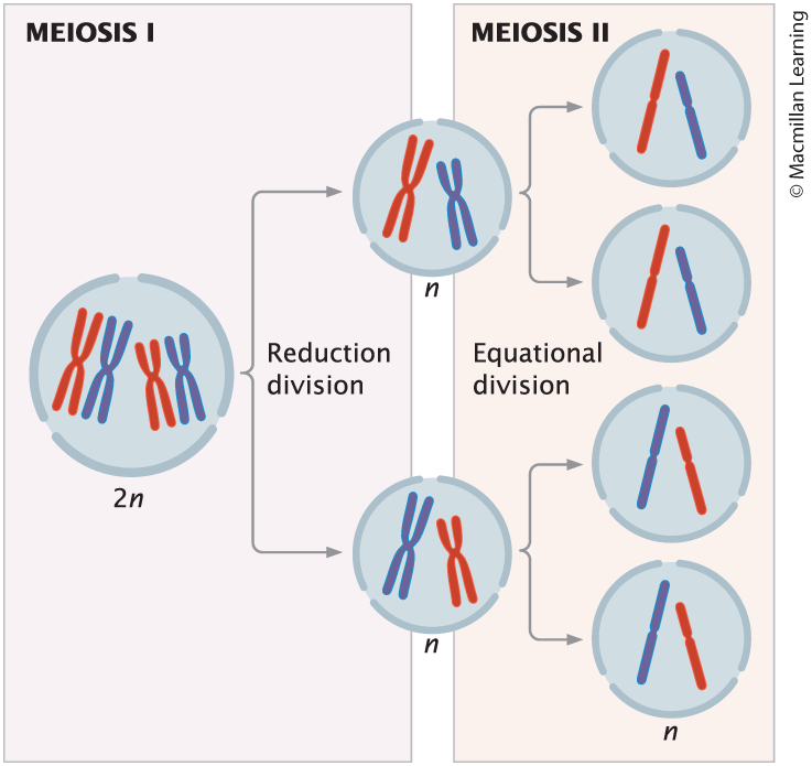
Meiosis and Consequences
Haploid gamete production; consists of meiosis I and meiosis II
Consequences:
Four haploid cells produced
Newly formed cells from meiosis are genetically different from one another and from the parental cell
What state precedes meiosis I but not meiosis II?
Interphase (same as before mitosis)
Fertilization
Fusion of Haploid Gametes
Meiosis I
separation of homologous chromosome pairs, and reduction of the chromosome number by half; reduction division (chromosomes number halves)
Prophase I, Metaphase I, Anaphase I, Telophase I, Cytokinesis, Interkinesis

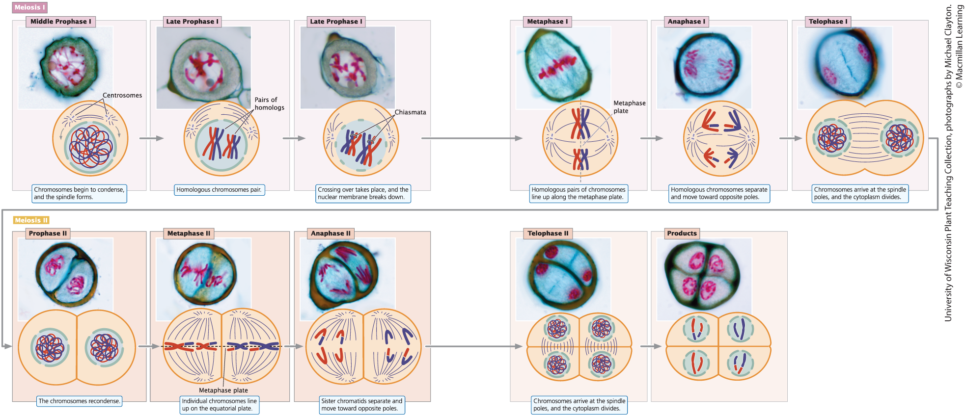
Prophase I
Chromsomes condense, homologous chromosomes synapse, crossing over takes place, the nuclear envelope breaks down, and the mitotic spindle forms
Synapsis
The close pairing of homologous chromosomes during Prophase I
Tetrad
The closely associated structure composed of four-sister chromatids of two homologous chromosomes during Prophase I
Crossing Over
The exchange of genetic information/chromosome segments between the sister chromatid of one chromosome and the sister chromatid of the other synapsed chromosome. This is the first mechanism of generating genetic variation in newly formed gametes.
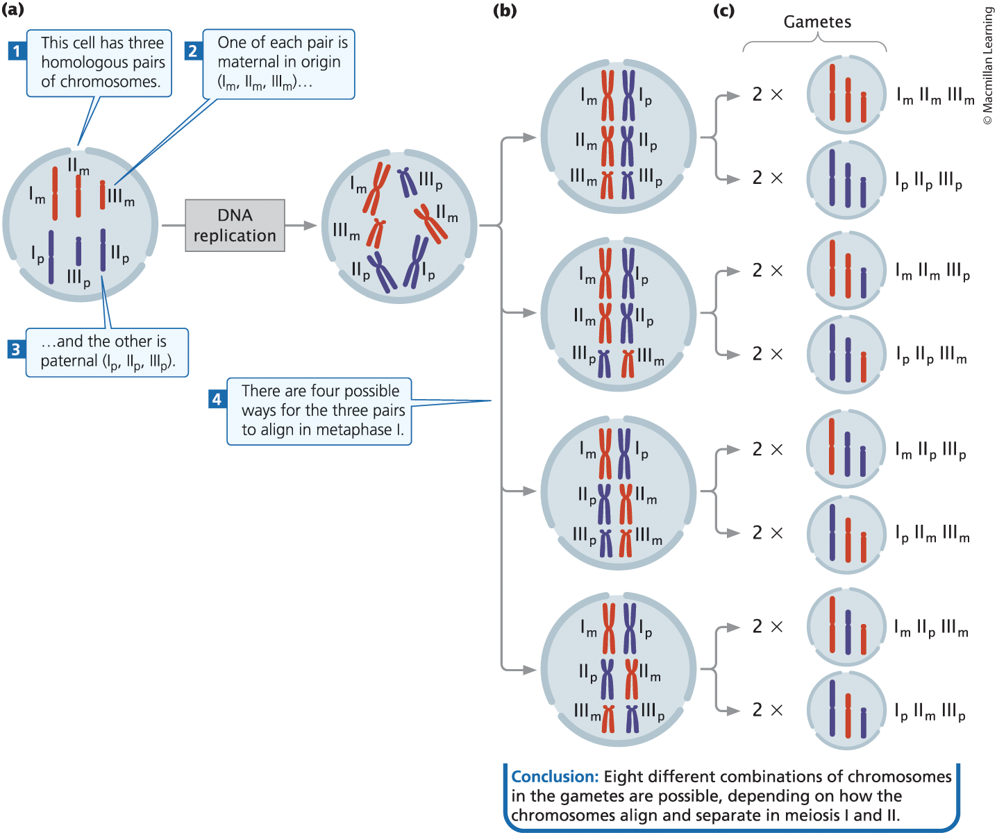
Metaphase I
Random alignment of homologous pairs of chromosomes along the metaphase plate
Anaphase I
Separation of homologous chromosome pairs and the random distribution of chromosomes into two newly divided cells. This is the second mechanism of generating genetic variation in the newly formed gametes.
Telophase I
chromosomes arrive at the spindle poles
Cytokinesis (Meiosis I)
cytoplasm is divided to produce two cells, each having half the original number of chromosomes
Interkinesis
In some type of cells, the spindle breaks down, chromosomes relax, and a nuclear envelope reforms, but no DNA synthesis takes place
Meiosis II
separation of sister chromatids, also known as equational division (chromosomes number remains equal)
Prophase II
Chromosomes condense, the spindle forms, and the nuclear envelope disintegrates
Only in cells in which the spindle has broken down after interkinesis, chromosomes have relaxed, and the nuclear envelope has reformed in telophase I. Other types of cells proceed directly to metaphase II after cytokinesis
Metaphase II
Individual chromosomes line up on the metaphase plate
Anaphase II
sister chromatids separate and move as individual chromosomes toward the spindle poles
Telophase II
Chromosomes arrive at the spindle poles; the spindle breaks down and nuclear envelope re-envelopes
Cohesin and Shugohisin
Cohesin: protein that holds the chromatids together and is key to the behavior of chromosomes in mitosis and meiosis
Shugohisin: protein that protects cohesin
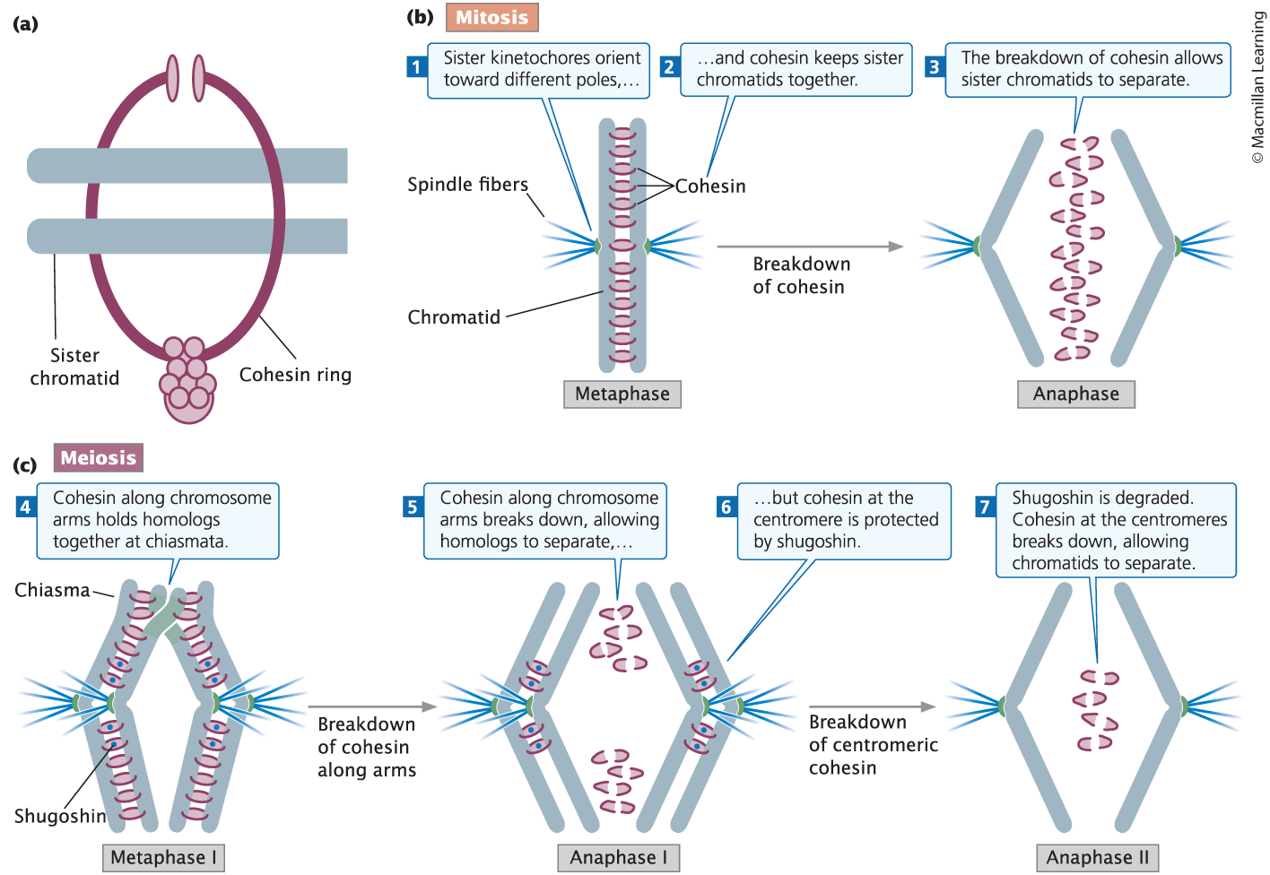
How does shugohisin affect sister chromatids in meiosis I and meiosis II?
In meiosis, cohesin is protected at the centromeres by shugohisin during anaphase I, so homologous chromosomes, but not sister chromatids, separate in meiosis I. The breakdown of shugohisin and thus centromeric cohesin allows sister chromatids to separate in anaphase II of meiosis.
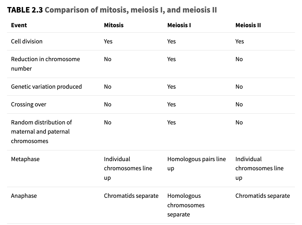
Compare Mitosis, meiosis I, and meiosis I.
Cell division, reduction in chromosome #, genetic variation produced, crossing over, random distribution of maternal and paternal chromosomes, metaphase, anaphase
Meiosis in animals
Spermatogenesis: male gamete production
Oogenesis: female gamete production
Spermatogenesis
The process of male gamete production

Spermatogonium
A diploid cell located in the testis that is capable of undergoing meiosis to produce sperm
Primary spermatocyte
A spermatogonium that has entered Prophase I, which is still diploid
Secondary spermatocyte
The haploid product of Meiosis I in male animals.
Spermatids
The immediate, haploid product of Meiosis II from secondary spermatocytes; these mature to become sperm.
Oogenesis
The process of female gamete production
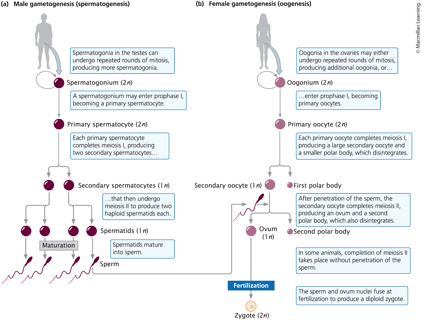
Oogonium
A diploid cell in the ovary capable of undergoing meiosis to produce an egg cell
Primary oocyte
An oogonium that has entered Prophase I
Secondary Oocyte
One of the products of Meiosis I in oogenesis; it is haploid and receives most of the cytoplasm; what fuses with sperm during fertilization
First Polar Body
One of the products of Meiosis I in oogenesis; it is the smaller cell, which is haploid and contains half the chromosomes but only a small part of the cytoplasm
Ovum
The mature female gamete; it is the larger cell that acquires most of the cytoplasm during oogenesis
Second Polar Body
One of the products of Meiosis II in oogenesis; it contains a set of chromosomes but little of the cytoplasm.
Meiosis in plants
one of the alternating life stages of plants
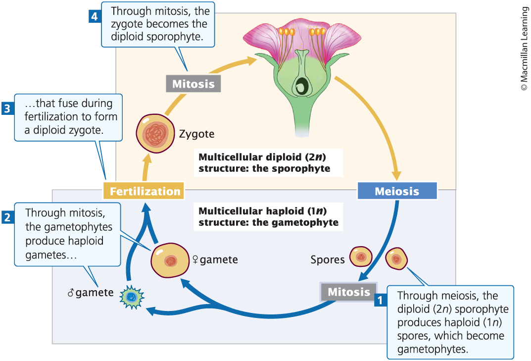
Microsporocyte
Diploid reproductive cell in the stamen of a plant; undergoes meiosis to produce four haploid microspores.
Microspore
Male haploid product of meiosis in plants.
Megasporocyte
Diploid reproductive cell in the ovary of a plant that undergoes meiosis to produce haploid megaspores.
Megaspores
Female haploid product of meiosis in plants.