Histology lab Quiz 1
 Call Kai
 Learn
Learn Practice Test
 Spaced Repetition
 Match
Match Flashcards
 Knowt Play
1/46

There's no tags or description
Looks like no tags are added yet.
Name | Mastery | Learn | Test | Matching | Spaced | Call with Kai | Chat |
|---|
No analytics yet
Send a link to your students to track their progress
47 Terms
Parenchyma
Functional cells of the tissue, make up the majority of a sample (blue)
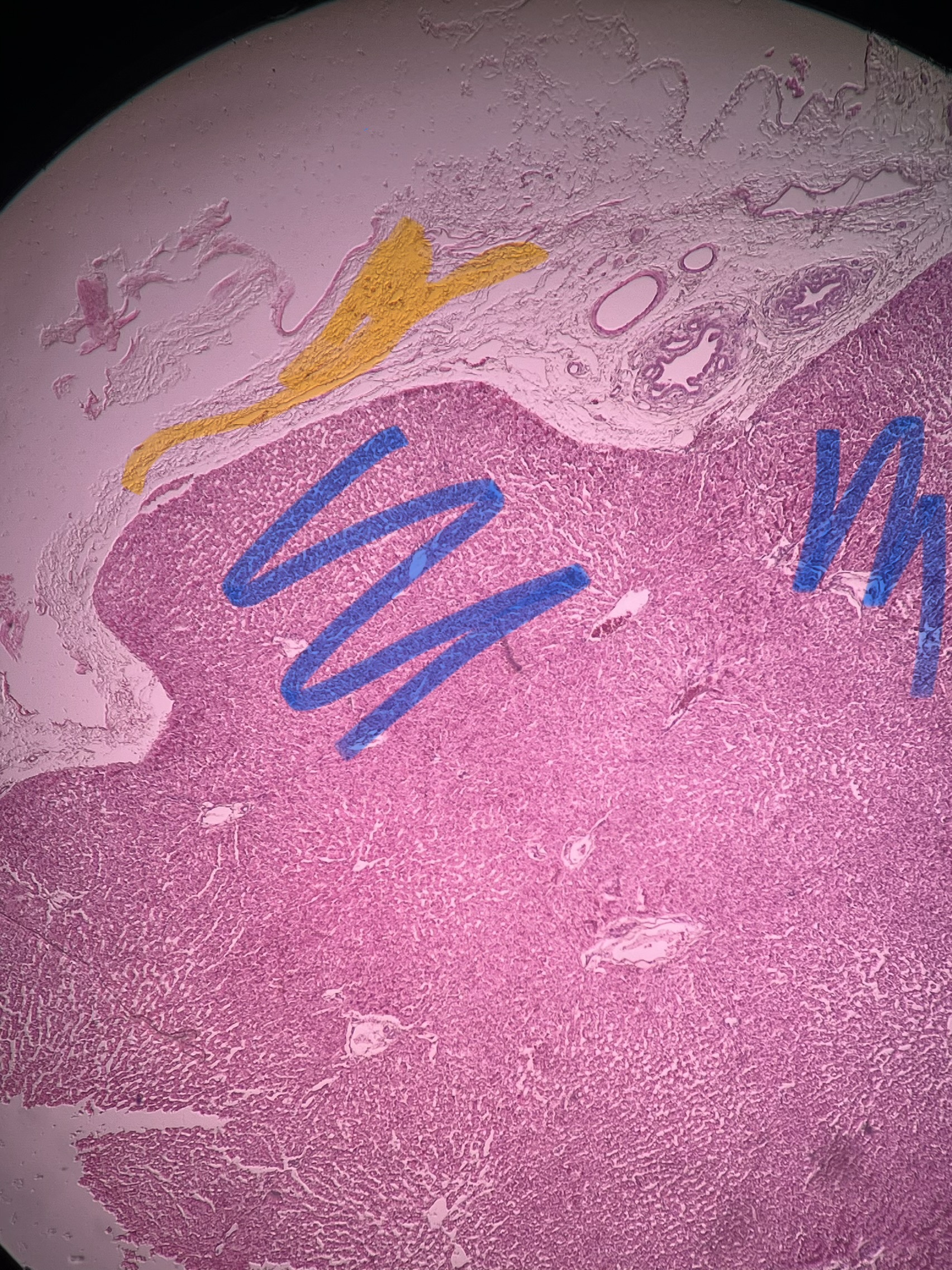
Stroma
Connective/structural cells of a tissue (yellow)

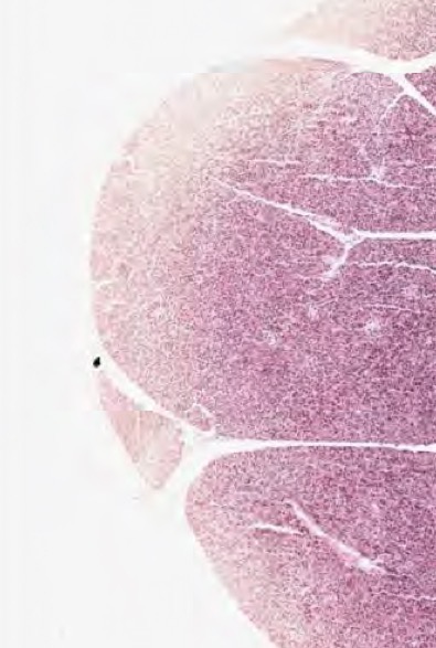
The edges of this sample are an example of what kind of artifact
Staining artifact
The edges of this sample are an example of what kind of artifact
Staining artifact
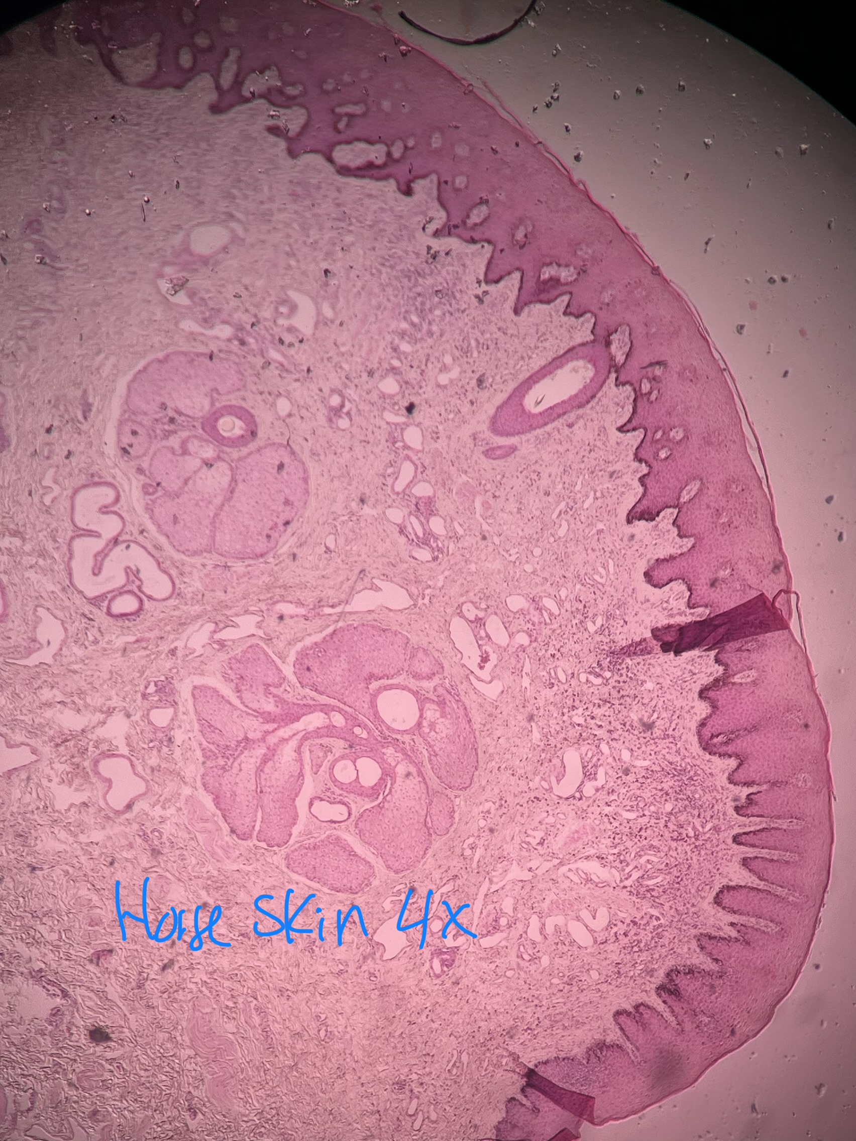
What artifact is present in this sample?
Folding artifact
Lobule
Grouping of tissue within an organ, typically separated by septa
Lobule
Grouping of tissue within an organ, typically separated by septa
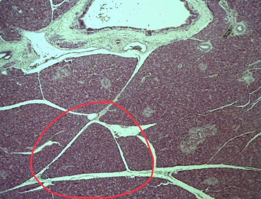
Septa (septum)
Category of stroma that separates lobules
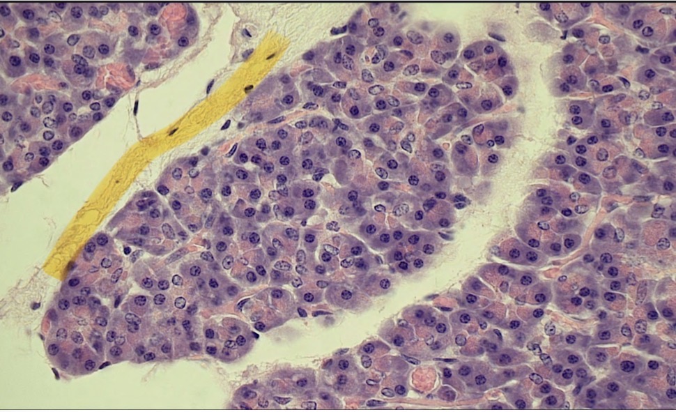

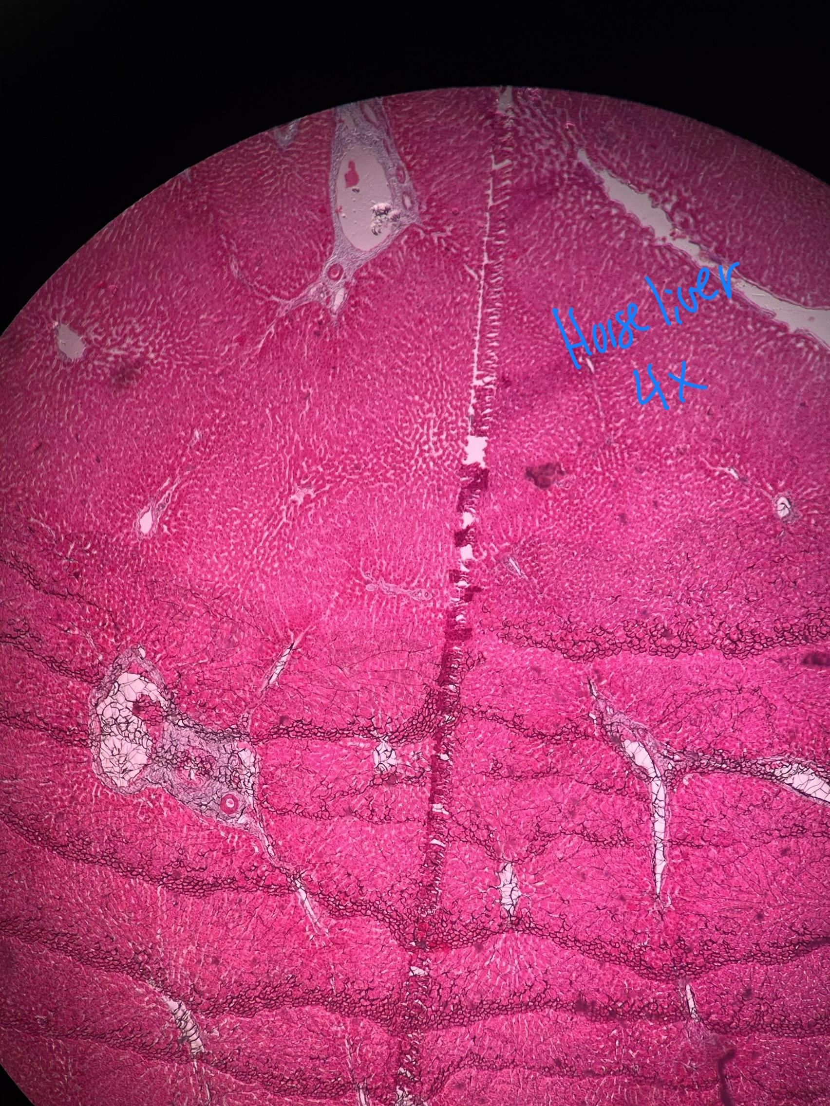
What artifact is present in this sample?
Shrinkage

What is circled in yellow?
Nucleolus

What is circled in green
Nucleus
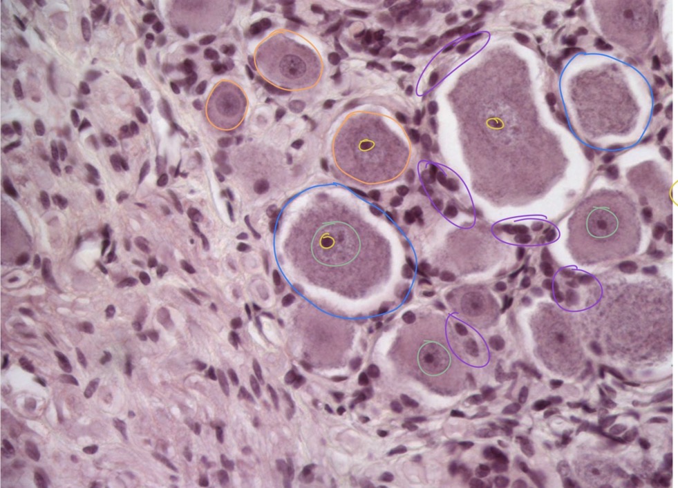
What is circled in blue
Cell membranes

What is circled in purple?
Satellite cells
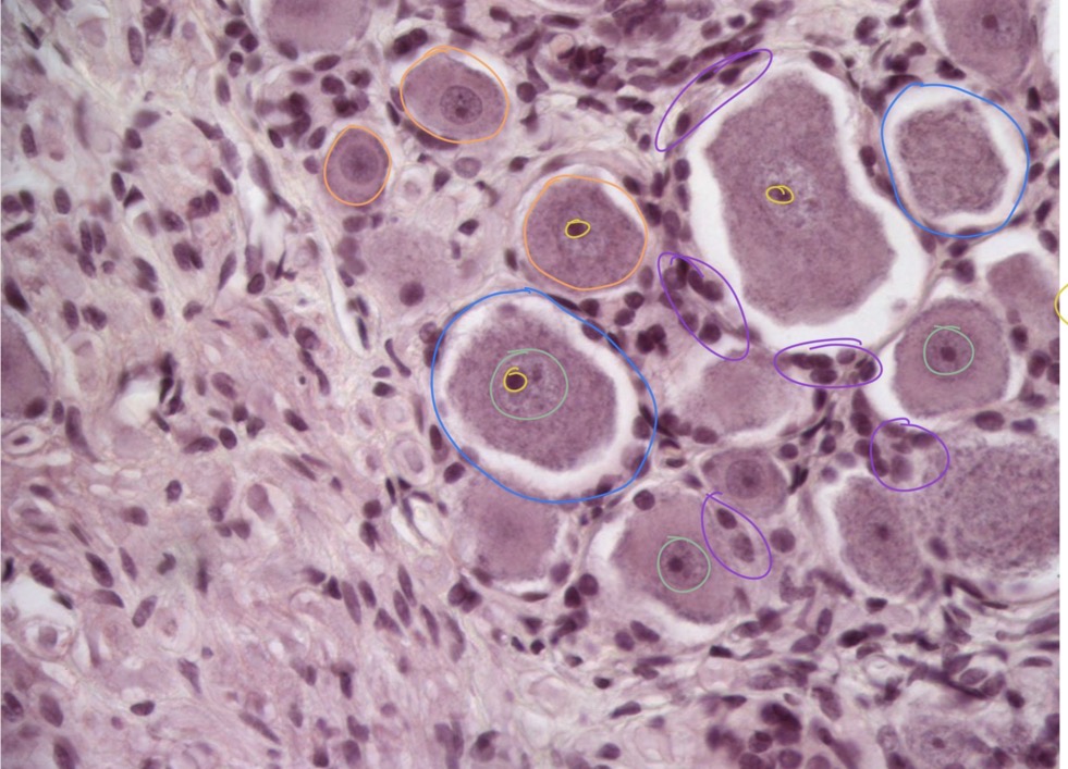
What is circled in orange?
Cytoplasms

What does the stain pattern of the cytoplasm tell you in this image?
This cell has a lot of rough endoplasmic reticulum and is making a lot of protein (Nissan Substance)

What is circled in green?
Closed faced nuclei
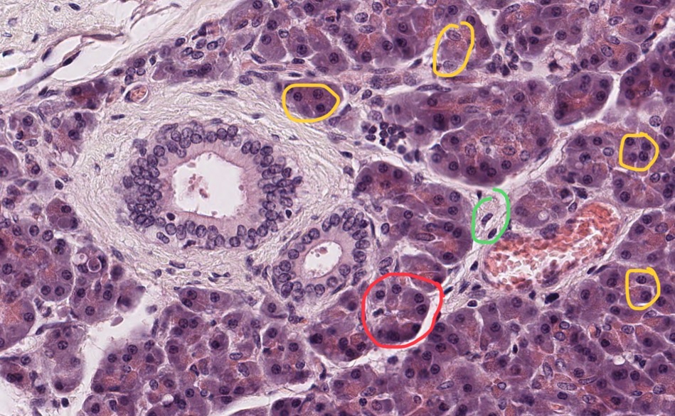
What is circled in red?
Acini

What is circled in yellow?
Open face nuclei
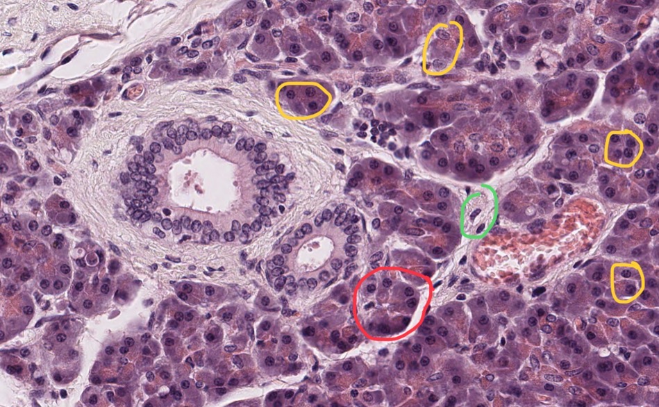
What cell inclusion can be found within the red circle?
Zymogen granules
What is a cell inclusion?
Something that the cell does not need to live but often is indicative classification and or pathology

Blue indicates the ____ side of the structure
Apical

Green indicates the ____ side of the structure
Basal
Acini
Specific cell arrangement that appears as pointed cells arranged in a circle, found commonly in the pancreas

The dark staining of this image represents a tissue that is _____ and is stained by a _____ stain
Acidic basophilic
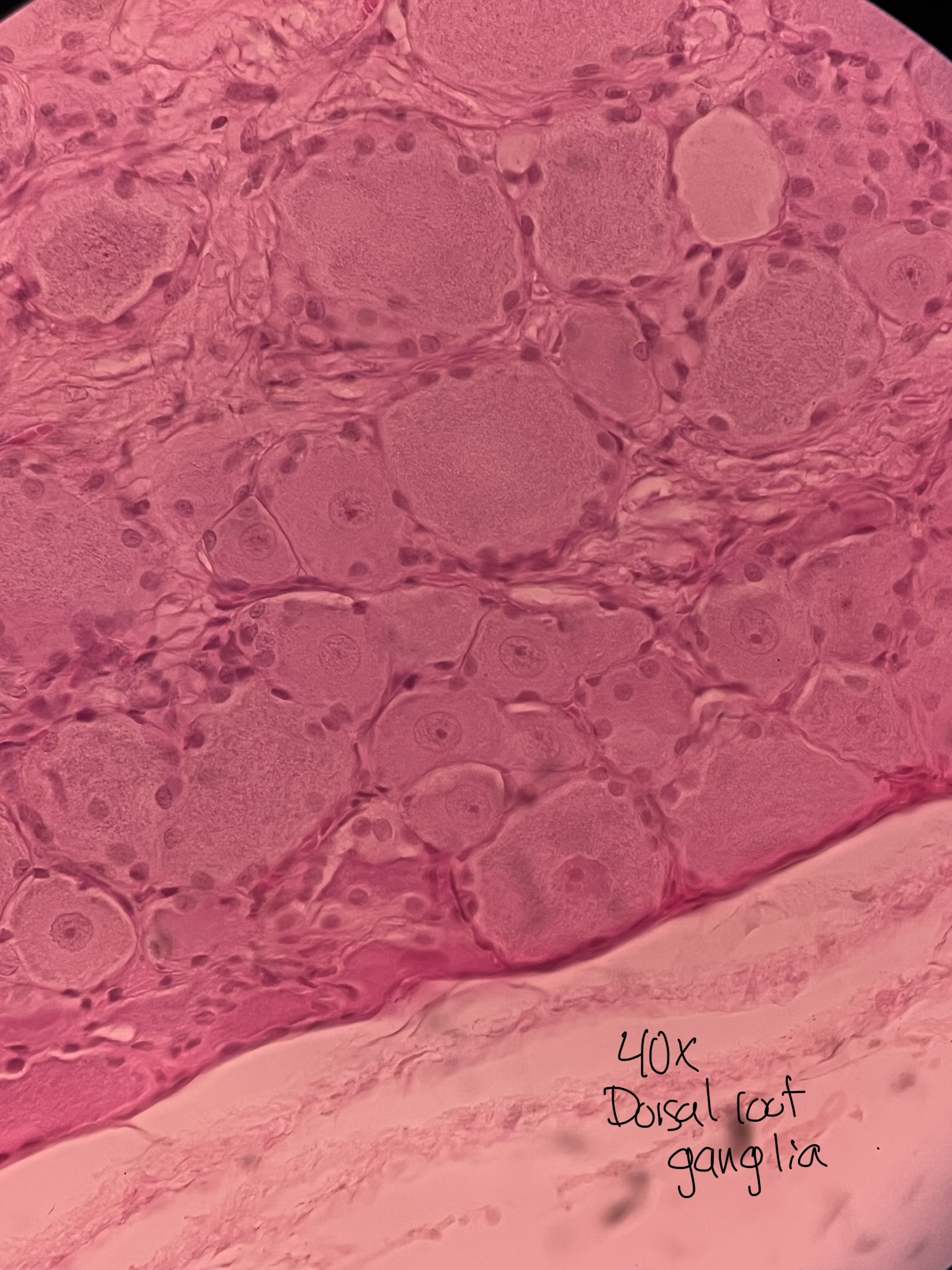
The lighter staining of this image represents a tissue that is _____ and is stained by a _____ stain
Basic acidophilic (eosinophilic)
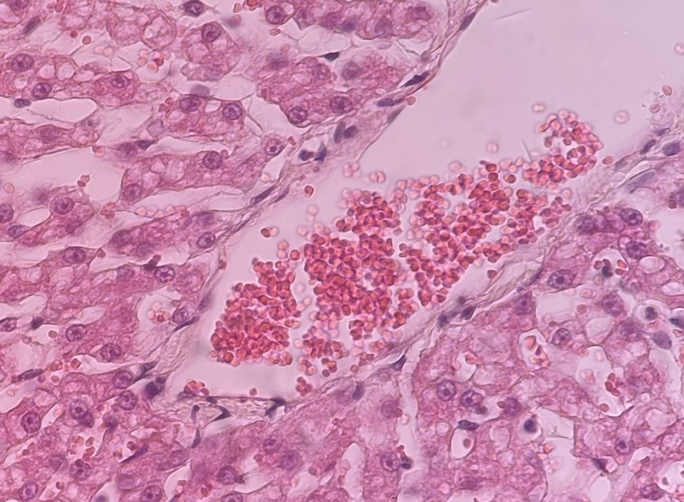
The cells stained brightest pink/red here are _____ and are great indicators on how well a eosinophilic stain is uptaking in a sample.
Erythrocytes (red blood cells)
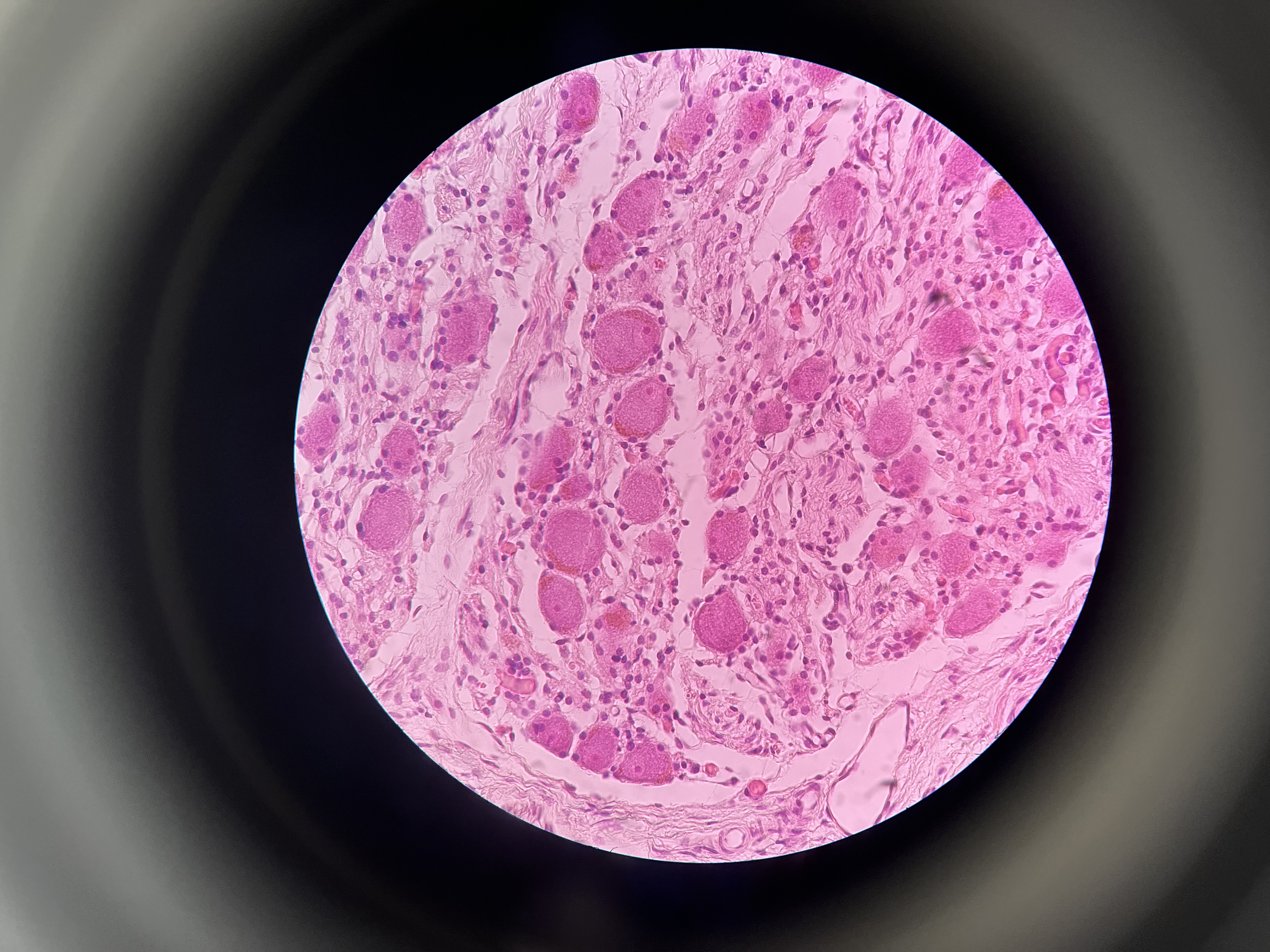
Why do some cells here present without a nucleus?
They have a nucleus that didn’t get grouped with the sample during preparation
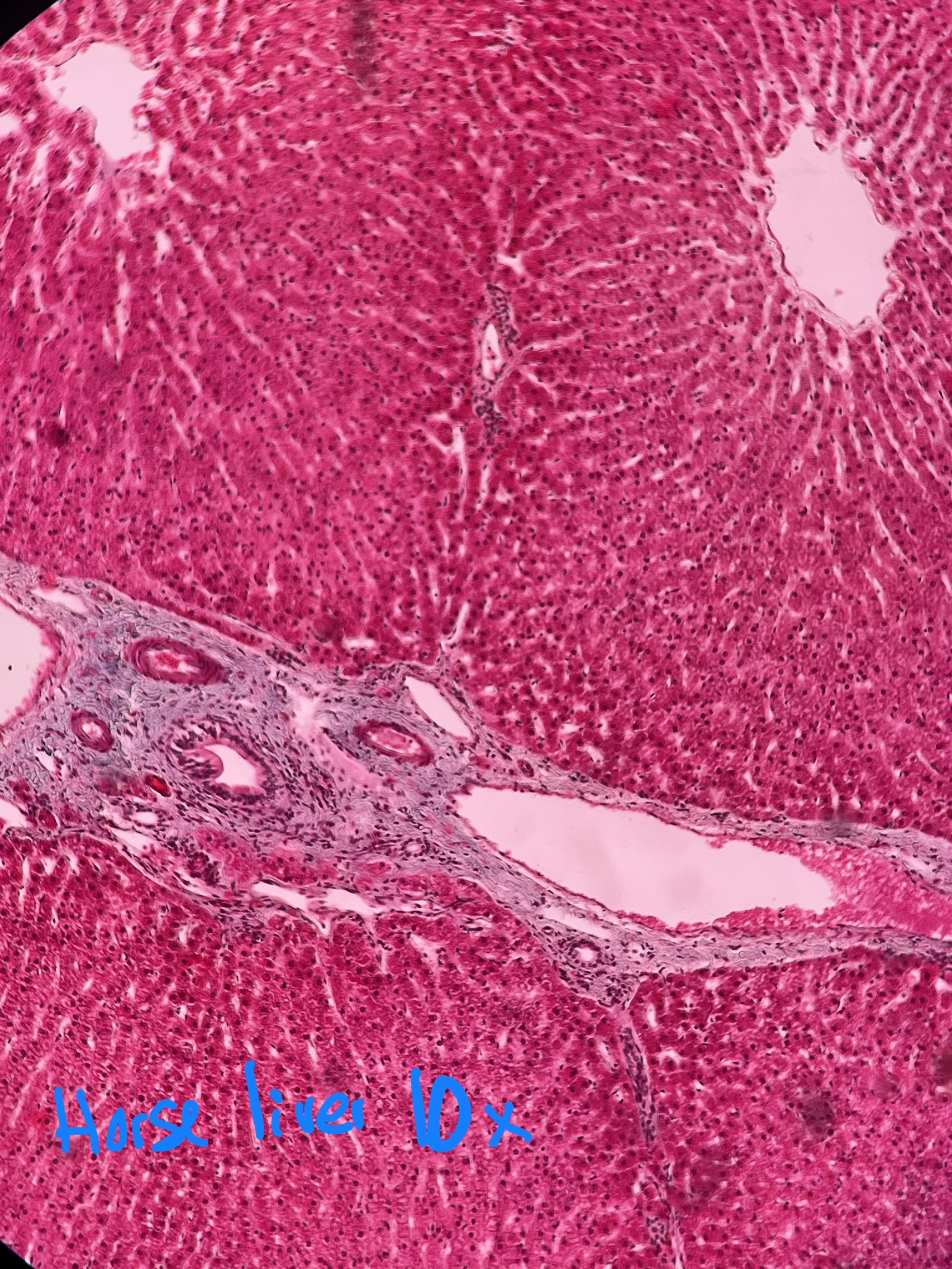
What is the white area between cells in this sample?
Modified blood vessels
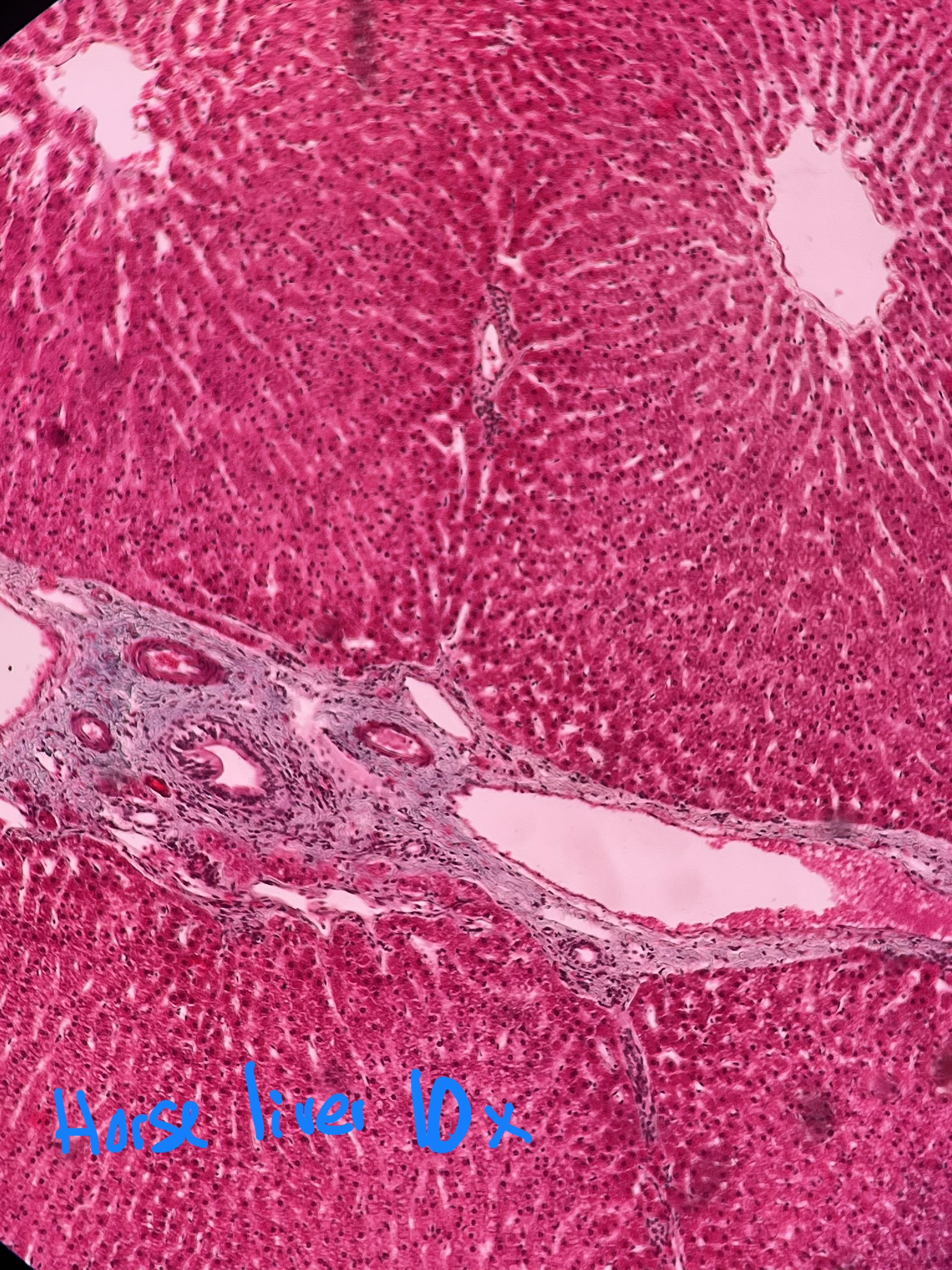
What kind of stain is present on this sample?
Masson Trichrome stain
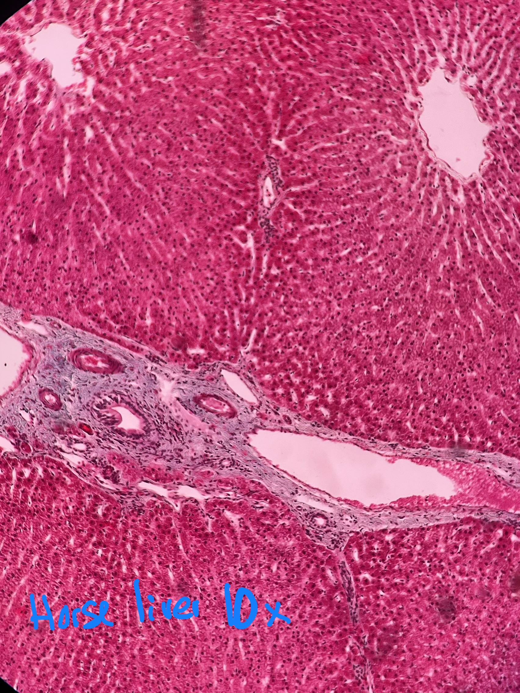
What kind of cell arrangement is present here?
Plate or cord
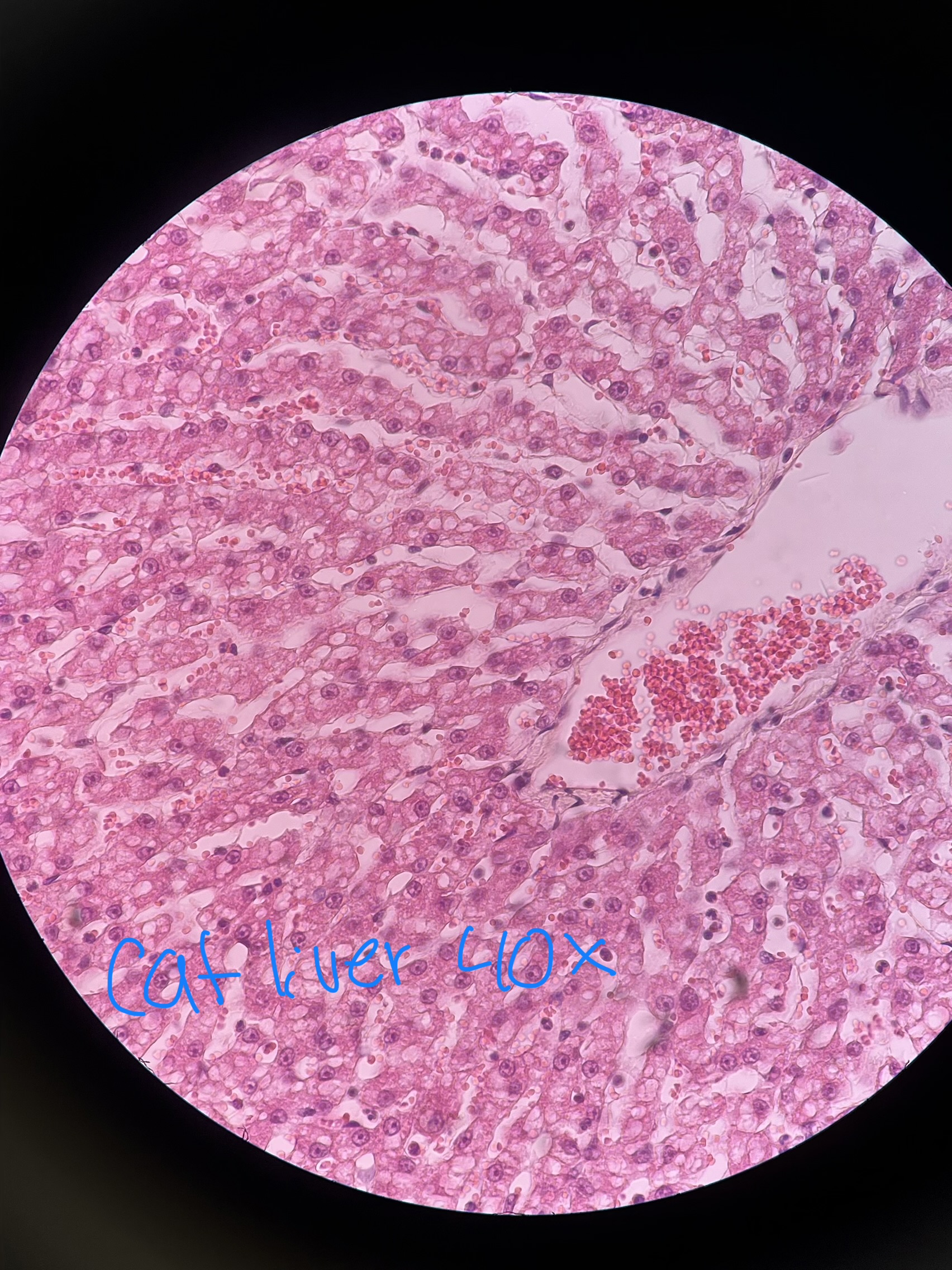
What cell inclusion is in this sample and why does it indicate?
Glycogen and lipid; Obese patient

What structure is circled here?
Neuronal cell body
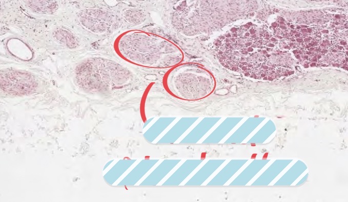
What structure is circled here?
Neuronal fiber bundle
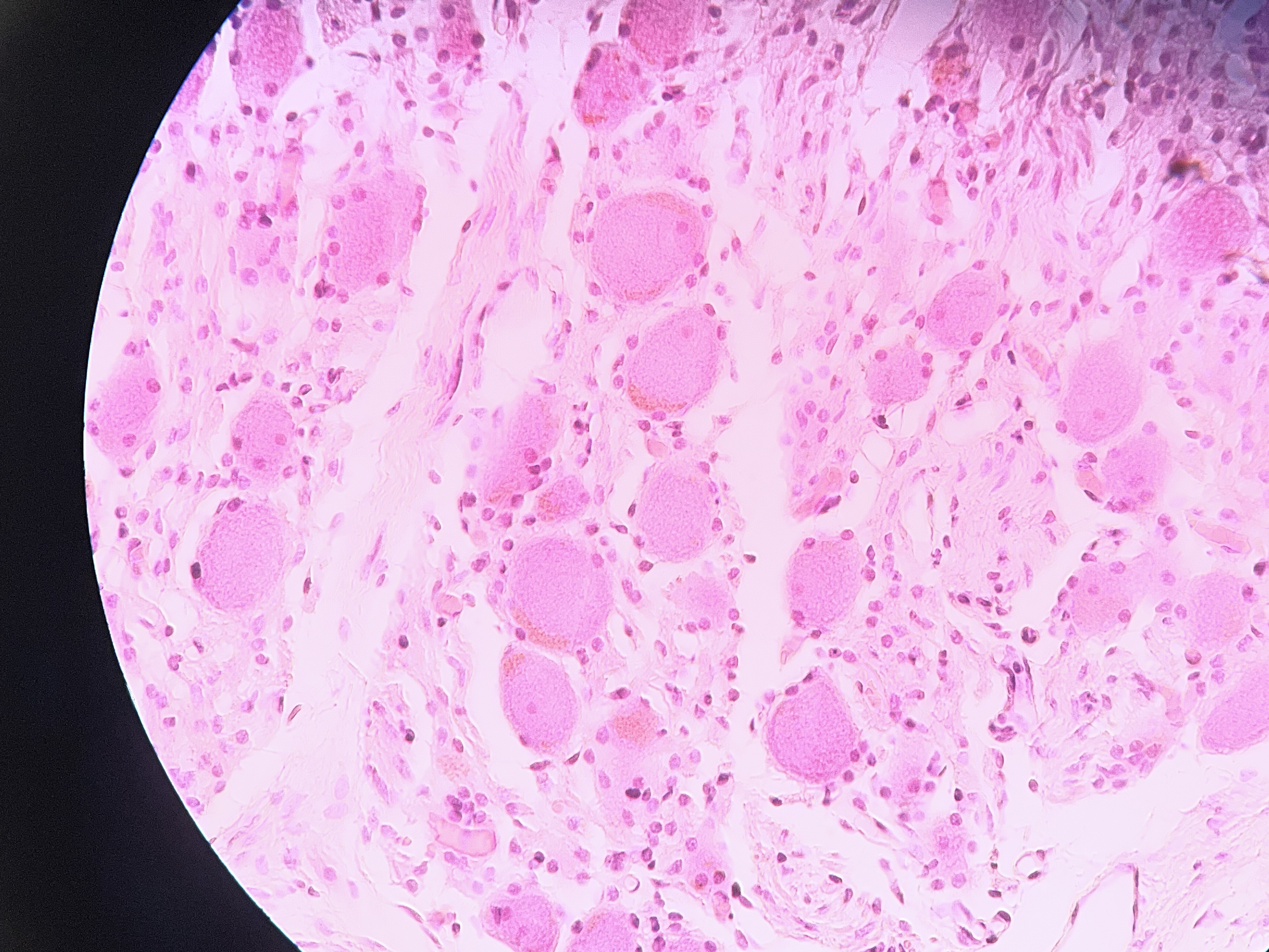
This sample shows evidence of what cell inclusion that is indicative of old age and or large stressors within the animals life?
Lipofuscin
Lipofuscin
Pigmented cell inclusion that is an indication of old age or large stressors within
Cell attempts to fuse items with a lysosome that fails and cell simply stores it in the corner
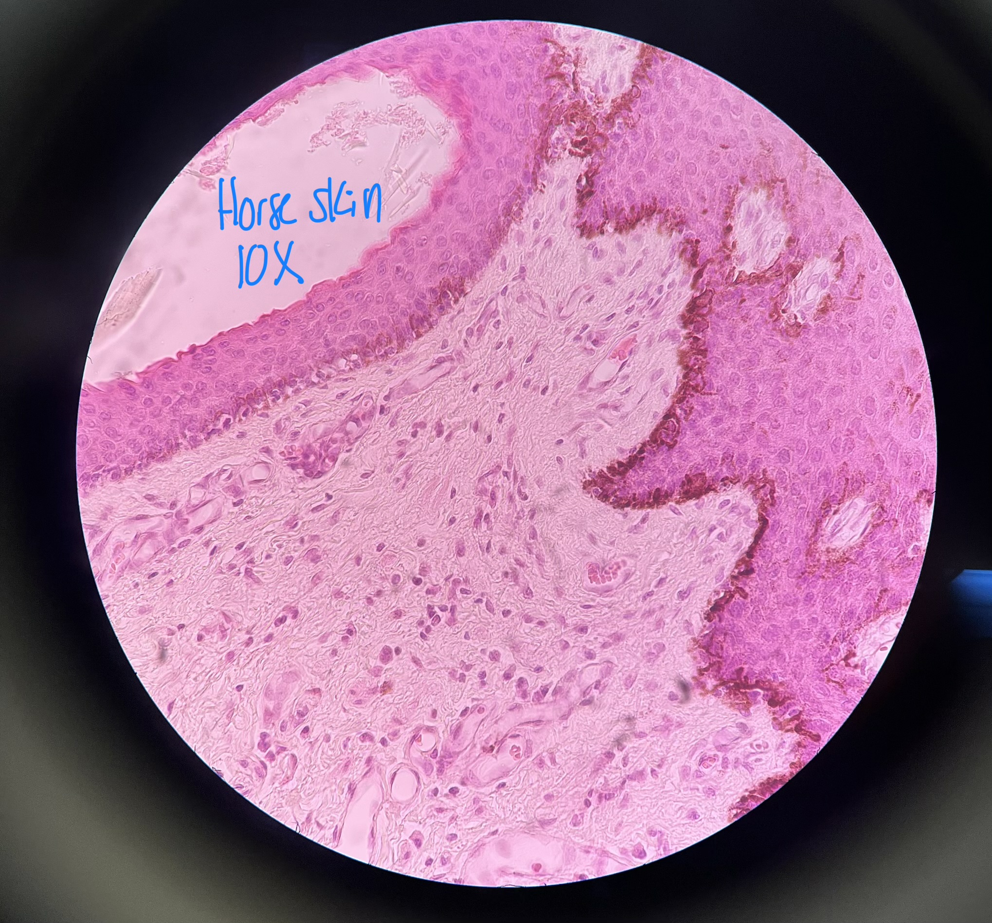
What cell inclusion is shown on the basement membrane of the dermis?
Melanin
Melanin
Pigment produced naturally by melanocytes in the body in response to UV DNA damage

Melanin tends to concentrate towards the ____ side of cells in order to maximize protective effects
Apical

What structures are circled here?
Uterine glands
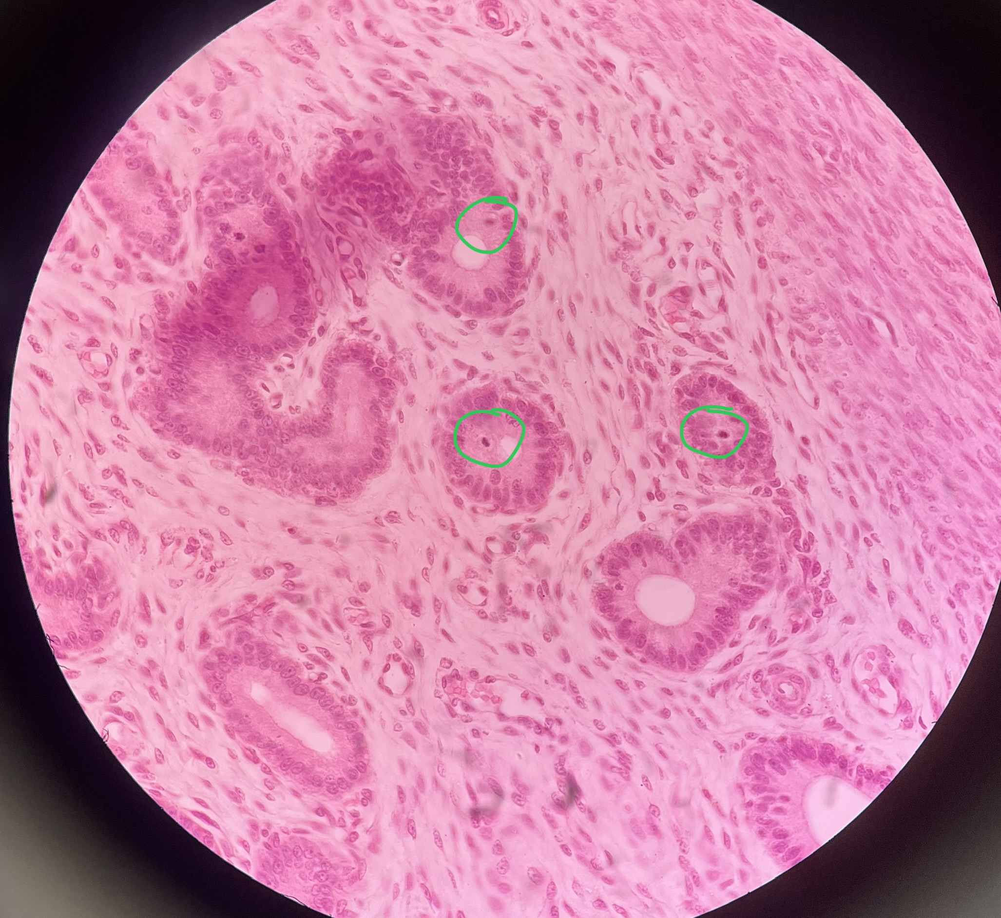
What cell inclusion is circled here?
Mitotic figures

What stage of mitosis is this cell in (green circle)
Anaphase
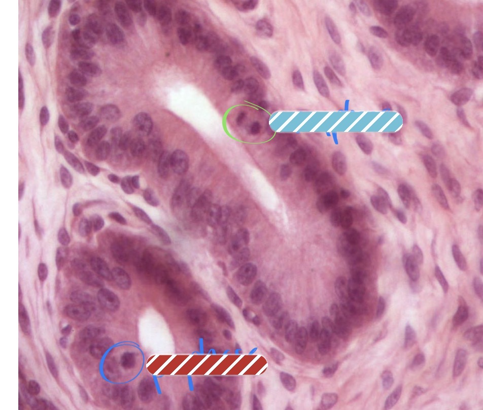
What stage of mitosis is this cell in (blue circle)
Prophase

What stage of mitosis is this cell in?
Metaphase

What stage of mitosis is this cell in?
Telophase
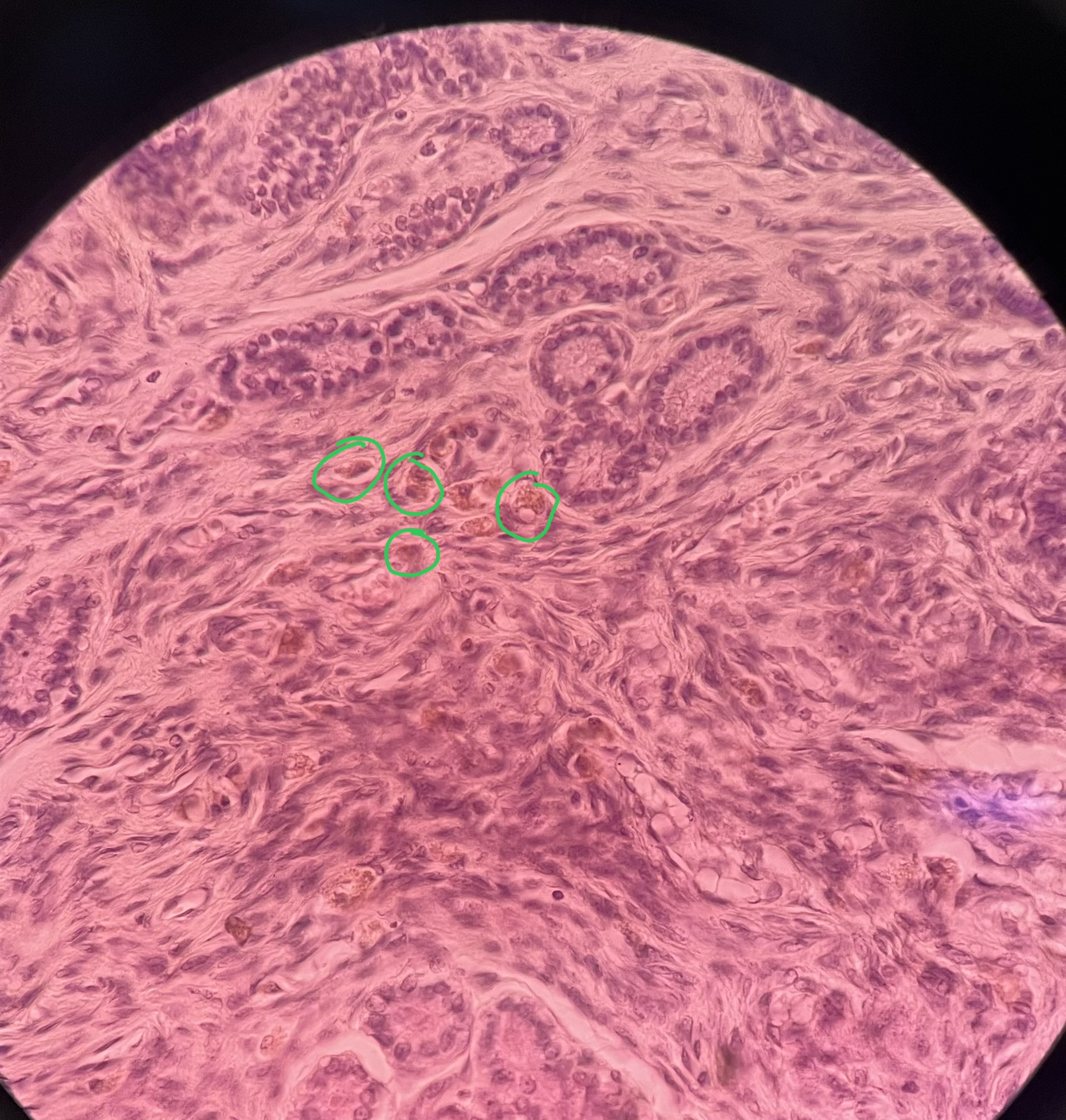
What cell inclusion is circled?
Hemosiderin
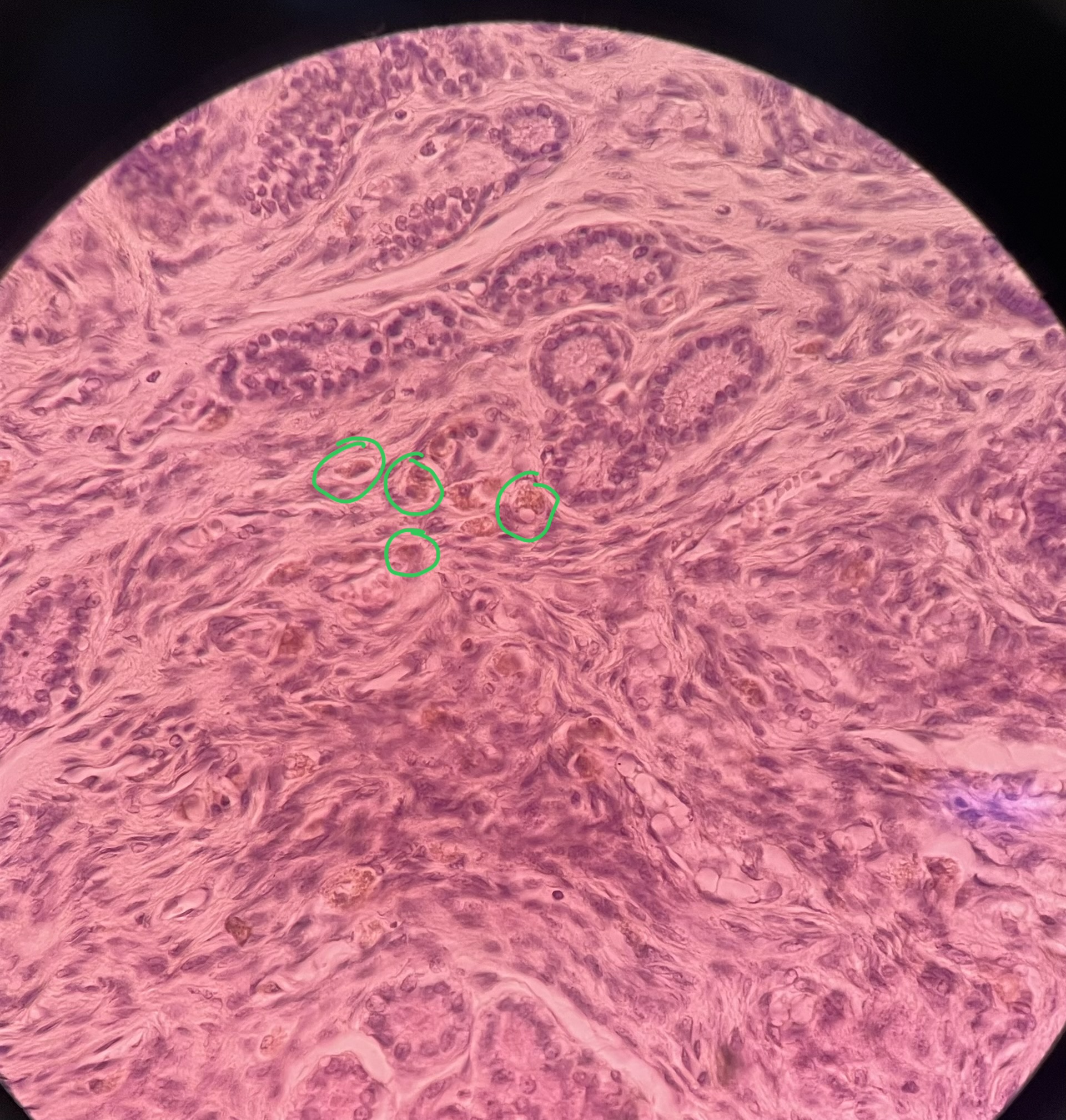
What cell type is circled here?
Macrophage
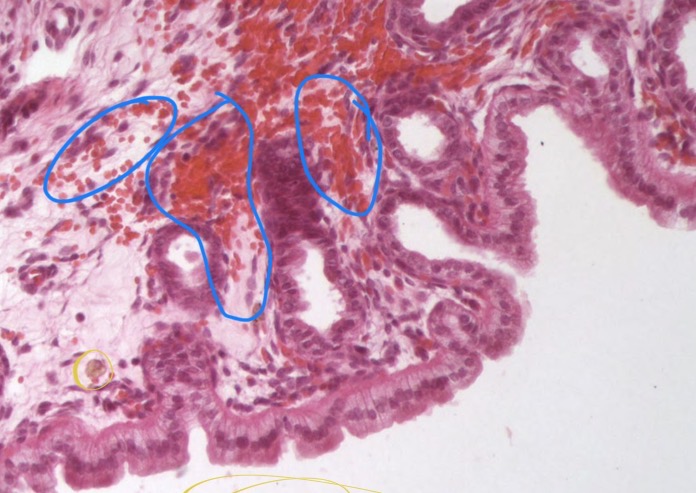
What is circled in this image (blue)?
Hemorrhaged red blood cells