Lab #2 - Dry Lab: Blood Supply & Brodmann's Areas
1/19
There's no tags or description
Looks like no tags are added yet.
Name | Mastery | Learn | Test | Matching | Spaced | Call with Kai | Chat |
|---|
No analytics yet
Send a link to your students to track their progress
20 Terms
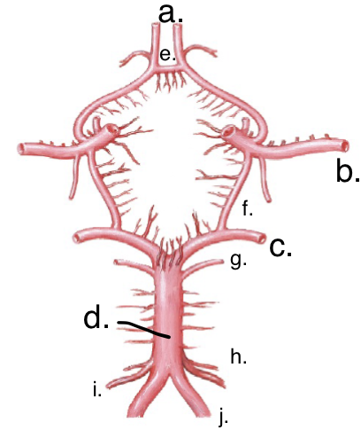
Label the cricle of willis
a. Anterior Cerebral Arteries (ACA)
b. Middle Cerebral Arteries (MCA)
c. Posterior Cerebral Arteries (PCA)
d. Basilar Artery (BA)
e. Anterior communicating artery
f. Posterior communicating artery
g. Superior cerebellar arteries (SCA)
h. Anterior Inferior Cerebellar Arteries (AICA)
i. Posterior Inferior Cerebellar Arteries (PICA)
j. Vertebral Arteries (VA)
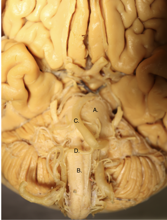
Identify these 2 structures and 2 arteries
a. pons
b. medulla
c. Basilar Artery
d. Vertebral Arteries

What artery supplies the blue region highlighted on the brain?
The ACA supplies the anterior region of the brain
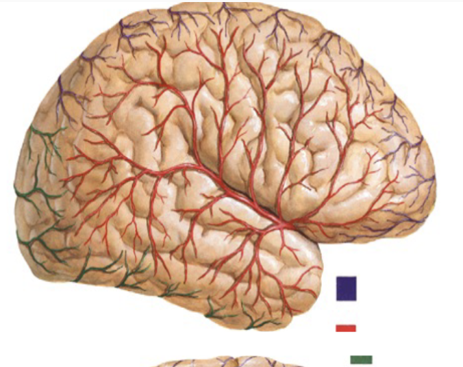
What artery supplies the red region highlighted on the brain
The MCA supplies the medial region of the brain
Lateral Frontal and Parietal Lobe

What artery supplies the green region of the brain
the PCA supplies the posterior region of the brain

What does the blue region represent
the ACA

What does the red region represent
the MCA
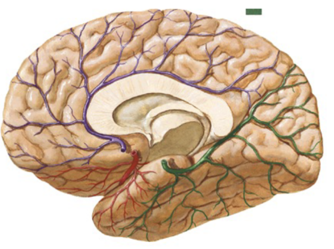
What does the green region represent?
the PCA

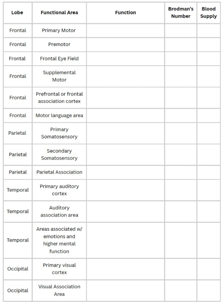
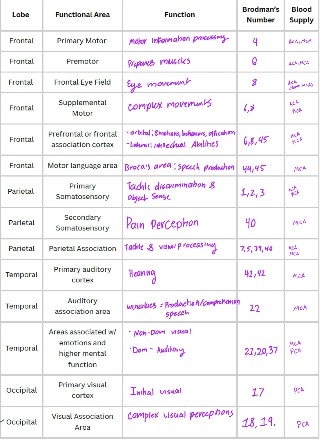
Identify the Brodmann’s areas for the frontal lobe
4 - Primary motor cortex
6 - Premotor cortex
8 - Frontal eye field
6 & 8 = Supplemental Motor
6, 8, 45 = Prefrontal/frontal association cortex
44/45- Broca’s Area

Identify the Brodmann’s Area for the parietal lobe
3,1,2 - Primary Somatosensory Cortex
40 - Secondary Somatosensory
7, 5, 39, 40 - Pariental Association

Identify the Brodmann’s Area for the Occipital Lobe
17 - Primary visual cortex
18, 19 - Visual Association Area
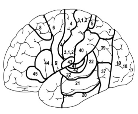
Identify the Brodmann’s Areas for the Temporal Lobe
41, 42 - Primary hearing cortex
22 - Auditory Association Area (Winerkies)
21, 20, 37 - Areas associated w/higher emotions and higher mental function
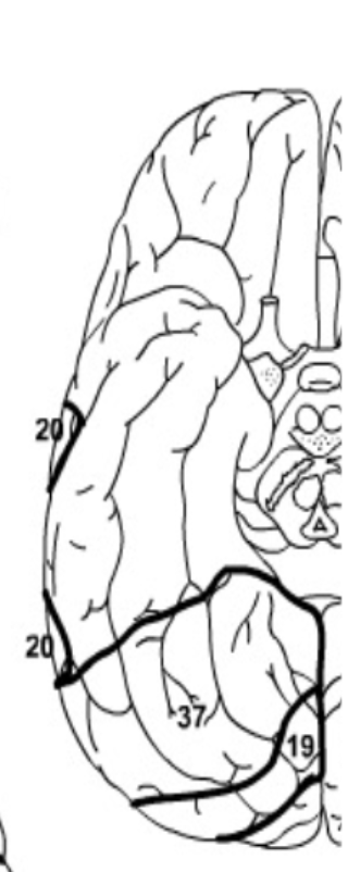
What Brodmann’s Areas can you see from this view?
20, 37 - Temporal lobe = Areas associated with higher emotions and higher mental functions
19 - Visual Association Area
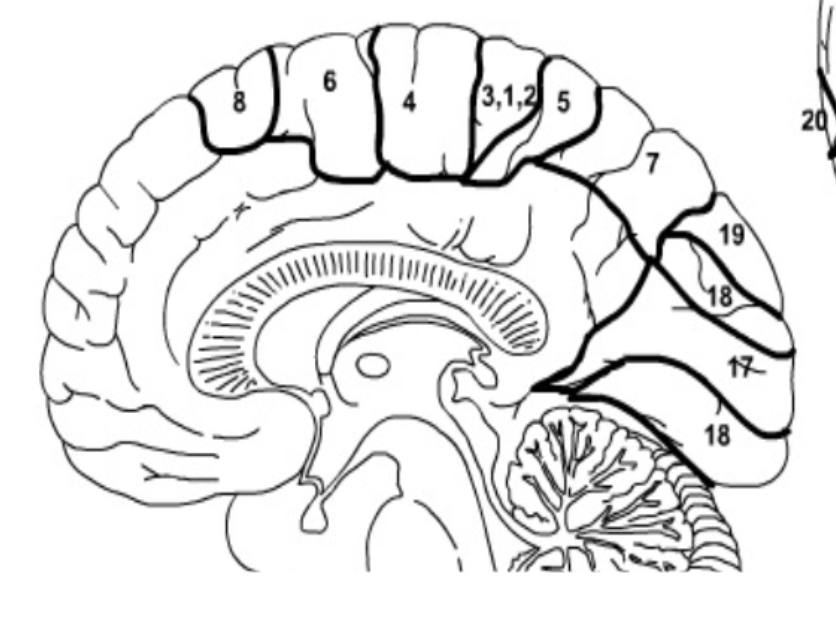
What Brodmann’s Areas can you see from this view?
8 - Frontal eye field
6 - Premotor
6, 8 = Supplemental Motor
4 - Primary Motor
3,1,2 - Primary Somatosensory
5,7 = Aspects of the Parietal Association region
19,18 = Visual Association Area
17 = Primary Visual Cortex
Fill this out:
Name the arteries in the anterior distribution of the Circle of Willis that we have identified in class.
Anterior Cerebral Arteries (2)
Anterior Communicating Artery
Medial Cerebral Arteries (2)
½ of the posterior communicating arteries (2)
Name the arteries in the posterior distribution of the Circle of Willis that we have identified in class.
½ Posterior communicating arteries (2)
Posterior Cerebral Arteries
Superior Cerebellar Arteries (2)
Basilar Artery
Anterior Inferior Cerebellar Arteries (AICA) (2)
Posterior Inferior Cerebellar Arteries (PICA) (2)
Vertebral Arteries (2)
List all the arteries responsible for providing blood to the following structures:
a. Frontal lobe
b. Parietal lobe
c. Temporal lobe
d. Occipital lobe
e. Cerebellum
a. = ACA & MCA
b. = ACA, MCA, & PCA
c. = PCA & MCA
d. = PCA, MCA
e. = Superior Cerebellar Arteries, AICA, & PICA
Name the cerebral artery responsible for providing blood to each of the following Brodmann’s areas
a. 44,45
b. 22
c. 17
d. 8
a. MCA
b. MCA
c. PCA
d. ACA, MCA
Name the associated functional deficits that may occur with a CVA impacting the Brodmann’s areas listed above.
If a CVA occurred that impacted the areas above I would expect to see difficulty with speech production (44,45), language comprehension (22), vision (17), and eye movement (8)