Measurement of Hearing Quiz 2 - Otoscopy
 Call Kai
 Learn
Learn Practice Test
 Spaced Repetition
 Match
Match Flashcards
 Knowt Play
1/146

There's no tags or description
Looks like no tags are added yet.
Name | Mastery | Learn | Test | Matching | Spaced | Call with Kai | Chat |
|---|
No analytics yet
Send a link to your students to track their progress
147 Terms

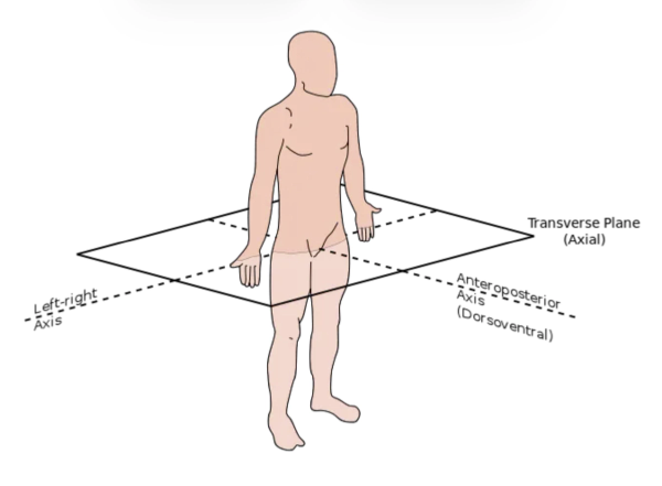
Direction
Anterior

Direction
Posterior

Direction
Inferior

direction
lateral
medial anatomical position

dorsal anatomical position

superior anatomical position

ventral anatomical position

coronal anatomical section

sagittal anatomical section

Transverse anatomical sections
peripheral system
-outer ear
-middle ear
-inner ear
-auditory nerve
proximal or cochlear end of the 8th cranial nerve
Central system
-Eighth cranial nerve/vestibulocochlear nerve
Distal/brainstem end
-Auditory brainstem nuclei (central)
-Auditory cortex (central)
What do systems do audiologists assess?
peripheral and central auditory system function and more!
Test battery goals
-Diagnose HL and, if HL is present, determine its degree, configuration, and type, and aid in determining a site of lesion and pathology
Primary audiology tests
-case history
-otoscopy
-immittance
tymps, acoustic reflexes, reflex decay
-Pure tone audiometry
air and bone conduction
-Speech testing
SRT and word rec
-Other physiological and behavioral measures
OAEs, stenger and tuning fork tests
Where is the bulk of the peripheral auditory system housed?
within the temporal bone

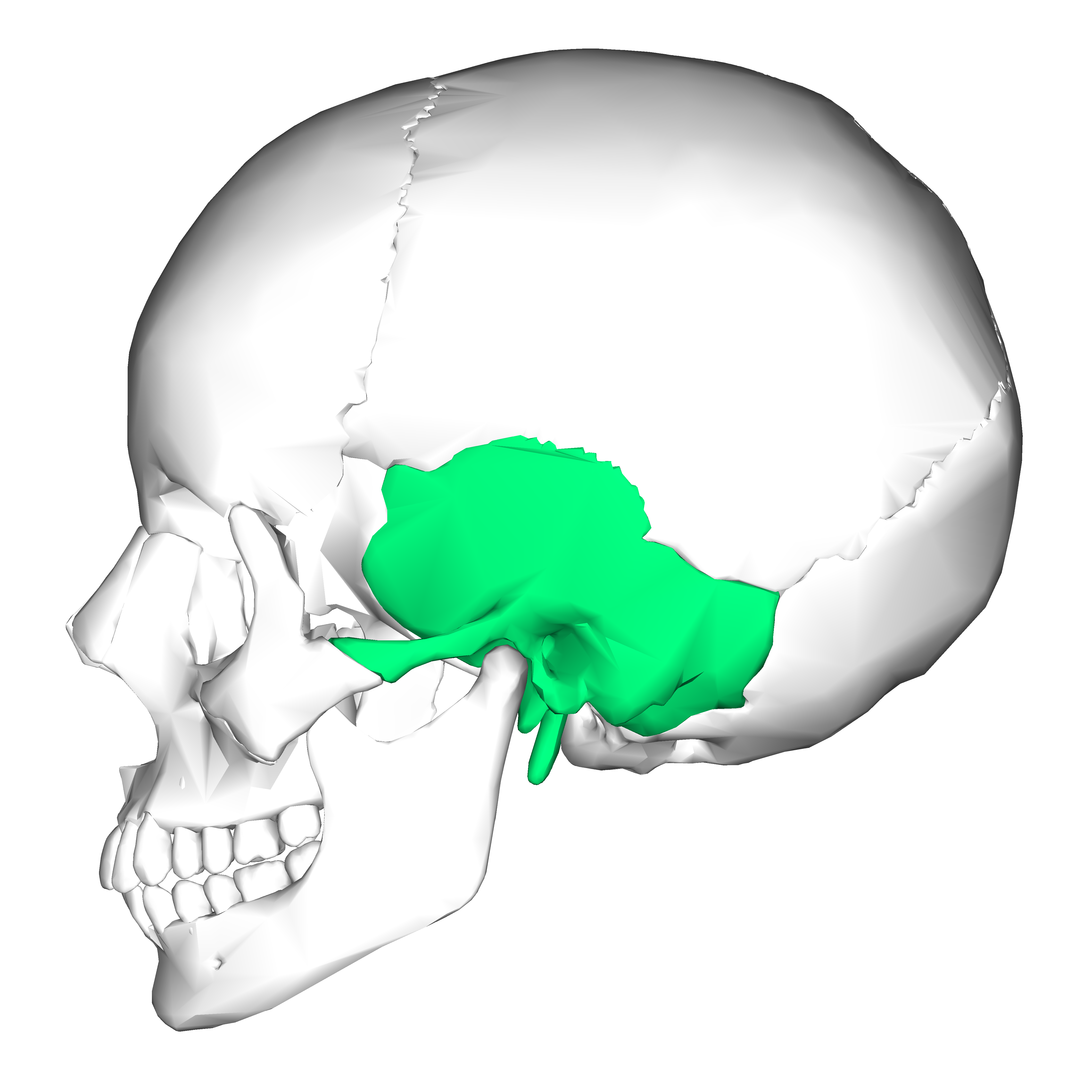
temporal bone
5 sections of the temporal bone
-squamous
-mastoid
-tympanic part/portion
-styloid process
-petrous (pyramid)

squamous portion of temporal bone


mastoid portion of temporal bone

mastoid
-forms portion of EAC
-used in BC testing
-honeycombed
prone to infection
tympanic part/portion of temporal bone


styloid process

Styloid process
attachment point for tongue and larynx muscles
Petrous pyramid
-very hard
-contains inner ear and internal auditory canal

petrous pyramid portion

outer ear consists of…
-pinna (auricle)
-External auditory meatus (ear canal)
-tympanic membrane?
Construction of the pinna
-primarily constructed from elastic cartilage
also contains some muscle tissues (most are non functioning)
-Fat pad in the lobule
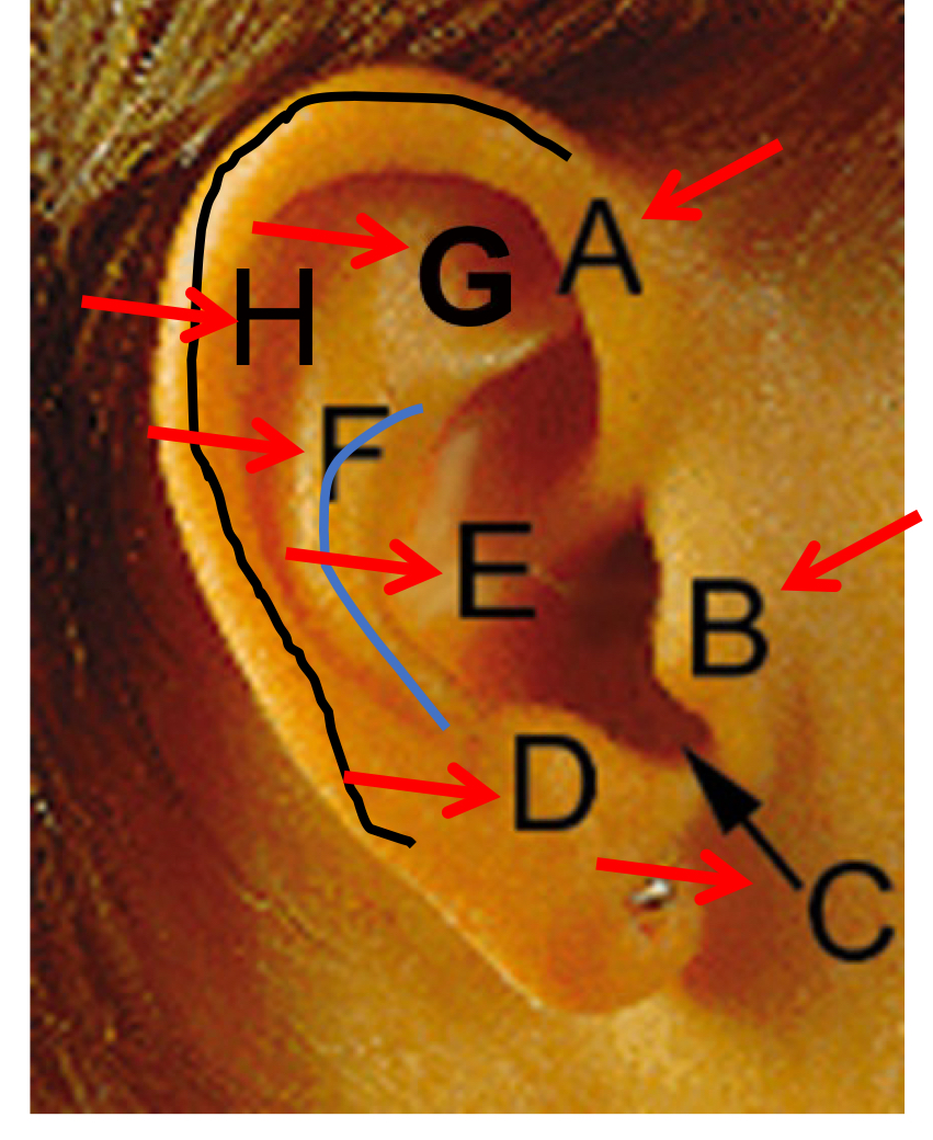
A
Helix

B
Tragus

C
Inter-tragal notch

D
Anti-tragus

E
Concha (bowl)

F
Anti-helix

G
Triangular fossa

H
Scaphoid fossa
External ear canal structure
-Cartilage support for outer 1/3 of length
contains hairs and sebaceous (oil) and veruminour (wax) glands
-Bony support for inner 2/3 of length
can be painful here
-In total ~2.5-3.5 cm long in adults
~1.4 cm at birth
-EAC is not straight!
S-Shaped with 2 bends in the canal
Seeing the eardrum can be a challenge
Functions of the outer ear
-protects more central structures
curvy canals protect the middle and inner ear
hair and wax impede access, keep skin moist and have antimicrobial protective characteristics
-Acts as a resonator
provides a boost in sound level at some frequencies
15-20 dB gain in the 2-5 kHz region for frontal sounds
The size of the boost varies with sound source location
can help little with horizontal localization and a lot with vertical plane localization
Middle ear system consists of…
-tympanic membrane
-ossicles
malleus
incus
stapes
-middle ear muscles
stapedius
tensor tympani
-eustachian tube
tympanic membrane function and structure
-function: convert sound waves in the air into vibrations of the middle ear ossicles
-structure:
~8-10 mm in diameter
~.074 mm thick (very thin)
Concave outward (not flat)
tilted at ~55 degrees
2 main sections of the tympanic membrane
-Pars tensa (has 4 layers)
-Pars flaccida (only 2 layers)
frequent site of pathologies

A
Pars flaccida

B
Pars tensa

C
Umbo (connection point of malleus

D
Light reflex (cone of light)
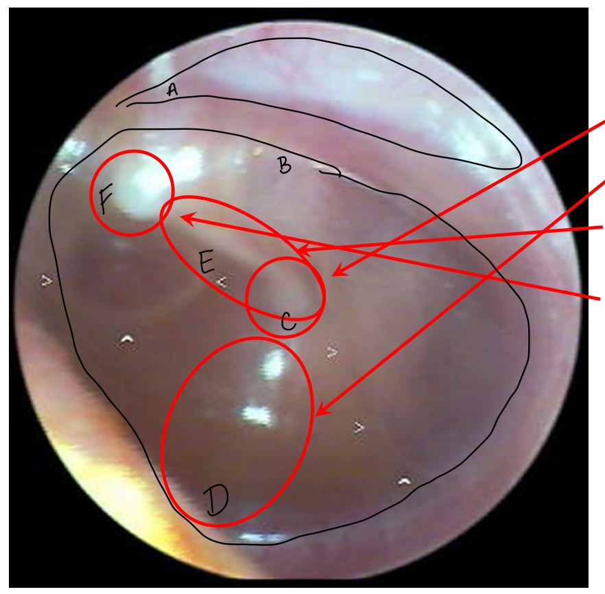
E
Long process of the malleus (e.g. handle/manubrium)

F
Lateral (short) process of the malleus
Quadrants of TM
-Umbo in the center
-handle of the malleus (manubrium) divides anterior and posterior sections
-line perpendicular to manubrium divides superior and inferior sections

1
Anterior-superior quadrant

2
anterior-inferior quadrant

3
posterior-inferior quadrant

4
posterior-superior quadrant
you need to be familiar with “normal” ears so you can:
-Identify conditions that warrant medical referral
-Identify conditions that may impact your diagnostic assessments
-Identify conditions that may impact your rehabilitative efforts
Purpose of otoscopy
-determine the need for medical referral
-identify conditions that may affect audiometric test results
Otoscopy results that lead to medical referral
-untreated pathology
trauma to or blood/drainage in EAC
-Obstructions
Foreign bodies or impacted cerumen in EAC
-Previously unidentified structural defects
pits/tags/stenosis/growths
perforations or other abnormalities of the TM
otoscopy results that lead to audiometric concerns
-collapsing EAC
-obstructions
foreign bodies or impacted cerumen in EAC
-Stenotic (narrow) or atrenic (closed) EAC
-Pressure equalization (PE) tubes
Otoscopy procedures
-Always do otoscopy before starting any audiologic procedure involving the EAC
Follow infection control procedures
hand washing and gloves if needed
clean specula (for each ear if needed)
make sure the otoscope light is bright
-explain what you are doing and start with good ear first (always ask “do you have a better hearing ear?”)
-start exam with the outer ear and surrounding areas, then examine the ear canal, and finally examine the TM
Examine the outer ear
-Inspect pinna and area around, behind and adjacent to the pinna
look for signs of ear surgery, inflammation, pits, and tags
-If possible, talk to your patient to explain what you are doing during this process
Pinnae and EAC deformities
-Pinnae and EAC deformities are often associated with conductive or mixed losses
always suspect HL in persons with these anomalies
-May be observed alone or as one of multiple craniofacial anomalies
often associated with genetic disorders
-If not previously identified - these finding warrant medical referral
particularly for children
Examples of pinnae deformaties
-Anotia
-microtia
-pre-auricular sinus (pit)
-pre-auricular “tag”
Anotia
no pinnae
microtia
-very small or deformed pinnae
-can vary from mild (grade I) to severe (grade III)
Pre-auricular sinus (pit) and pre-auricular “tag”
-Pit: small depressions in the skin around the pinna that are suggestive of abnormal development during crucial time periods in utero
-Tag: additional skin protrusions
-in isolation, pits and tags may not warrant a medical referral
if not previously identified:
note them in your report and encourage the patient to speak with their physician about the findings, particularly if identified in a child
if found in conjunction with other flags, (eg. HL) then refer.
Otoscopy procedure for checking the ear canal and TM
-Use the pencil grip if feasible and brace fingers against the patient’s head
-begin with the outer ear then move to EAC
Check for collapsing canal first, then
gently pull up and back (on adults) on the pinna to straighten the EAC
pull pinna down and back for young children
systematically check EAC and all TM quadrants
Ear canal disorders
-stenosis: narrow EAC
may or may not cause HL
-atresia: absent EAC
blockage may be in the cartilaginous and/or bony portions of the canal
results in conductive HL
skin disorders in the ear canal
-otitis externa
acute or chronic
-Osteoma and exostoses
-excessive cerumen/wax
-collapsing ear canals
otitis externa/external otitis
-can be acute or chronic
may be due to viral, bacterial, or fungal infection
-Painful with foul smelling discharge
-can spread to middle and inner ear if left untreated
may cause HL if severe swelling occurs in the EAC
-Warrants medical referral!
Variations in cerumen
-a normal product of a healthy ear
but in excess it can impact audiologic procedures
effect on hearing varies from non to a large conductive loss (if ear is completely occluded)
-Only warrants referral is excessive and/or impacting your ability to complete testing
Foreign bodies
potentially warrants a referral. solid objects can become embedded in the skin and lead to infection and/or need for surgery
Collapsing canals
-more prevalent in elderly and infants/newborns
typically benign
if using supra-aural phones can lead to a “false” ABG (air bone gap) in audiometric testing
gaps up to 15-30 dB often high frequency conductive loss
-When using supra-aural headphones, check for collapsing canals during otoscopy
-Typically no referral unless collapsed all the time
Cysts
-benign sacs beneath the skin
-no referral
Tumors
-can be benign or malignant
-vascular or bony
-no referral
Osteomas and exostoses
-Most common bony outgrowths (tumor) in the EAC
-Often seen in cold water swimmers
-Usually benign, may need removal if causing EAC blockage or recurrent external otitis
-Typically no referral
Tympanosclerosis
-scarring on the ear drum
-Very common following ear disease (e.g. otitis media)
-May or may not affect hearing thresholds
-Typically no referral needed
TM perforation
-sometimes they are easy to see, other times not so much.
-if unidentified and/ active drainage, refer
-if longstanding, no referral
Middle ear fluid
-May or may not have active otitis media or hearing loss
-if causing pain, refer
-if unidentified and not sick, refer
-if unidentified and sick with treatment, no referral
Monomeric scar
-previous infection or perforation that is partially healed
-no HL and normal tests, no referral
ballooning monomers
without fibrous regions, skin can balloon on the TM
Atelctatic eardrum
-Extreme negative pressure, Eustachian tube dysfunction
-Not always necessary referral
PE tubes
-In and of itself do not require referral but underlying conditions may
Cholesteatoma
-middle ear pathology that may spread to the EAC and be visualized via otoscopy
very serious
absolutely needs referral
Ossicles
-Purpose: transmit TM vibrations to the cochlea via the oval window
-Consists of the malleus, incus, stapes
-smallest bones in the body
stapes ~3.5 mm (1/8th in)
-Held in space by ligaments & tendons of two ME muscles
stapedius muscle
tensor tympani muscle
Middle ear muscles
-Our focus is on the stapedius muscle
smallest muscle in the body
connects to the stapes
innervated by the VII (facial cranial nerve)
-Both ME muscles may contract reflexively, in response to acoustic and nonacoustic stimuli
loud sounds, puff of air, tactile stimulation
-Stapedius (acoustic) reflex-
This reflex provides important diagnostic information
middle ear space
-the brain, arteries, veins, and several cranial nerves are in or near this space
V (Trigeminal)
tensor tympani
VII (facial)
stapedius muscle
chorda tympani branch
sensory information from tongue
IX (glossopharyngeal)
tympanic branch
Middle ear functions
-Convert pressure variations in the air (sound waves) to mechanical vibration to stimulate the inner ear
-optimize energy transmission
impedance matching function
focuses sound energy on the oval window
-pressure equalization - eustachian tube
-protective mechanism (?)- acoustic reflex
Energy transmission in the middle ear
-We lose 99.9% of sound energy (~30dB) as sound waves are transmitted from air (ear canal) to fluid medium (cochlea)
due to the “impedance mismatch”
-middle ear system compensates for this via:
areal ratio difference (oval window is smaller so pressure is greater
lever action
curved membrane buckling
-and by focusing energy directly into the cochlea
Eustachian tube
-connects middle ear space to nasopharynx
3.5-4 cm long
45 degrees downward tilt in adults
near horizontal in children
-provides aeration, drainage, and pressure equalization for ME
-opened by contraction of tensor palatini muscle
innervated by the Vth (trigeminal) cranial nerve
-eustachian tube dysfunction may lead to middle ear effusion (fluid)
particularly in children
Acoustic (stapedius) reflex
-acoustic contraction of stapedius muscle in response to a loud sound (>85 dB SPL)
This stiffins the ossicles and attenuates sound flow
largest effect on low frequency sounds
-purpose is unclear
protect the cochlea from loud sound?
adjust ossicular chain to
maximize sound transmission at different levels?
aid in sound localization?
2 major components of the inner ear
-osseous labyrinth
bony outer casing carved within the temporal bone
-membranous labyrinth
encased within the bony labyrinth
3 main sections of the inner ear
-semicircular canals
vestibular (balance) portion
-vestibule
connects the cochlea and semicircular canals
-cochlea
auditory portion
Membranous labyrinth
-size of a flattened pea (approx. 5 mm (h) x 9 mm (w) at base
-2 ¾ spiral turns in humans
-about 35 mm long if uncurled
-internally divided into 3 fluid filled sections

1
Scala media
reissner’s membrane
basilar membrane
contains sensory organ of hearing (organ of corti)

2
Scala vestibuli
stapes/oval window connection

3
Scala tympani
round window connection

4
spiral ganglion

5
auditory nerve
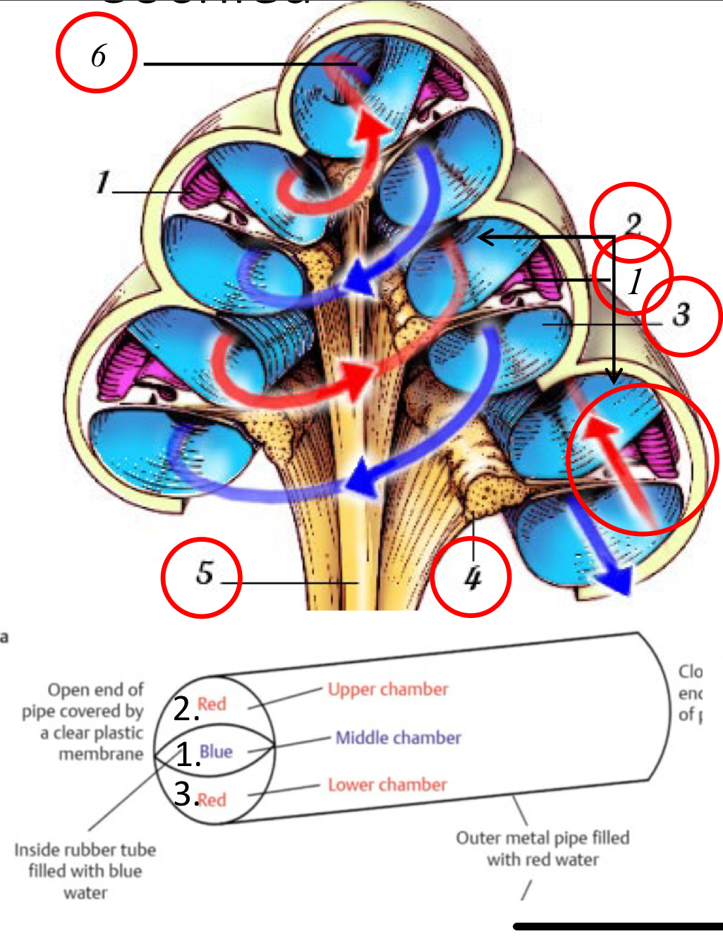
6
helicotrema

1
scala vestibuli

2
scala tympani