Biology: Nucleotides and Nucleic acids
1/35
There's no tags or description
Looks like no tags are added yet.
Name | Mastery | Learn | Test | Matching | Spaced |
|---|
No study sessions yet.
36 Terms
3.1 Nucleic Acids
3.1 Nucleic Acids
Nucleotide structure
Nucleotides are the building blocks of nucleic acids such as DNA and RNA. Nucleotides are monomers and can join together to form dimers (dinucleotides) and polymers (polynucleotides).
A nucleic acid is the functional molecule made of one or more polynucleotide chains.
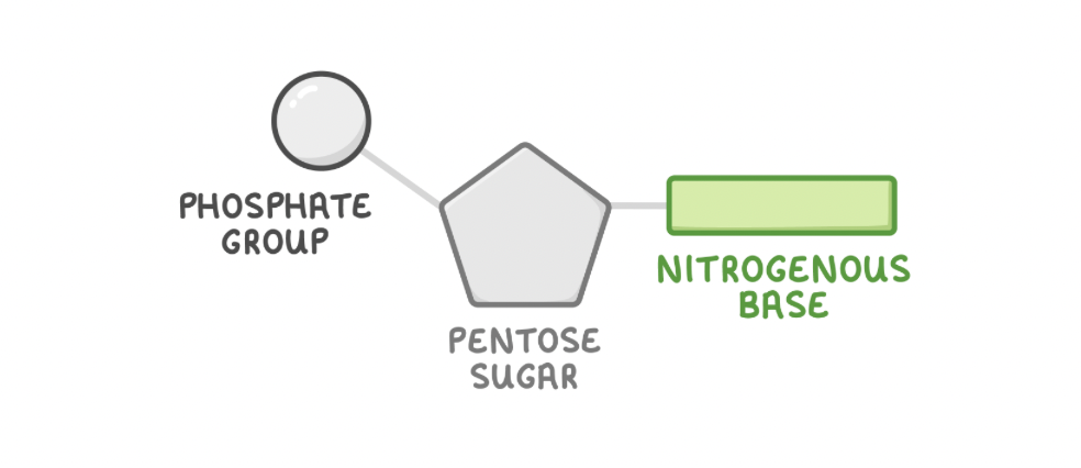
Nucleotides are made up of three components:
A pentose sugar - Contains 5 carbon atoms.
A nitrogenous base - Contains carbon and nitrogen.
A phosphate group - Contains phosphate.
Polynucelotides
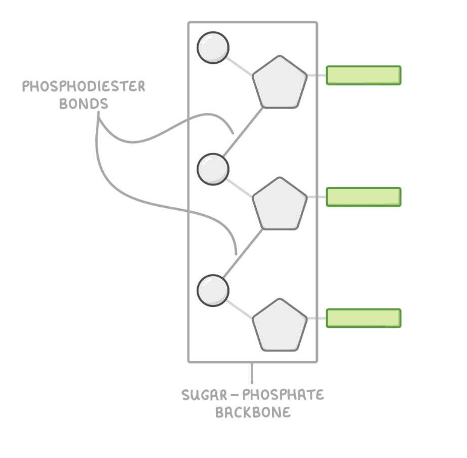
Nucleotides are joined together via condensation reactions to form a polynucleotide. The phosphate group of one nucleotide forms a covalent bond with the sugar of another. This forms a phosphodiester bond.
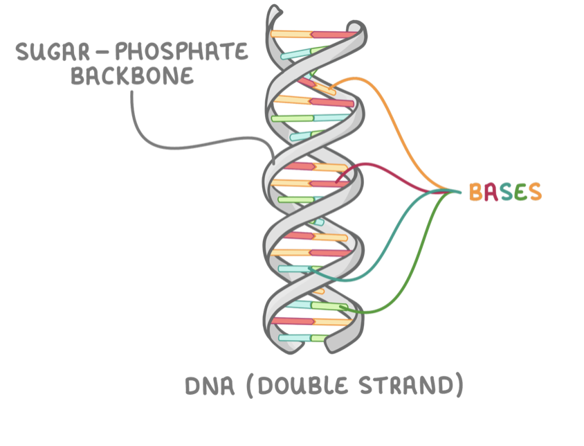
Many nucleotides can join in this way to create a chain of phosphates and sugars known as the sugar-phosphate backbone.
Phosphodiester bonds can be broken via hydrolysis reactions, releasing the nucleotide monomers
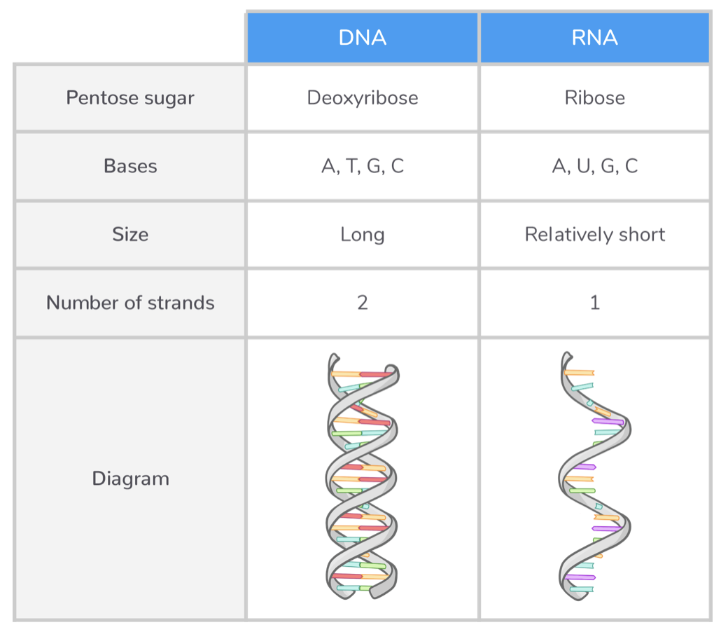
DNA
Deoxyribonucleic acid (DNA) is a type of nucleic acid that contains the instructions needed to make proteins.
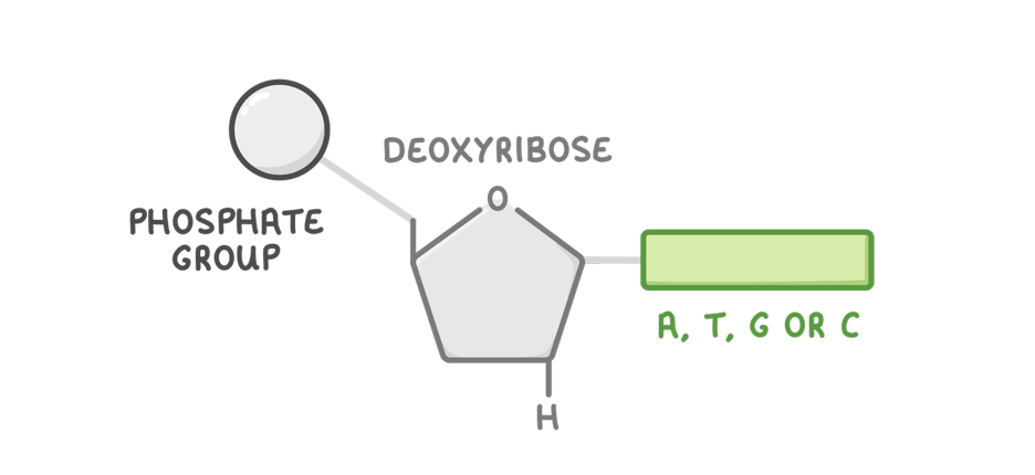
Each DNA nucleotide is made up of three components:
Deoxyribose - A pentose sugar.
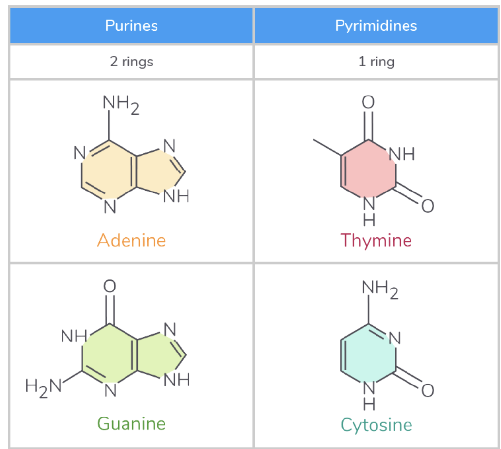
A, T, G, or C base - Adenine, thymine, guanine, or cytosine.
A phosphate group
DNA structure
DNA is made up of two polynucleotide strands wound around each other to form a double helix.
The following features allow DNA to pass genetic information from one generation to another:
Sugar-phosphate backbone - This protects coding bases on the inside of the helix.
Double stranded - This allows strands to act as templates in DNA replication.
Large molecule - It stores lots of information.
Double helix - This makes the molecule compact.
Complementary base pairing - This allows accurate DNA replication.
Weak hydrogen bonds - This allows strands to separate in DNA replication.
Purines and pyramidines
+Uracil in pyramidine column
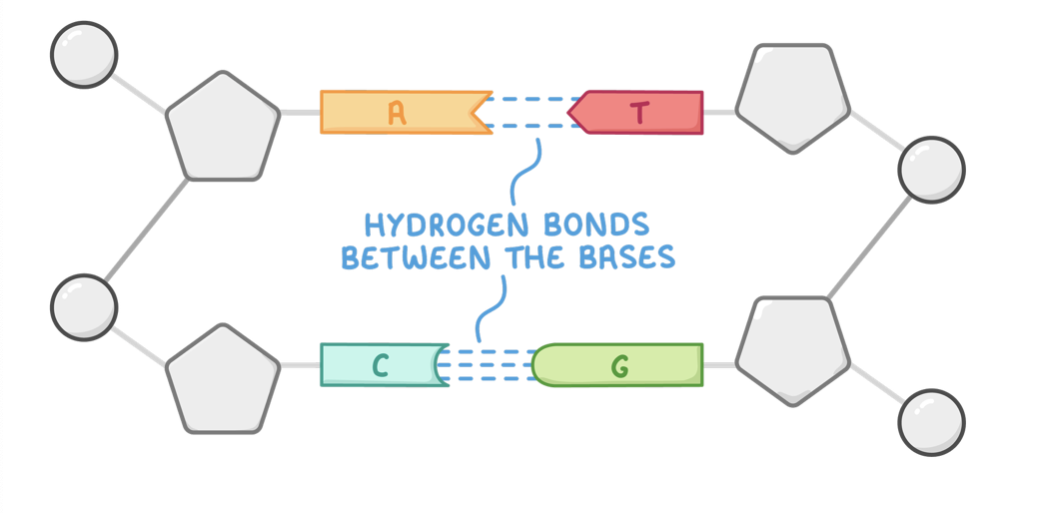
Complementary base pairing
The two DNA strands are held together via hydrogen bonding between bases.
Each base only joins with one other specific base:
Adenine pairs with thymine via 2 hydrogen bonds.
Cytosine pairs with guanine via 3 hydrogen bonds.
This is known as complementary base pairing.
A smaller pyrimidine base always binds to a larger purine base. This arrangement maintains a constant distance between the two sugar-phosphate backbones.
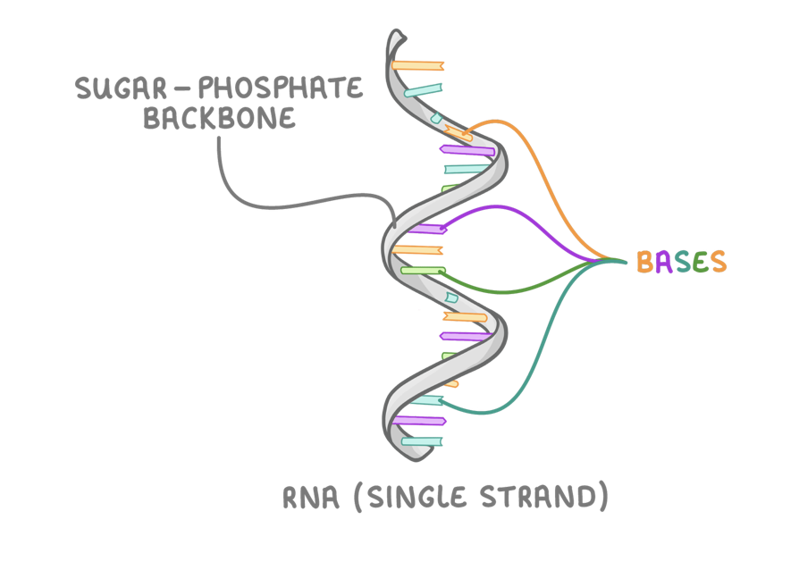
RNA
Ribonucleic acid (RNA) is a type of nucleic acid that uses information from DNA to synthesise proteins.
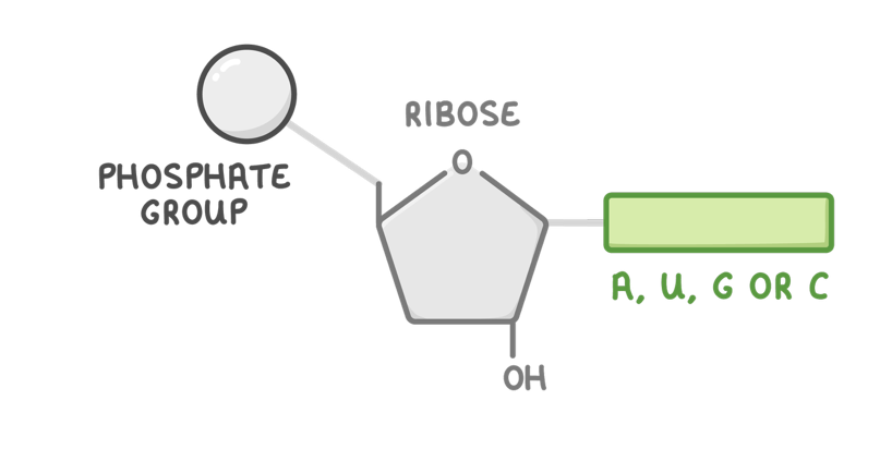
Each RNA nucleotide is made up of three components:
Ribose - A pentose sugar.
A, U, G, or C base - Adenine, uracil, guanine, or cytosine.
A phosphate group
RNA Structure
Unlike DNA, RNA nucleotides contain the sugar ribose rather than deoxyribose. They also contain the base uracil in place of thymine (uracil pairs with adenine).
RNA is a single stranded molecule made up of just one polynucleotide strand. These strands are much shorter than DNA strands.
Comparing DNA and RNA
3.2 DNA Replication
3.2 DNA Replication
DNA Replication
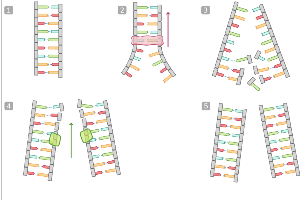
DNA is copied via a process known as semi conservative replication.
This produces DNA molecules consisting of one original DNA strand and one newly synthesised DNA strand.
A DNA molecule ready to be replicated.
The enzyme DNA helicase breaks hydrogen bonds between complementary bases. This unwinds the double helix and separates the strands
Each strand acts as a template as free nucleotides attract to their complementary bases. Adenine pairs with thymine, while cytosine pairs with guanine.
The enzyme DNA polymerase joins the free nucleotides together via condensation reactions in the 5' to 3' direction. This forms phosphodiester bonds to create the sugar-phosphate backbone of the new DNA strand.
Two identical copies of DNA are made. Each copy is made up of one original DNA strand and one new DNA strand (semi-conservative replication).
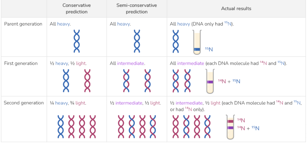
Meselson and Stahl experiment on conservative and semi conservative DNA replication
Their experimental design was based on three main principles:
DNA contains nitrogen.
Two different nitrogen isotopes can be used to mark DNA strands and track them during replication:
(which is lighter).
(which is heavier).
Bacteria use nitrogen from their surroundings to build new DNA molecules.
The experimental process:
Bacteria were grown in a medium containing 15N , so all their DNA is ‘heavy’.
The bacteria were transferred to a medium with 14N for one round of replication, so the lighter nitrogen was incorporated into any new DNA strands they made.
The DNA was extracted and centrifuged.
Steps 2-3 were repeated for another round of replication.
The distribution of heavy and light DNA was analysed to track how the DNA was replicating:
The heavier bands sink lower in the test tube.
The intermediate bands, made of DNA with one heavy strand and one light strand, are in the middle of the test tube.
The lighter bands are higher up in the test tube.
The results
The experiment showed that DNA replicates through a semi-conservative process.
In the first replication:
The original, heavy DNA strands separate.
Each heavy strand acts as a template for a new complementary strand.
New, light strands form alongside the original strands.
Each resulting DNA molecule consists of one old, heavy strand and one new, light strand.
In the second replication:
Both original strands and the two new strands act as templates.
New, light strands form alongside all four templates.
Half the resulting molecules have one, original heavy strand and one new light strand.
The other half are completely made of light strands.
3.3 Genetic code
3.3 Genetic code
How DNA is condensed into chromosomes
DNA molecules are incredibly long and must be tightly packed up to fit within the nucleus of a eukaryotic cell.
DNA molecules are wound around proteins known as histones to form a DNA-histone complex.
These complexes coil further to form chromatin, which helps pack the DNA into chromosomes. Each chromosome contains just a single molecule of DNA.
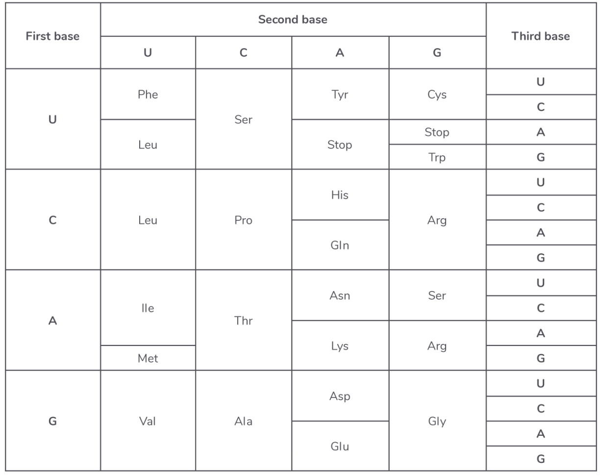
How to interpret tables that show codons for amino acids
The genetic code can be summarised into a table as shown below. The code is often shown as mRNA bases rather than DNA, so the base uracil (U) is shown instead of thymine (T).
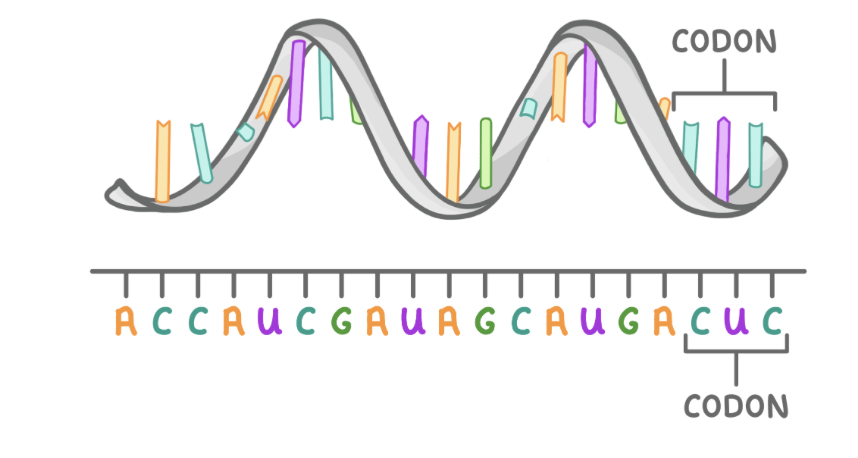
Each amino acid is coded for by a sequence of three mRNA bases known as a codon (similar to a DNA triplet).
We can see from the table above, that many amino acids are coded for by multiple codons. For example, the amino acid phe (phenylalanine) is coded for by codons UUU and UUC. This means that they are degenerate
The genetic code
A gene is a short section of DNA that codes for a polypeptide (a protein). Each gene is located at a specific position along a chromosome known as a locus.
The complete set of genes within a cell is known as the genome. The full range of proteins that a cell is capable of producing is known as the proteome.
The genetic code refers to the sequence of bases that code for amino acids.
Scientists found that each amino acid is coded for by a sequence of three DNA bases, known as a triplet.
Features of genetic code
The genetic code has the following features:
Universal - Each DNA triplet codes for the same amino acid in all organisms (with a few minor exceptions).
Non-overlapping - Each base in the DNA sequence is only read once (e.g. CGTATC is read as CGT and ATC).
Degenerate - Most amino acids are coded for by more than one triplet (e.g. ACA and ACG both code for cysteine).
3.4 mRNA and tRNA
3.4 mRNA and tRNA
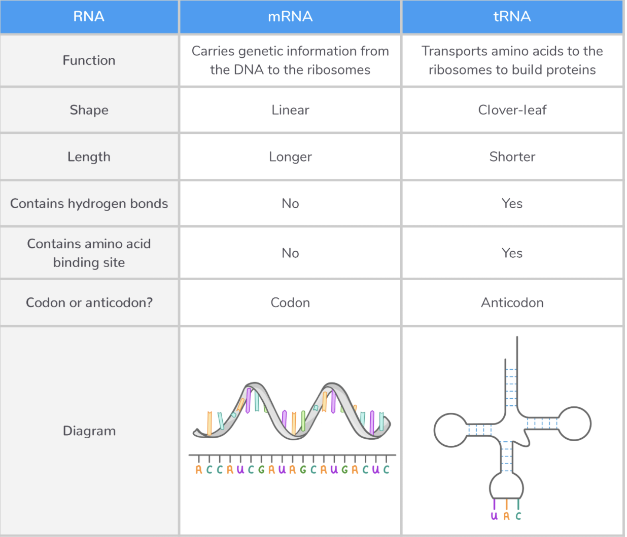
mRNA
Messenger RNA (mRNA) is a type of RNA synthesised during the process of transcription.
Its role is to carry genetic information from the DNA to the ribosomes (where proteins are made).
Features of mRNA:
Single-stranded, linear molecule.
Contains a base sequence complementary to a DNA sequence.
Contains codons, which are sets of three bases that code for an amino acid.
Small enough to leave the nucleus.
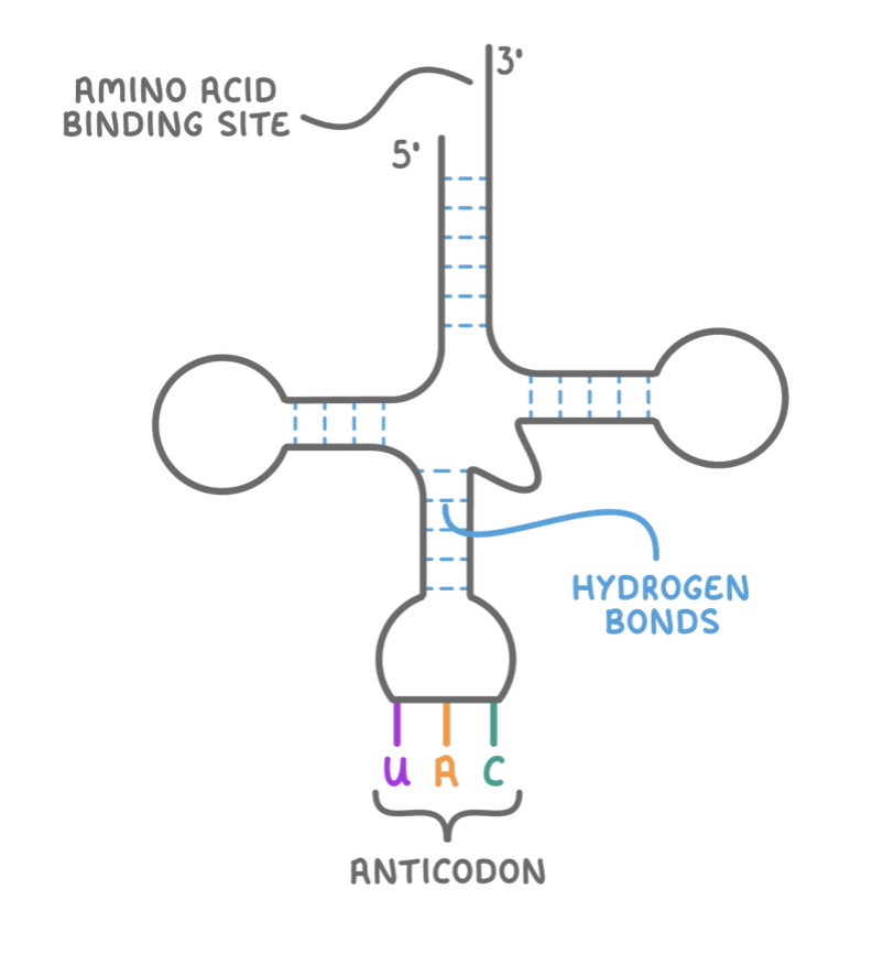
tRNA
Transfer RNA (tRNA) is a type of RNA used in the process of translation.
Its role is to transport amino acids to ribosomes to build up a polypeptide chain.
Features of tRNA:
Single-stranded molecule folded into a clover-leaf shape.
Uses hydrogen bonds between complementary base pairs to hold it in shape.
Contains a specific sequence of three bases at one end, known as the anticodon.
Contains an amino acid binding site at the opposite end.
Comparing mRNA and tRNA
3.5 Protein synthesis: Transcription
3.5 Protein synthesis: Transcription
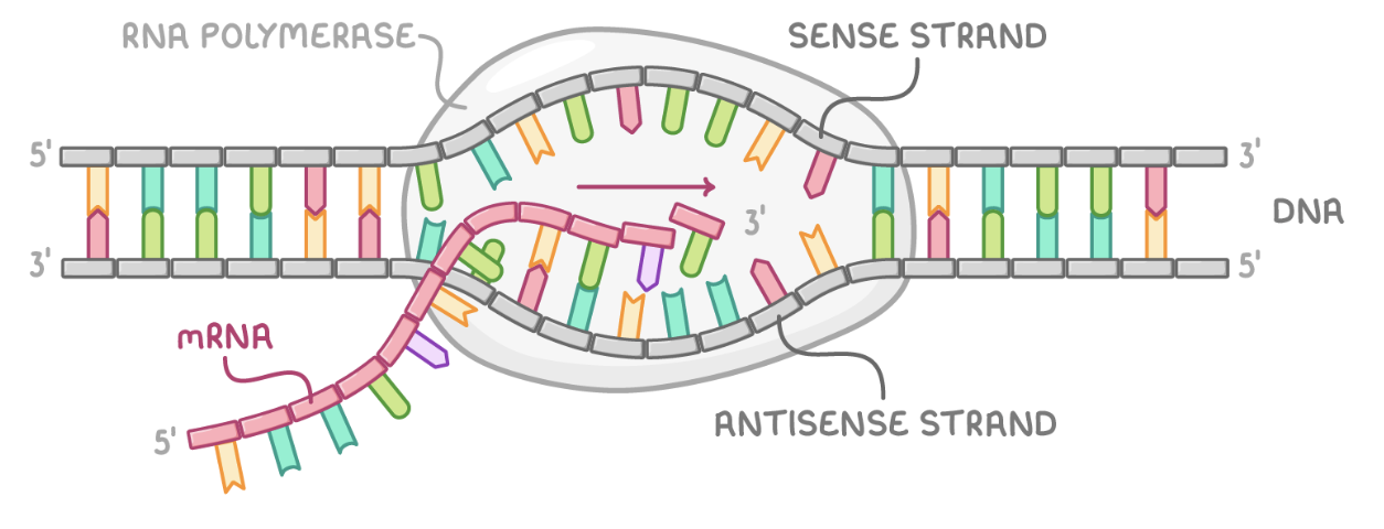
Transcription
Transcription is the initial step in the synthesis of proteins. It involves creating an mRNA copy of a gene from the DNA template.
In eukaryotic cells, transcription takes place within the nucleus. This mRNA then exits the nucleus and enters the cytoplasm, where translation occurs.
Key events in transcription:
Transcription begins with the RNA polymerase enzyme binding to DNA.
The hydrogen bonds between the DNA bases break, and the two strands of the double helix separate.
The antisense strand acts as the template for mRNA synthesis.
Free RNA nucleotides align with the DNA template through complementary base pairing.
In the RNA molecule, uracil pairs with adenine, while adenine pairs with thymine, and cytosine pairs with guanine.
RNA polymerase catalyses the formation of phosphodiester bonds between adjacent RNA nucleotides.
A complementary mRNA strand is formed, carrying the same base sequence as the DNA sense strand.
The process ends when RNA polymerase reaches a stop codon, detaches from DNA and terminates transcription.
mRNA is released, detaches from DNA, and DNA rewinds into its double helix structure.
Transcription
3.6 Protein synthesis: Translation
3.5 Protein synthesis: Translation
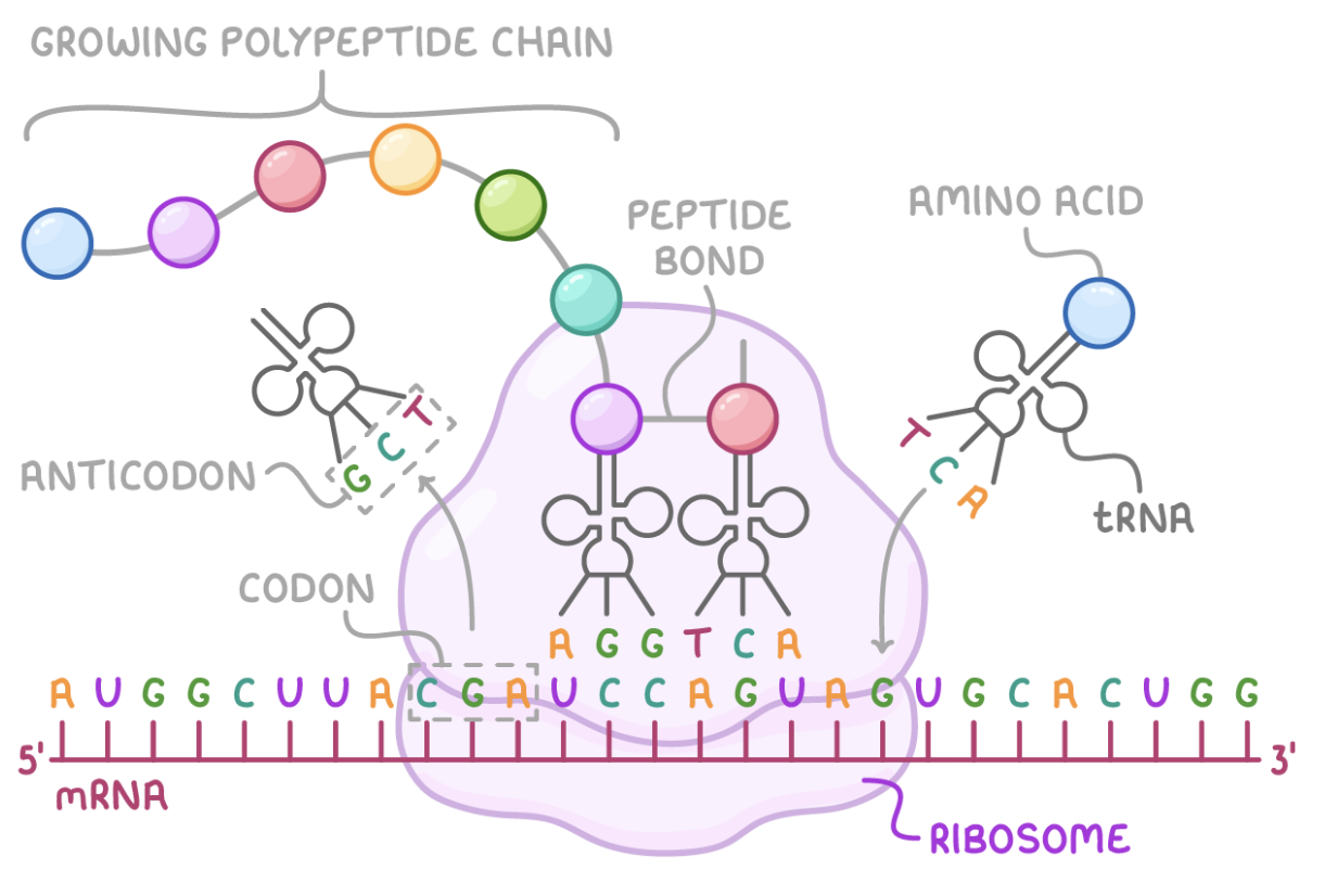
Translation
Translation occurs in the cytoplasm, specifically on the ribosome in both eukaryotic and prokaryotic cells.
Translation is the process of decoding the information in messenger RNA (mRNA) to synthesise a polypeptide chain, with the help of transfer RNA (tRNA). This chain then folds into a functional protein.
Translation involves several key steps:
The ribosome attaches to the mRNA strand at a start codon (AUG).
A tRNA molecule, carrying a specific amino acid and with an anticodon (UAC) that is complementary to the start codon, binds to the mRNA.
A second tRNA molecule with an anticodon complementary to the next mRNA codon, and also carrying a specific amino acid, attaches to mRNA.
The amino acids carried by the first two tRNA molecules are linked together via a peptide bond using ATP.
The first tRNA molecule detaches from mRNA and is free to collect another amino acid for future use.
The ribosome moves along mRNA, allowing another tRNA molecule, which carries the next amino acid, to bind to the next codon on mRNA.
The process from step 4 to 6 is repeated, which elongates the polypeptide chain.
At any point during this process, two tRNA molecules can be attached to the ribosome.
The sequence continues until the ribosome reaches a stop codon on mRNA.
The completed polypeptide chain detaches from the ribosome.
Translation
3.7 ATP
3.7 ATP
What is ATP?
ATP is an important molecule involved in energy transfer within cells and is often referred to as the 'energy currency' of the cell.
You can think of it like a battery, temporarily holding energy and transferring it from one part of the cell to another.
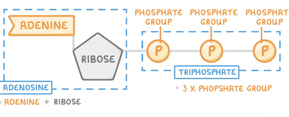
Structure of ATP
ATP is a nucleotide derivative. It has a similar structure to the nucleotides (monomers) found in DNA and RNA.
ATP stands for adenosine triphosphate.
ATP consists of 3 parts:
Adenine - Nitrogenous base
Ribose - 5-carbon sugar
Phosphates - 3 phosphate groups
Uses of ATP in the body
ATP is used for a variety of energy-requiring processes such as:
Movement, such as muscle contraction or for sperm cells to swim.
Active transport of molecules against the concentration gradient, such as ions entering plant roots.
Synthesis of large molecules, such as DNA and proteins.
Secretion of substances from cells, such as releasing hormones from glands.
ATP can also activate molecules by phosphorylating them. When ATP is hydrolysed, the phosphate can be added to other molecules (such as enzymes) to make them more reactive.
ATP Reactions
ATP is broken down using a hydrolysis reaction and re-synthesised using a condensation reaction:
Hydrolysis - The addition of water to break a chemical bond between two molecules.
Condensation - The removal of water to form a chemical bond between two molecules.
The breakdown and re-synthesis of ATP is an example of a reversible reaction.
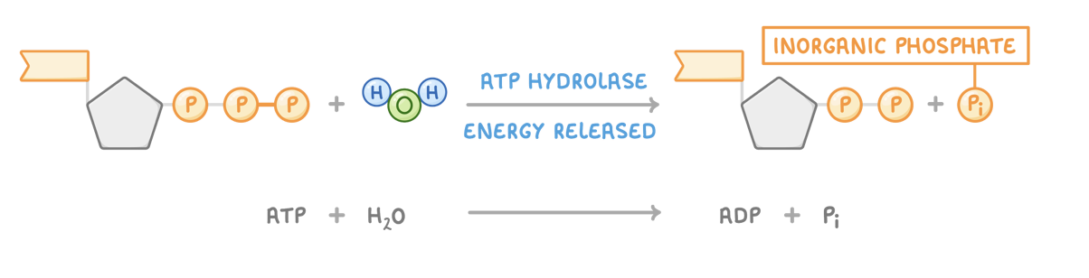
Hydrolysis of ATP to release energy
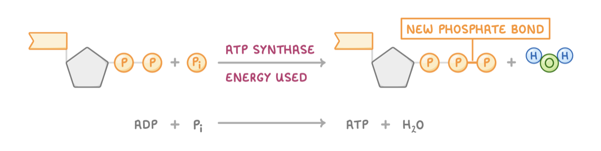
When water is added to ATP, it breaks down into adenosine diphosphate (ADP) and inorganic phosphate (Pi). This reaction is catalysed by the enzyme ATP hydrolase and releases energy for use in cells.
Condensation reaction to reform ATP
When a phosphate group and ADP join, a water molecule is released. This reaction is catalysed by the enzyme ATP synthase. This process requires energy and traps chemical energy in the bond.