The Nervous System: Structure and Function
 Call Kai
 Learn
Learn Practice Test
 Spaced Repetition
 Match
Match Flashcards
 Knowt Play
1/50

There's no tags or description
Looks like no tags are added yet.
Name | Mastery | Learn | Test | Matching | Spaced | Call with Kai | Chat |
|---|
No analytics yet
Send a link to your students to track their progress
51 Terms
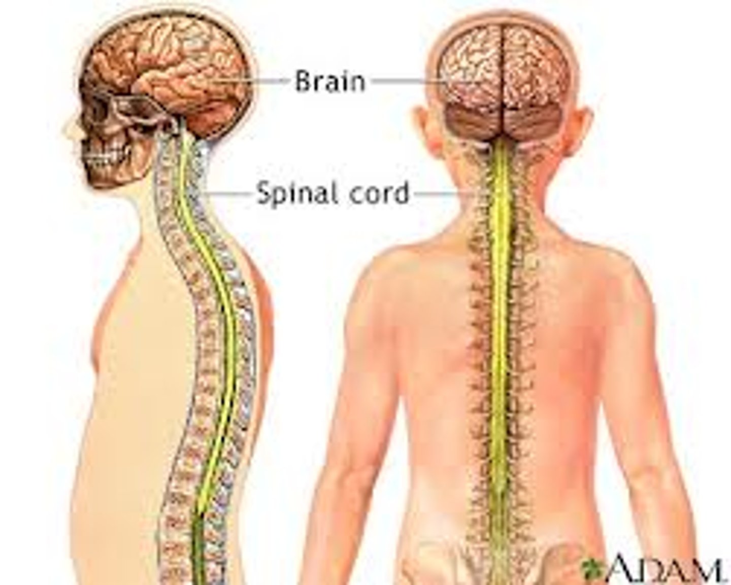
Central Nervous System
located within the skull and spine
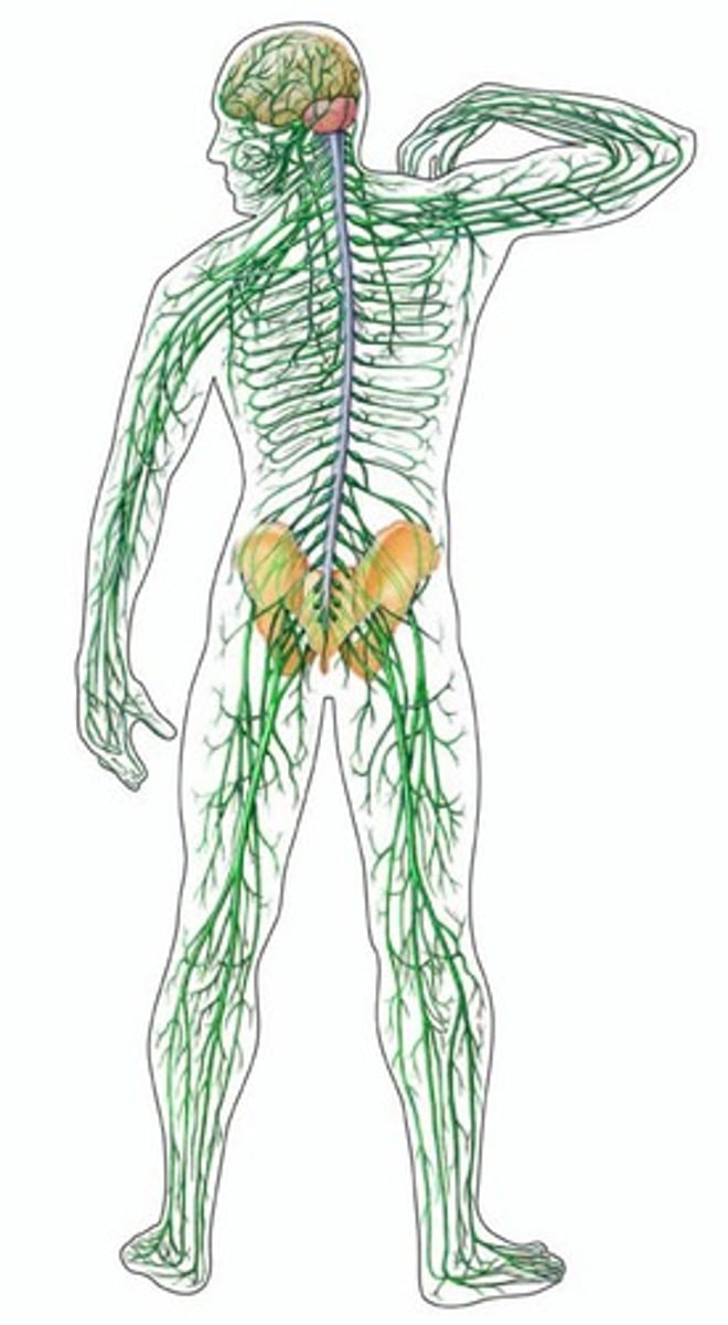
Peripheral Nervous System
outside the skull and spine
Somatic Nervous System
-division of the PNS
-interacts with environment
-efferent and afferent nerves
Autonomic Nervous System
-division of the PNS
-regulates body's internal environment
-two types of efferent nerves (sympathetic and parasympathetic)
Sympathetic Nerves
-ANS efferent nerve
-Stimulate, organize, mobilize energy
-Arousal
-Synapse far from target organ
-Project from CNS in lumber and thoratic region of spinal cord
-two stage neural path
Parasympathetic
-ANS efferent nerve
-conserve energy, relaxation
-synapse close to target
-project from brain and sacral
-two stage neural path
Three Important Principles of Para and Sym Nerves
1.) Para = conserve energy
Sym = stimulate/use
energy
2.) Target organs get input from both types to regulate functioning
3.) Para = relaxation
Sym = arousal
Cranial Nerves
12 pairs of nerves that extending from the brain instead of the spine like the rest of PNS efferent nerves
Vagus Nerves
longest cranial nerves and contain both motor and sensory nerves traveling two and from the gut
Afferent Nerve
carry sensory info from sensory organs to the CNS
Efferent Nerves
care motor signals from CNS to skeletal muscles
Anterior
Head: towards the face
Body: towards head
(contrast = posterior)
Posterior
Head: back of head
Body: towards the booty
(contrast = anterior)
Dorsal
Head: top of skull
Body: towards the back
(contrast = ventral)
Ventral
Head: towards brain stem
Body: towards chest
(contrast = dorsal)
lateral
From the center of the body out
Medial
Going towards the midline of the body
Ipsilateral
belonging to or occurring on the same side of the body
Contralateral
On the opposite side of the body
Spinal Cord Structure
Composed of two diff areas:
1.) inner H shaped core of gray matter
2.) surrounding area of white matter
Gray Matter
composed largely of cell bodies and unmyelinated interneurons
White Matter
composed largely of myelinated axons
Meninges
3 protective membranes that encase brain and spinal cord (dura mater, archnoid, pia mater)
Dura Mater
outer most, tough membrane of meninges
tough mother
Arachnoid
fine, spider like membrane in the meninges that follows the dura mater
Superarachnoid Space
space under arachoid containing large blood vessels and cerebrospinal fluid
Pia Mater
delicate membrane that adheres to surface of CNS and innermost layer of meninges
pious mother
Cerebrospinal Fluid
protects brain and spinal cord
fills central canal and cerebral ventricles of the brain
Ventricles
four large chambers in the brain
5 Major Brain Divisions
1.) Telencephalon
2.) Diencephalon
3.) Mesencephalone
4.) Metencephalon
5.) Myelencephalon
*T on top and the rest in alphabetical order
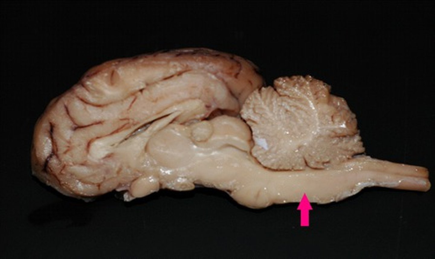
Myelencephalone or Medulla (hindbrain)
Most posterior of brain regions
Composed of:
-Large tracts carrying signals bw rest of brain
-Reticular Formation
-Role in sleep, arousal, attention, movement, muscle tone, reflexes
Reticular Formation
-complex network of approx. 100 tiny nuclei that occupy central core of brain stem from posterior of myelencephalon to anterior of midbrain
-role in arousal, sleep, attention, movement, maintenance of muscle tone, and various reflexes
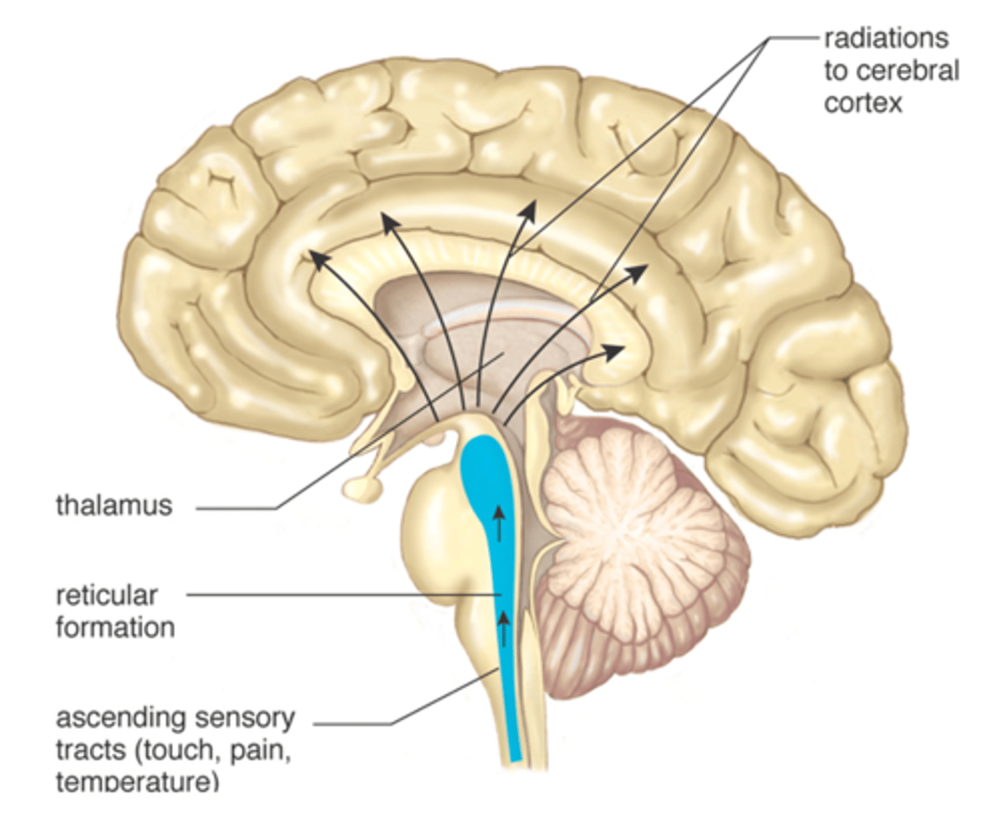
Metencephalon (hindbrain)
Composed of:
-ascending/descending tracts
-Pons
-Cerebellum
-helps regulate breathing, heart and blood vessel function, digestion, sneezing, and swallowing.
-center for respiration and circulation.
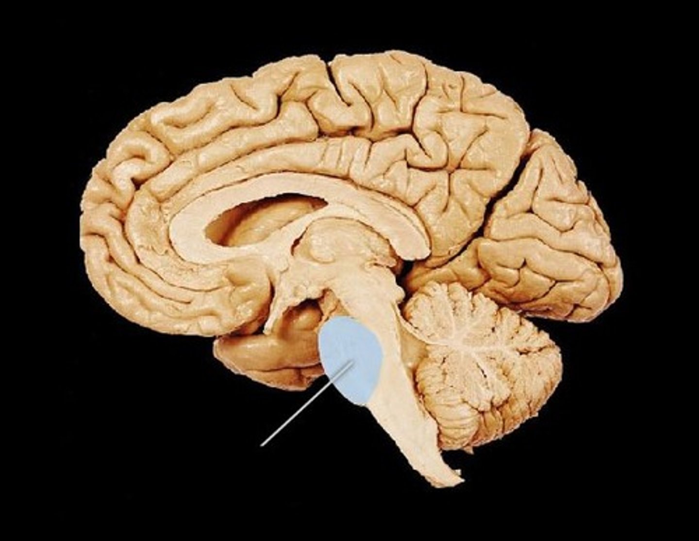
Pons
-anterior bulge of metencephalon
- communications and coordination center between the two hemispheres of the brain
-helps in the transferring of messages between various parts of the brain and the spinal cord
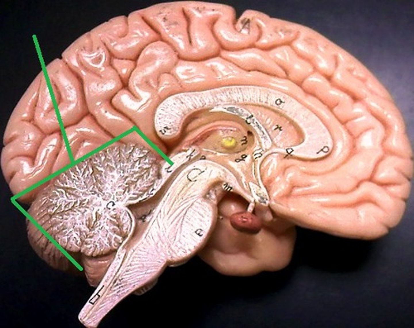
Cerebellum
-posterior bigger bulge and its important to sensorimotor functions
-damage = loss precise movements, adapting to changing conditions, cognitive deficits, use of language
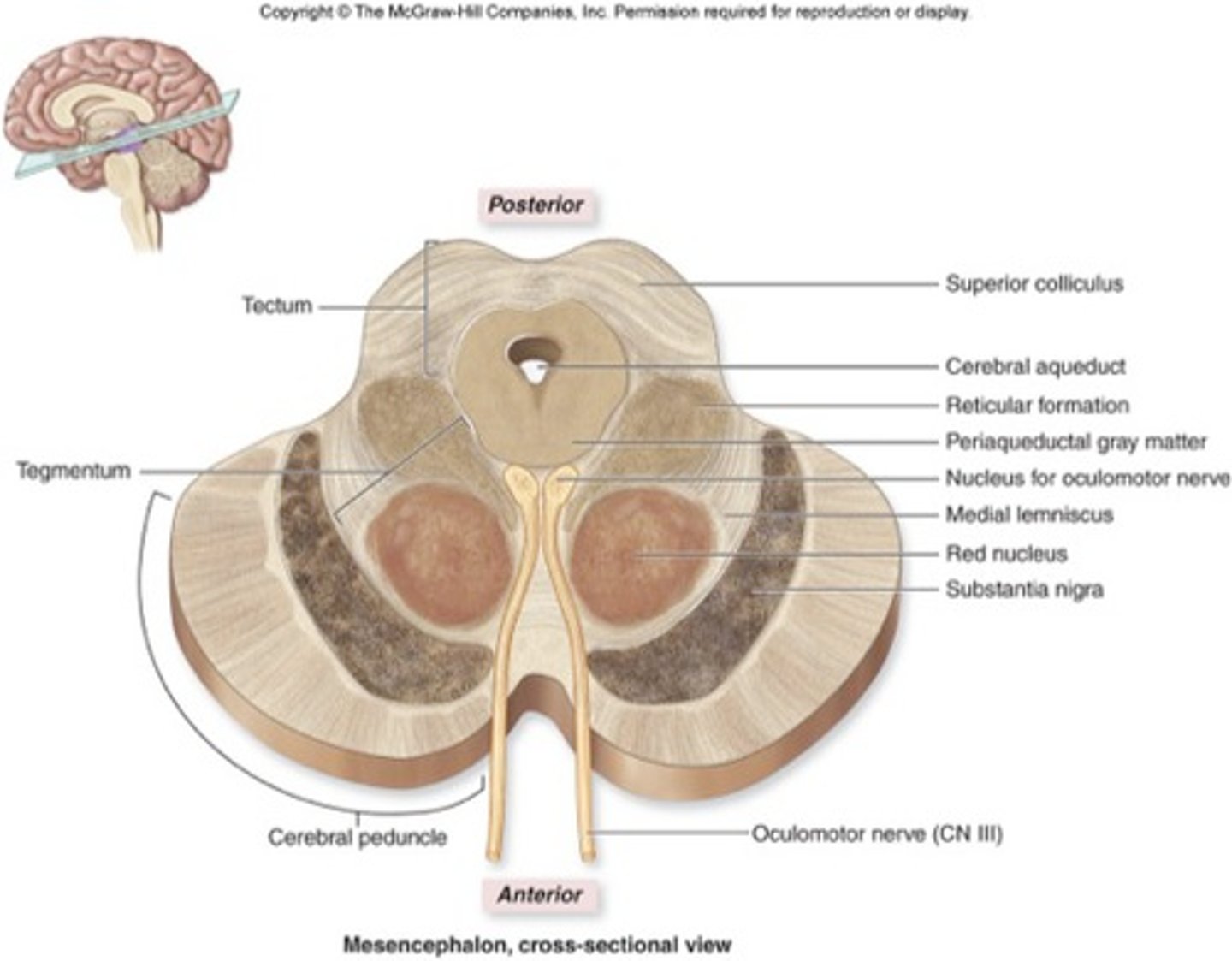
Mesencephalon (midbrain)
two major divisions:
-tectum
-tegmentum
Tectum (roof)
dorsal surface roof of midbrain
audio and visual-motor functions
composed of two bumps
-inferior colliculi
-superior colliculi
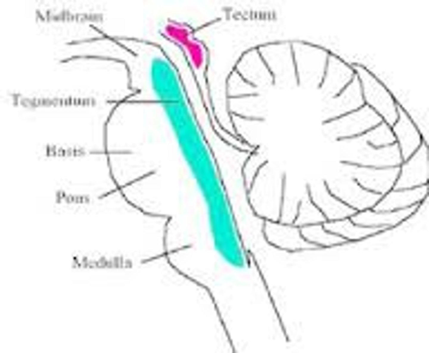
Tegmentum
ventral to tectum
contains reticular formation, tracts, periaqueductal gray, substantia nigra, red nucleus
Diencephalon (forebrain)
Thalamus
Hypothalamus
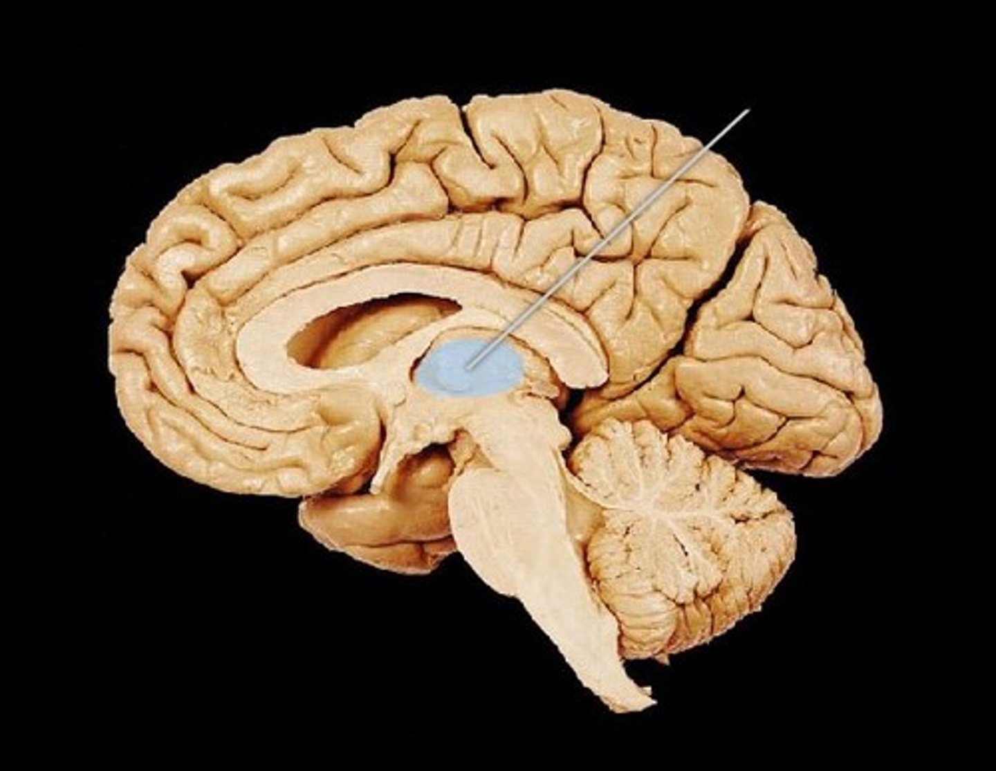
Thalamus
large, two lobed structure in brain stem
lobes joined by massa intermedia
white lamina of myelinated axons cover surface
Hypothalamus
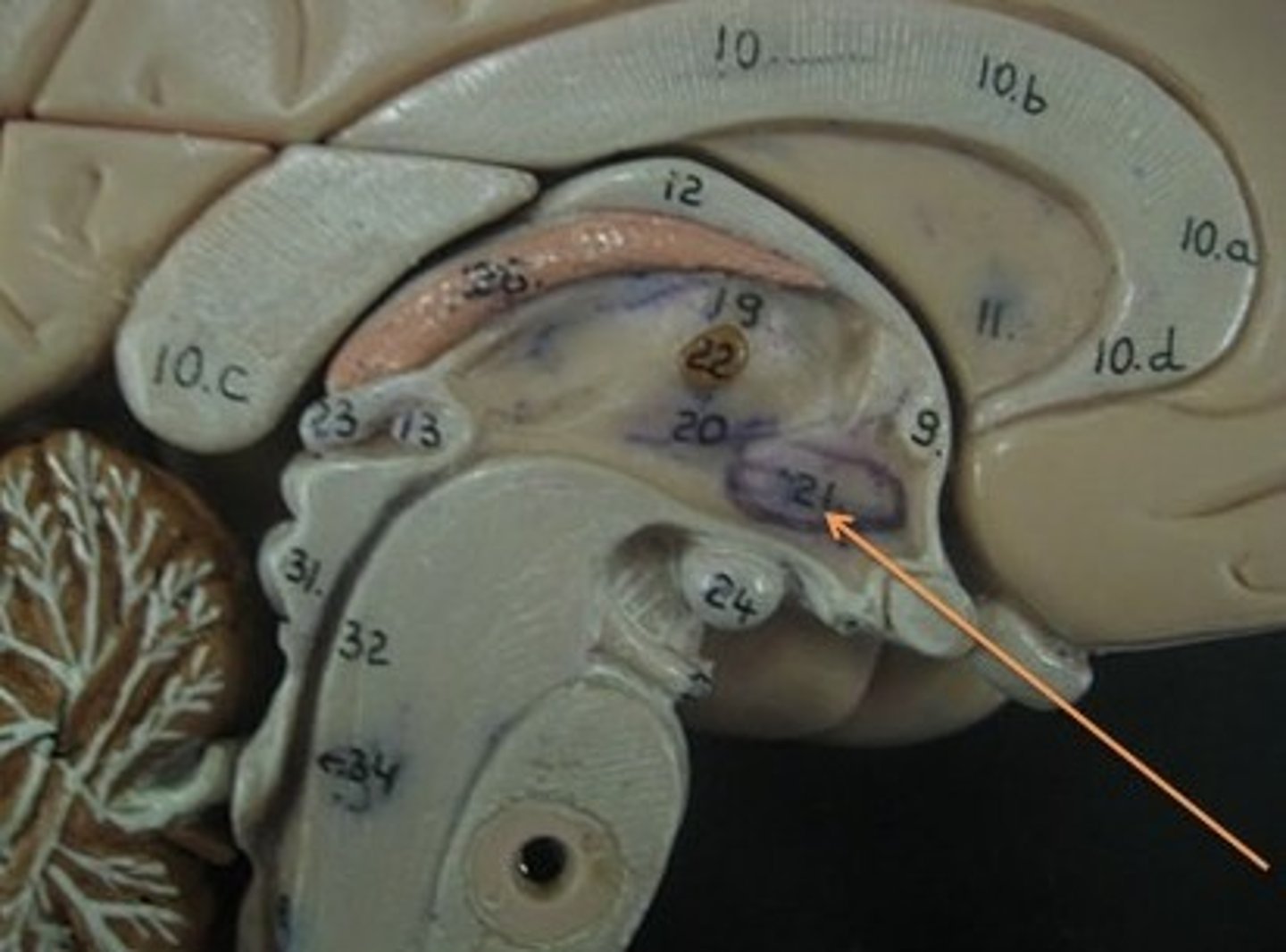
just below thalamus
major role in regulation of motivation behaviors (eat, sleep, sex)
Telencepholon (forebrain)
largest brain division
mediates most complex functioning (voluntary movement, sensory input, learning, speaking, problem solving)
crebral cortex
diff lobes
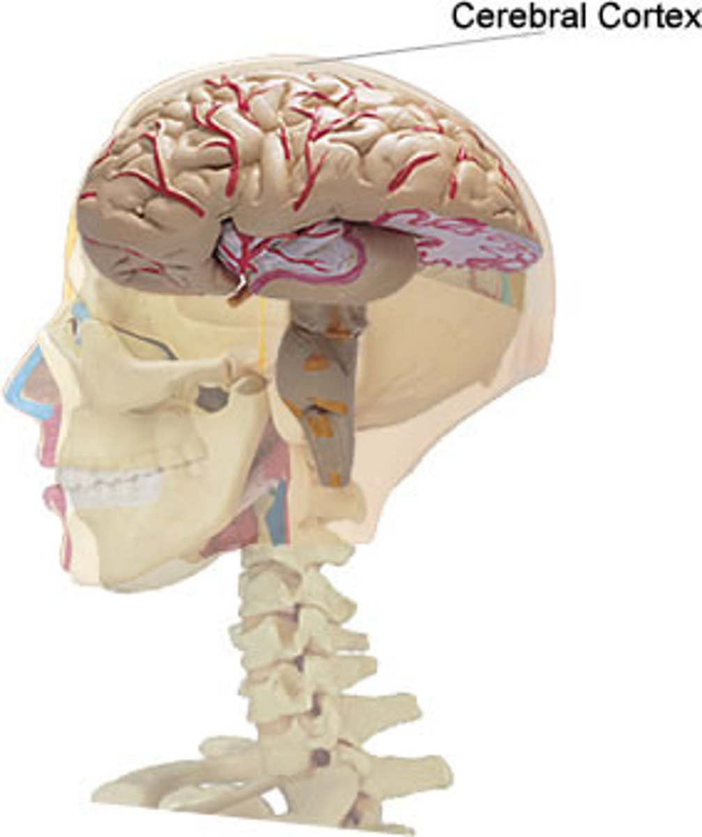
Cerebral Cortex
layer of tissue that covers cerebral hemisphere
gray matter
deepy convoluted (furrowed)
Corpus Callosum
largest cerebral commissure (tract that directly connects cerebral hemispheres)
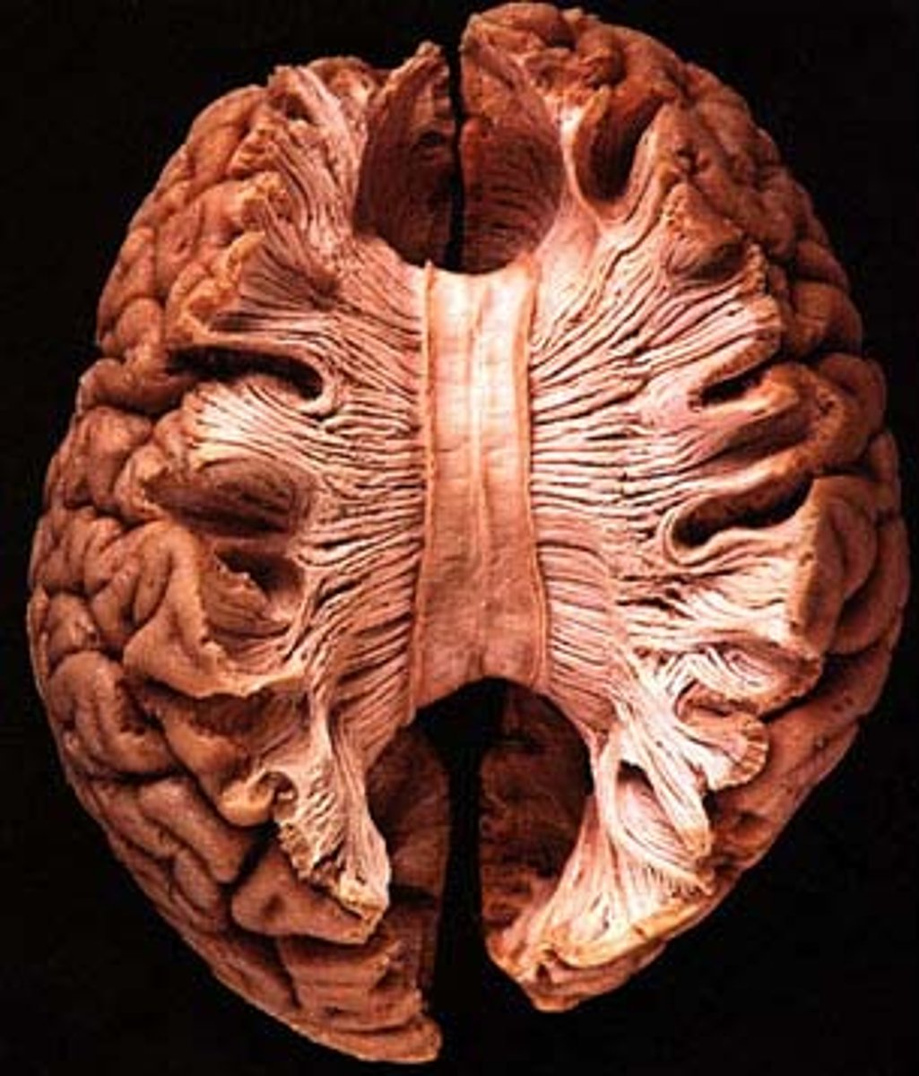
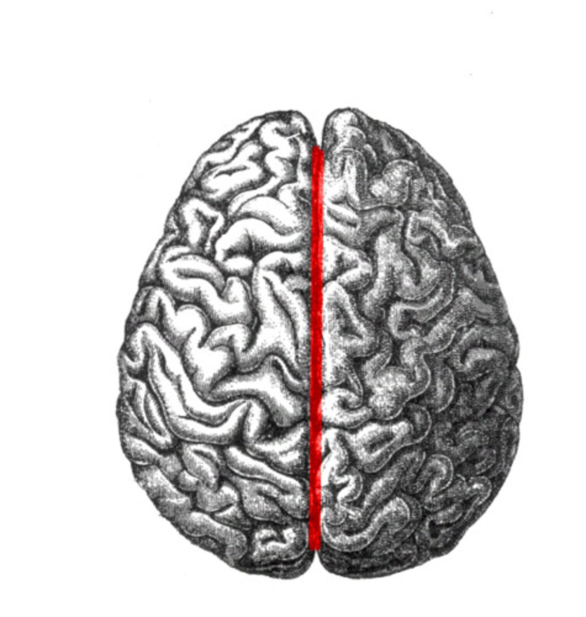
Longitudinal Fissure
separation of cerebral hemispheres
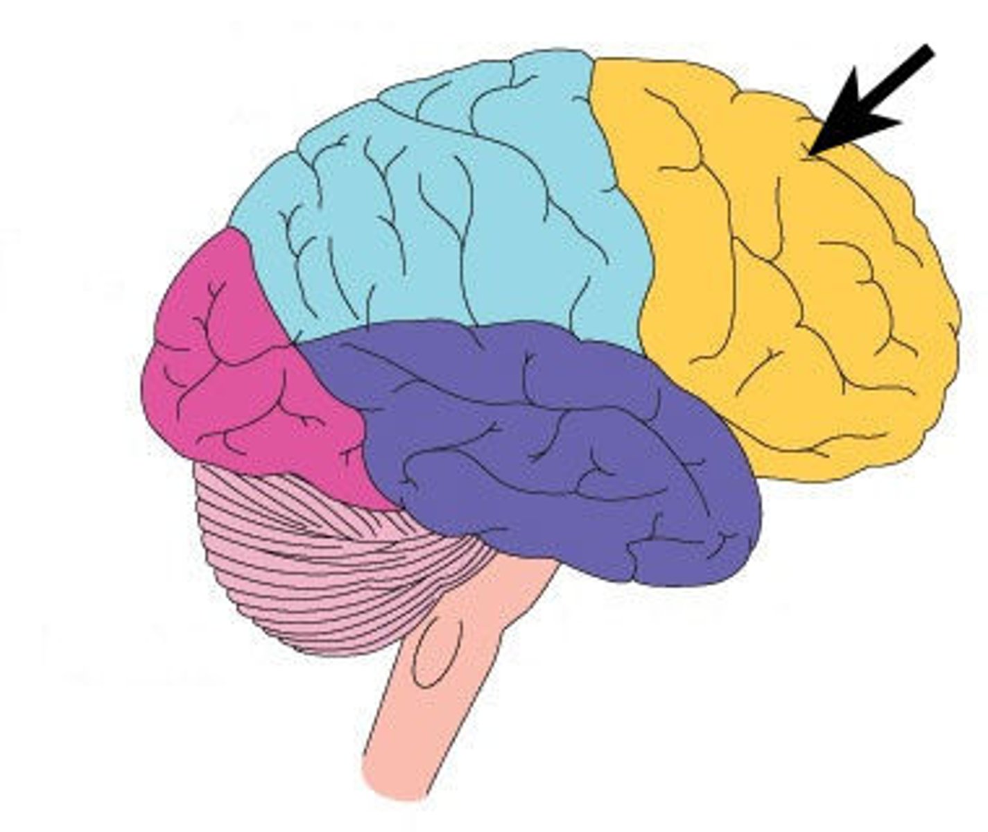
Frontal Lobe
primary motor cortex
planning, executive functioning, language/grammar, working memory
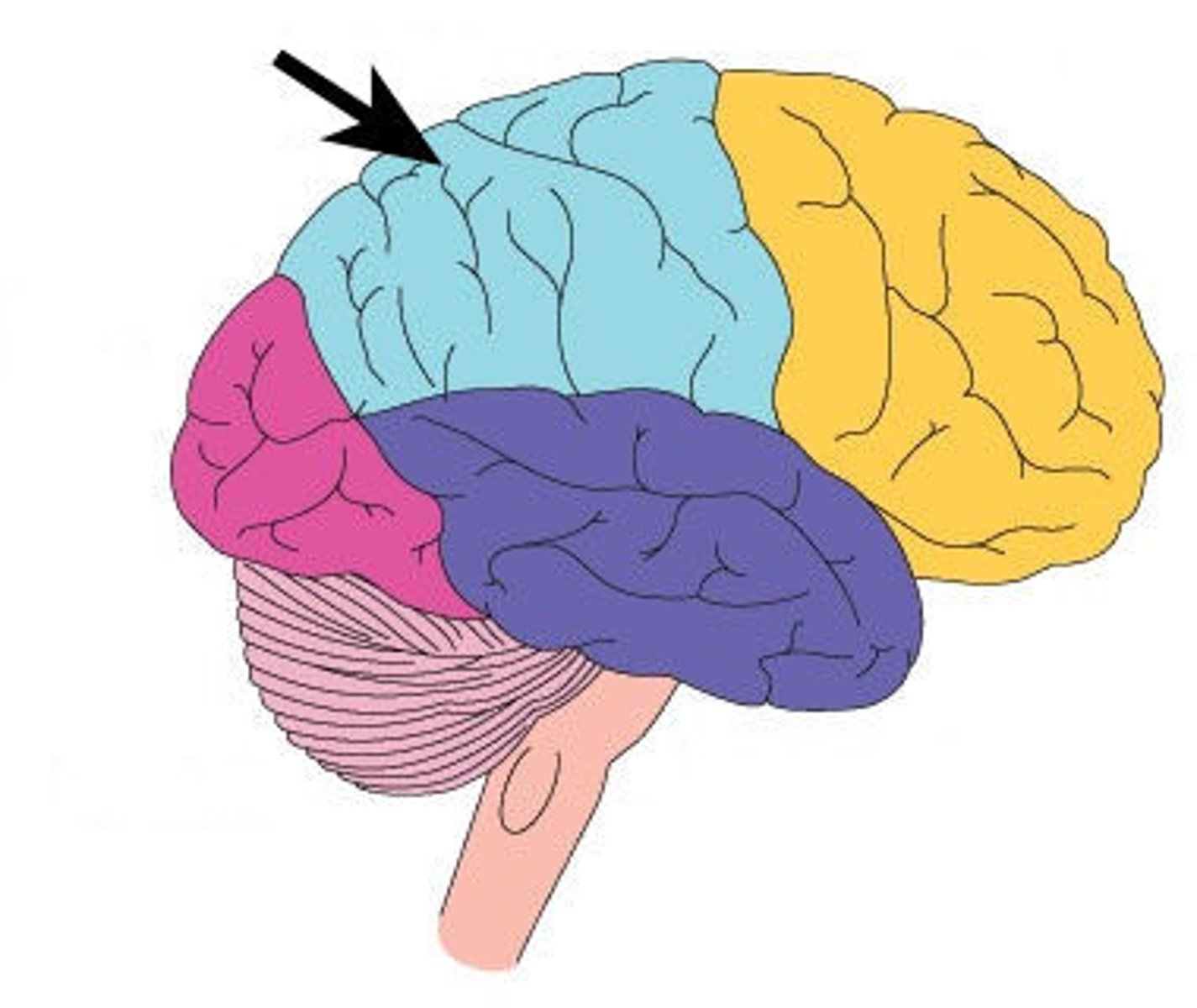
Parietal Lobe
somatosensory cortex
space/body awareness, math, reading/writing
Postcentral Gyrus
analyzes sensations from the body
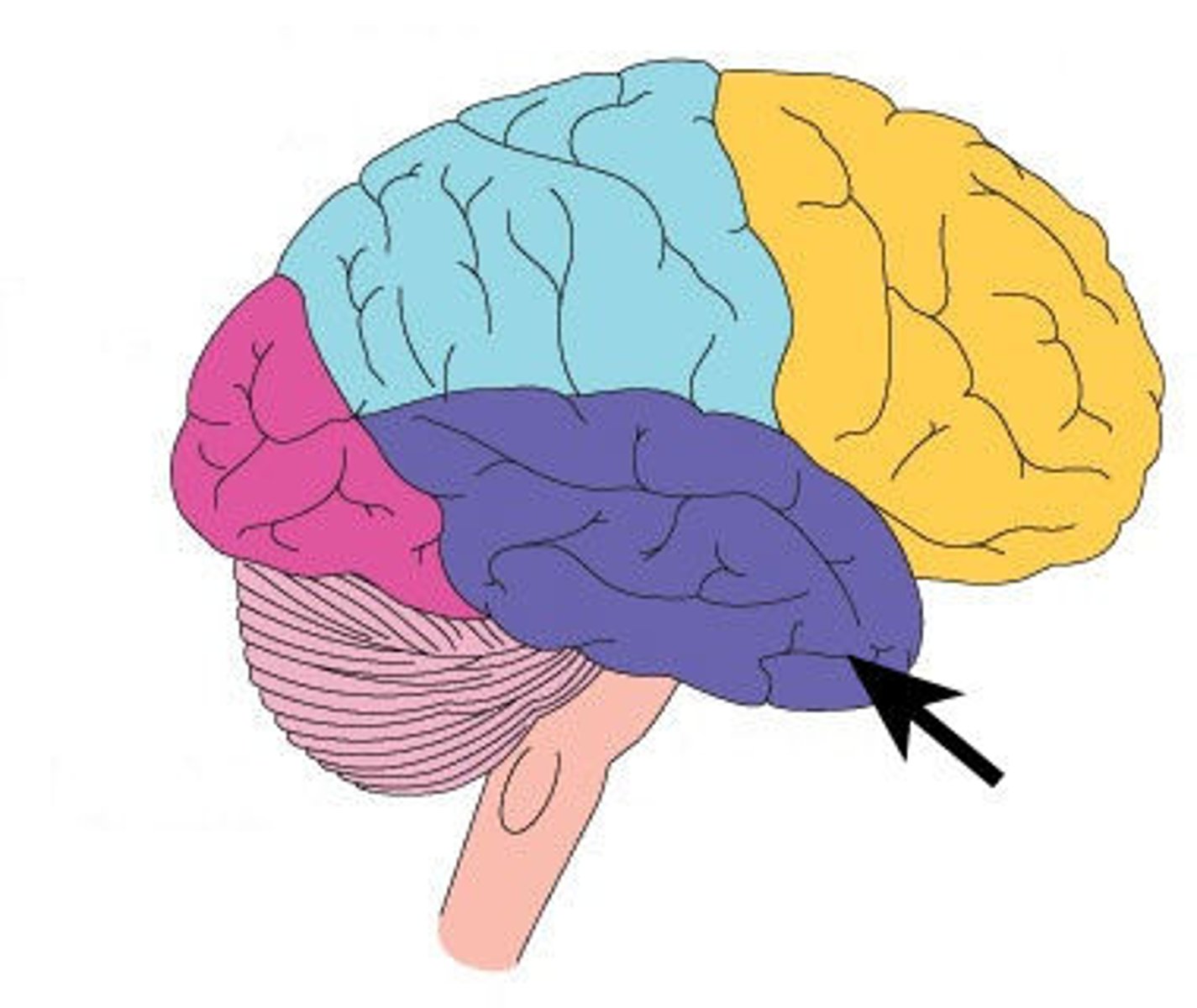
Temporal Lobe
auditory cortex
object recognition, face recognition, audio processing, word processing
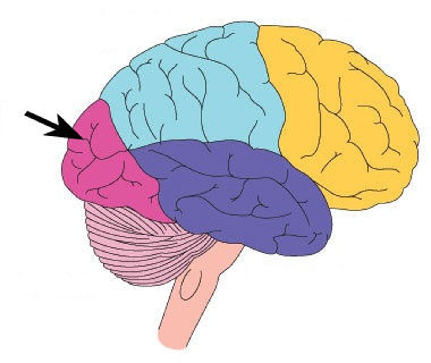
Occipital Lobe
visual cortex
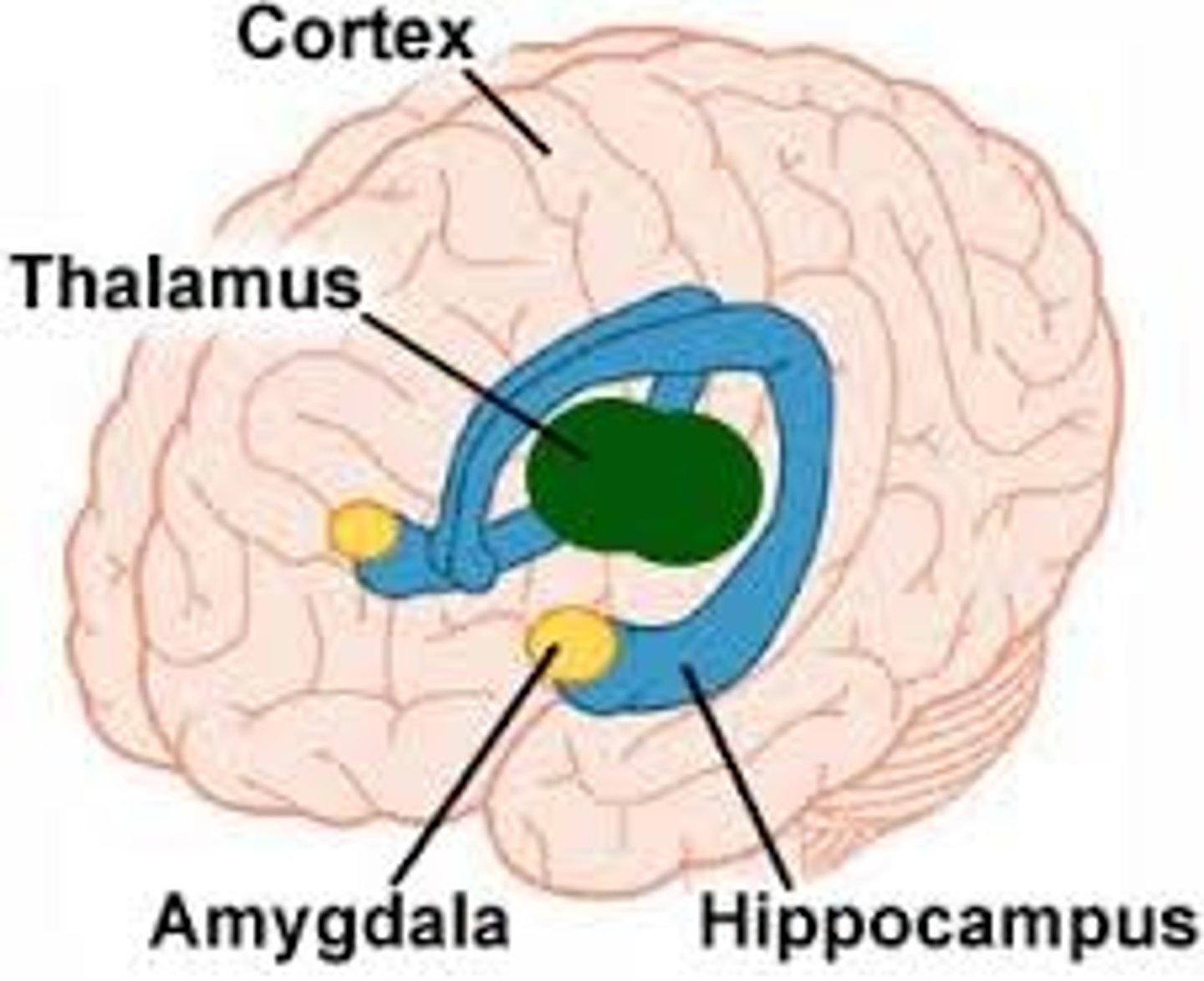
Limbic System
circuit of midline structures that circle the thalamus
regulation of motivation (fleeing, feeding, fighting, ****ing)