PHAS 7050 Exam 2
 Call Kai
 Learn
Learn Practice Test
 Spaced Repetition
 Match
Match Flashcards
 Knowt Play
1/396

There's no tags or description
Looks like no tags are added yet.
Name | Mastery | Learn | Test | Matching | Spaced | Call with Kai | Chat |
|---|
No analytics yet
Send a link to your students to track their progress
397 Terms
liver, gallbladder, pylorus, duodenum, head of pancreas, hepatic flexure, R kidney, ascending and transverse colon
RUQ contents
L lobe of liver, spleen, stomach, body and tail of pancreas, splenic flexure, L kidney, transverse and descending colon
LUQ contents
cecum, appendix, ascending colon, R ovary and fallopian tube, R ureter
RLQ contents
sigmoid colon, descending colon, L ovary and fallopian tube, L ureter
LLQ contents
costovertebral angle
angle formed by the 12th rib and the vertebral column on the posterior thorax, overlying the kidney; allows for assessment of kidney pathology
inferior to renal arteries
most aneurysms of the abdominal aorta occur?
iliac arteries
are sometimes palpable in the lower abdomen
femoral arteries
palpable in the groin/inguinal area, palpation dependent on habitus
xiphoid to symphysis pubis
full exposure of the abdomen requires exposure of
supine, knees flexed, feet flat on table, draped appropriately; good lighting
proper approach and positioning for abdominal exam
inspection, auscultation, percussion, palpation, abdominal vasculature
proper sequence of abdominal exam
scars, striae, dilated veins, rashes, lesions, color; pulsations, contour, umbilicus
noted with inspection of abdomen
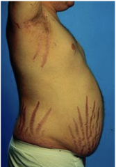
Cushing's disease
pink-purple striae
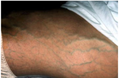
portal hypertension (cirrhosis, IVC obstruction)
dilated veins
visible aortic pulsation
can indicate abdominal aortic aneurysm or small size
scaphoid abdomen
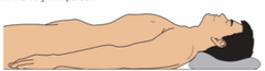
flat abdomen

rounded abdomen
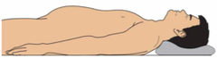
protuberant abdomen
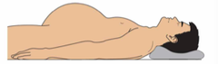
ascites
may be indicated by bulging flanks due to free fluid
umbilical hernia
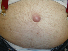
ventral hernia
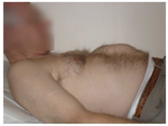
auscultation
performed first during abdominal exam as bowel sounds may be altered if listening after percussion and palpation; use diaphragm
borborygmi
loud, gurgling bowel sounds signaling increased motility or hyperperistalsis; occurs with early bowel obstruction, gastroenteritis, diarrhea
absent bowel sounds
may be secondary to bowel obstruction, abdominal surgery, paralytic ileus, or peritonitis; listen for minimum 1-2 min/quadrant
bruits
abnormal "swishing" sounds; may indicate vascular stenosis or AAA
flat
heard on percussion over bone or heavy muscle
dull
heard over normal liver or heart, over a lung or abdominal mass, or over a lung area with accumulated fluid
resonant
normal lung sound
hyperresonant
heard when there is air trapping in lung; COPD or pneumothorax
tympanitic
heard on percussion over gastric air bubble
percussion
used to assess amount and distribution of gas, identify masses that are solid/fluid filled, estimate liver size
right mid clavicular line
followed along chest and abdomen when percussing for liver size
tympany to dullness
percussed with lower liver border
resonance to dullness
percussed with upper liver border
light palpation
used to note tenderness, muscular resistance, guarding, and superficial organs and masses
deep palpation
used to delineate masses (location, size, shape, consistency, tenderness, pulsations)
pregnant uterus
may be palpated above the level of the symphysis pubis in the midline; physiologic mass
diverticulitis, AAA, colon cancer, distended bladder, dilated loop of bowel
pathologic/concerning masses found on deep palpation
left hand under the abdomen, right hand over into RUQ
bimanual technique for palpating liver edge
hooking technique
helpful for liver assessment with obese patients
two handed technique
use both hands to palpate edge of liver
right lateral decubitus
used for assessment of spleen
CVA tednerness
indicator for pyelonephritis
size of abdominal aorta
McBurney's Point
RLQ point (1/3 of the way from the ASIS to the umbilicus) tenderness indicative of appendicitis
guarding
contraction of abdominal wall
voluntary guarding
may diminish if patient is distracted
involuntary guarding
will persist, suggests peritonitis
rigidity
involuntary contraction of the abdominal wall from peritoneal inflammation, usually firm, stiff
rebound tenderness
peritoneal inflammation causes increased pain when the examiner takes away his/her hand abruptly
Rovsing's sign
pain in RLQ with palpation of LLQ indicative of appendicitis
referred rebound sign
complaint of pain in RLQ after palpating LLQ and taking hand away abruptly
psoas sign
pain in RLQ with contraction or stretch of psoas muscle
obturator sign
RLQ pain with stretch of obturator (hip flexion and internal rotation)
rectal exam, pelvic exam
always performed on patients with abdominal pain
PID, adnexal inflammation/mass/cysts/abscess
should be considered in F patients
shifting dullness
percuss from area of tympany to area of dullness, turn to R side and percuss again, dullness should shift (dullness indicates fluid presence)
Murphy's Sign
used to test for acute cholecystitis; deeply palpate the RUQ and ask patient to take a deep breath; catching breath or stopping due to pain is positive finding
C7
vertebra prominens, easily palpable at back of neck
ribs 7,8
at level of inferior angle of scapula
just above the clavicle
location of apex of lungs
6th rib, midclavicular line; 8th rib, midaxillary line
location of inferior border of lungs anteriorly
T10
inferior border of lungs posteriorly
T3 spinous process
level of spine of scapula, medial border of oblique fissures
6th rib, midclavicular line
lateral border of oblique fissures
4th rib anteriorly, 5th rib (midaxillary line) posteriorly
horizontal fissure of R lung
Sternal Angle (Angle of Louis)
anterior thoracic landmark where the body of the sternum meets the manubrium; site of tracheal bifurcation into L and R bronchus
T4 spinous process
location of bifurcation of trachea posteriorly
pleurae
serous membranes lubricated with pleural fluid; allow lungs to move easily within ribcage during breathing
visceral pleura
covers outer surface of lung
parietal pleura
line the inner ribcage and upper surface of diaphragm
pleural space
space between visceral and parietal pleura
diaphragm
primary muscle of respiration
scalenes, sternocleidomastoids, pectoralis minor, intercostals
accessory muscles of respiration; indicate respiratory distress
inspection, palpation, percussion, auscultation
order of examination of the thorax and lungs
skin colors, nails (clubbing), tracheal position, shape of chest, thoracic deformities
noted on inspection of thorax
less than
A:P diameter should be (more than/less than) the width of the transverse diameter
barrel chest
increased A:P diameter, indicator of COPD
pectus excavatum
a chest that is hollowed out/funnel chest; congenital
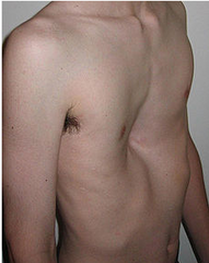
pectus carinatum
forward protrusion of the sternum; pigeon chest; congenital
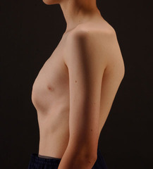
kyphosis
excessive outward curvature of the spine, causing hunching of the back.
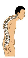
kyphoscoliosis
combination of kyphosis and scoliosis, which may produce a severe restrictive lung defect as a result of poor lung expansion
rate, rhythm, chest wall movement, effort
noted on inspection of breathing
retractions
inward movement of the chest wall (intercostals) with inspiration; usually a sign of respiratory distress
tachypnea
rapid breathing; more than 20 cycles/minute
hyperpnea/hyperventilation
rapid deep breathing
bradypnea
slow breathing; less than 12 breaths/minute
apnea
absence of breathing
Cheyne-Stokes Breathing
hyperpnea alternating with apnea; may be normal in infants
tactile fremitus
palpable vibrations transmitted through bronchopulmonary tree to chest wall; use the ulnar surface of hands/bony palmar surface at base of fingers
increased
fremitus with pneumonia (fluid in lungs)
decreased/absent
fremitus with air trapping (COPD/pneumothorax)
pneumothorax
air in the pleural cavity; may cause unilateral changes when assessing chest expansion
resonant
percussion of healthy lung
flat
percussion of pleural effusion
dull
oercussion of lobar pneumonia
hyperresonant
percussion of COPD, pneumothorax
tympanitic
percussion of large pneumothorax
diaphragmatic excursion
distance that the diaphragm moves with inhalation; percuss on deep breath and exhalation