Micturition and urinanalysis
 Call Kai
 Learn
Learn Practice Test
 Spaced Repetition
 Match
Match Flashcards
 Knowt Play
1/15

Earn XP
Description and Tags
Describe the micturition process in a normal animal Explain behavioural relevance of micturition Evaluate the principles of performing a urinalysis Discuss the relevance of urine examination based on normal renal function
Name | Mastery | Learn | Test | Matching | Spaced | Call with Kai | Chat |
|---|
No analytics yet
Send a link to your students to track their progress
16 Terms
what is micturition?
Micturition is the formal medical term for the act of urination, or the process by which the urinary bladder expels urine through the urethra.
It is a complex physiological event involving both voluntary and involuntary muscle contractions coordinated by the nervous system.
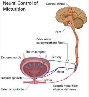
what are the two phases of micturition?
Storage Phase: The bladder acts as a reservoir. The detrusor muscle (bladder wall) remains relaxed, while the internal and external urethral sphincters are contracted to prevent leakage.
Voiding Phase: Once the bladder reaches a certain volume, stretch receptors trigger the micturition reflex. This causes the detrusor muscle to contract and the sphincters to relax, allowing urine to flow out
this pic is acc a video
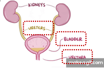
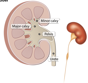
ureter-urine transport from kidney to bladder
Renal pelvis → urine composition
Urine leaving the renal pelvis is largely unchanged in composition
Exception (horse): mucus secretion in renal pelvis/upper ureter → viscous/cloudy urine
Ureters → transport system
Act as conduits for urine flow
Transport driven by peristaltic contractions originating from the renal pelvis
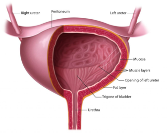
Entry into bladder (trigone region)
Ureters enter the bladder at the proximal angles of the trigone
Follow an oblique intramural course through the bladder wall
Functional significance
Oblique entry creates a valve-like mechanism
Prevents urinary reflux (vesicoureteral reflux)
Protects kidneys from backflow pressure and infection
bladder
1. Bladder wall architecture
Smooth muscle layers (detrusor muscle) → enable contraction for voiding
Transitional epithelium (urothelium) → specialised for stretch
Forms rugae → allows expansion as bladder fills
Helps accommodate volume without large pressure increase
→ prevents back-pressure on kidneys and preserves Glomerular Filtration Rate
2. Sphincters and outlet control
•Internal urethral sphincter (IUS)
•Thickened smooth muscle at bladder neck
•Involuntary control
•External urethral sphincter (EUS)
•Striated muscle
•Voluntary control
3. Innervation (overview)
•Controlled by the autonomic nervous system (details later lecture)
•Coordinates storage vs voiding phases
4. Ureter–bladder interface
•Ureters pass through the detrusor muscle at the trigone
•Entry supports one-way flow into bladder (anti-reflux mechanism)
5. Sensory function
•Bladder wall contains stretch-sensitive receptors
•Detect filling → initiate micturition reflex
6. Clinical/functional relevance
•Bladder position varies with filling state → important during examination
•Proper compliance prevents:
•↑ pressure transmission to kidneys
•↓ renal function (GFR compromise)
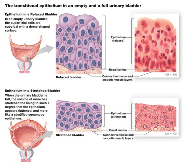
bladder characteristics in different states
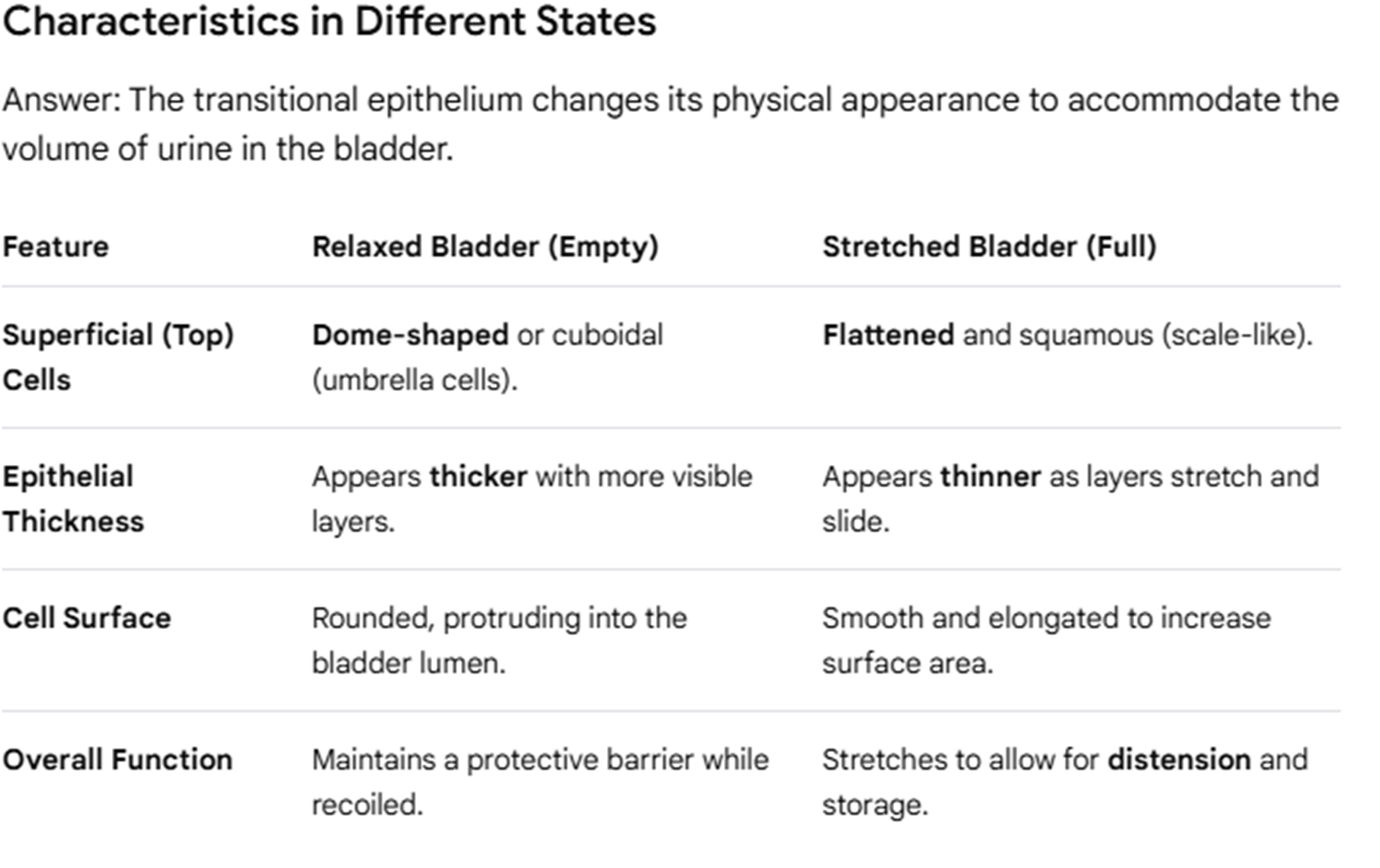
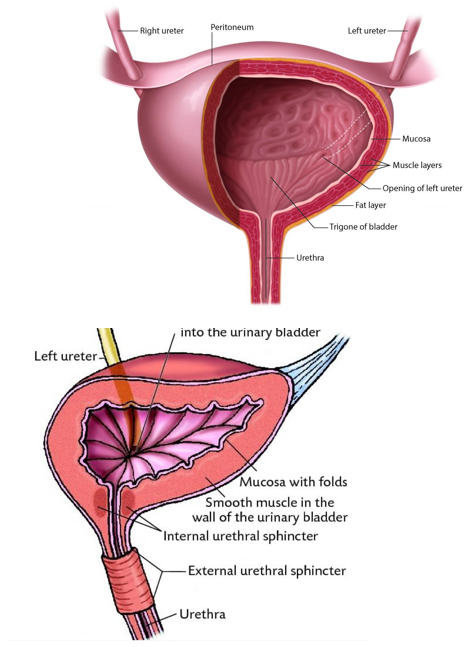
bladder emptying
§Contractions of the muscle cells in the wall of the renal pelvis force - peristaltic contractions- urine produced out to the ureters into the bladder
§The bladder slowly fills up as there is no sphincter.
§As bladder pressure fills up, ureters get compressed at the point they enter bladder.
§IUS keeps urethral opening closed as long as pressure in bladder is not high.
§When bladder fills, stimulates stretch sensitive nerve endings – due time, detrusor muscle contracts
§+ve feedback - detrusor contracts, pressure in bladder increases.
§IUS opens, more tension at neck of bladder – stops the contraction of EUS
§+ve feedback – urine flow stimulates sensory cells in urethra, increases activity of parasympathetic fibers to detrusor muscle -> to increase contraction
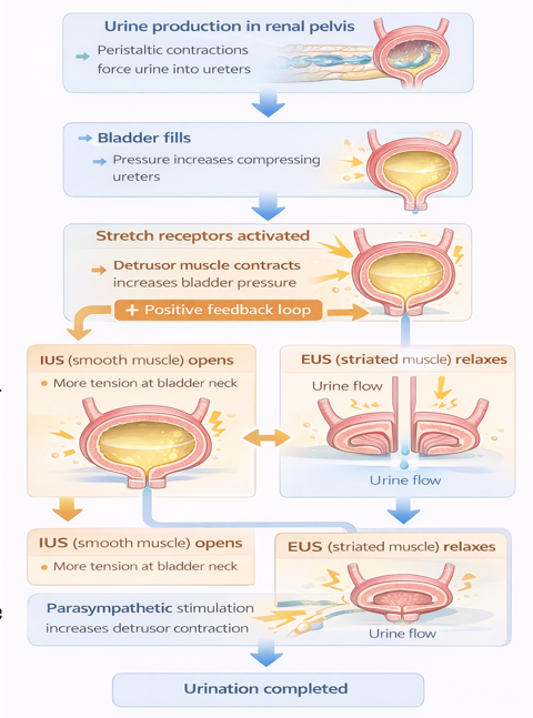
behavioural importance
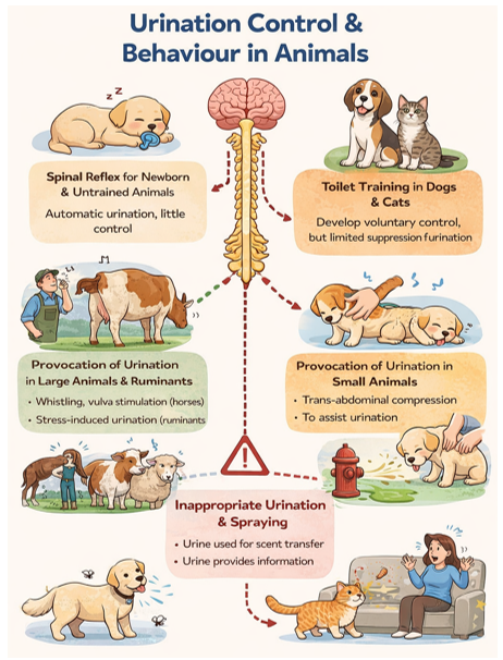
§Newborn and untrained animals – urination is largely a spinal reflex
§Dogs and cats – toilet training means to develop voluntary control. Remember: voluntary suppression is only to a certain level.
§Provocation of urination
Large animals - whistling, stimulation of vulva
Ruminants - stress induced urination – very common
Small animal – trans-abdominal compression
§Important to recognise inappropriate urination and spraying
§Urine used for scent transfer
§Urine also a way to provide more information
urinalysis
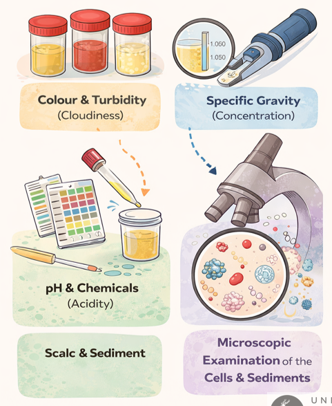
Urinalysis is a diagnostic investigation that involves the systematic examination of urine to assess renal function, urinary tract integrity, and overall metabolic and hydration status.
§Assessment of colour and turbidity (cloudiness).
§Measurement of specific gravity (concentration).
§Measurement of pH (acidity) and chemical components.
§Microscopic examination of the cells and sediments
urine collection and storage
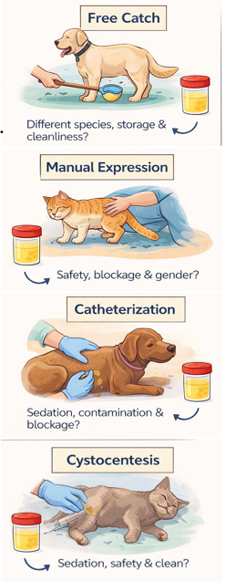
1. Free catch
Free-catch urine collection is non-invasive and suitable for many species, but the method is prone to contamination from skin, hair, litter, or the environment. Fresh midstream samples are preferred, and delays in analysis or improper storage can alter pH, cell integrity, and bacterial counts.
2. Manual expression
Manual expression involves external pressure on the bladder and should only be performed when urethral obstruction has been ruled out to avoid bladder rupture. It is generally easier in small animals, may be uncomfortable, and should be used cautiously with consideration of patient size, temperament, and sex.
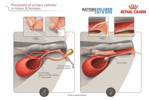
3. Catheterization
Catheterization allows relatively clean urine collection and is particularly useful when bladder emptying is required, but it may require sedation and technical skill.
4. Cystocentesis
Cystocentesis involves percutaneous needle aspiration of urine from the bladder and provides the cleanest sample for culture and sensitivity testing. It may require sedation in some patients
urine collection and storage cont. on gender
urine colour species variation
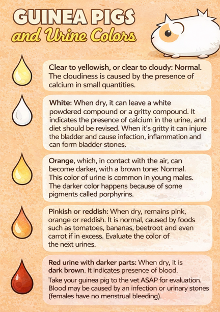
§Rabbits, horses, guineas have naturally occurring cloudy urine
§Rabbit/ GP urine can be discoloured but be normal
§Diet/ stress/ dehydration can cause discolouration.
urine composition - dipstick analysis
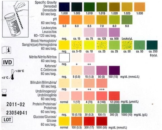
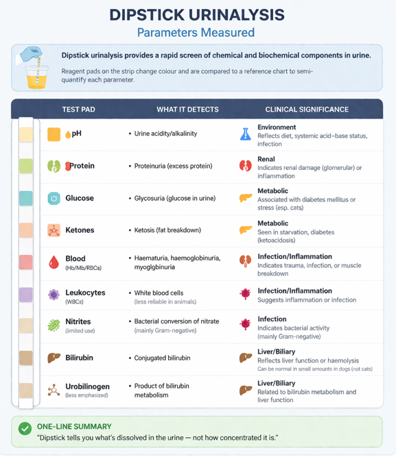
§Using urine strip, drip with pipet or dip into urine specimen.
§Test pathological colour changes vs normal colour change.
§Read results based on the colours *Discussed more in practical
urine specific gravity - refractometer
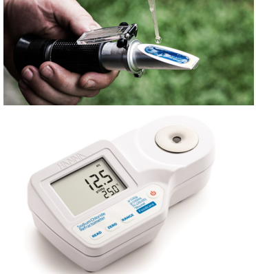
§Measured clinically using a Refractometer.
1. Evaluates renal function by assessing whether water is being excreted or conserved appropriately, according to need.
2. Provides an approximate guide to urinary solute concentration that is sufficiently accurate for clinical purposes.
§Concentrated urine: USG >1.030 (dog) or >1.035 (cat)
§Dilute urine: USG <1.008
§Moderately concentrated urine: USG 1.013 to 1.029 (dog) or 1.034 (cat)
§Inappropriately dilute urine: USG <1.030 (dog) or <1.035 (cat) in a dehydrated animal
§Isosthenuria: USG 1.008 to 1.012
To know if concentration is inappropriate, you must know hydration status!!!! (PCV/TP of patient’s blood)
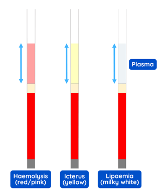
urine specific gravity - PCV vs TP
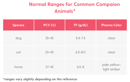
Packed Cell Volume (PCV) and Total Protein (TP) are two of the most valuable, rapid "bedside" blood tests used in veterinary medicine.
While they are blood tests, they are often used alongside urinalysis (specifically Urine Specific Gravity - USG) to immediately assess hydration, blood loss, and protein balance.
microscopic examination of urine
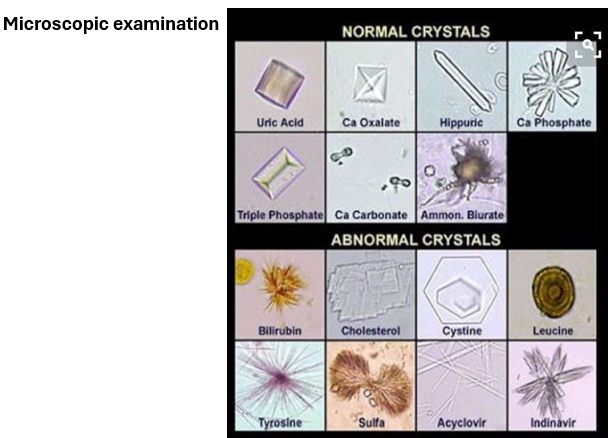
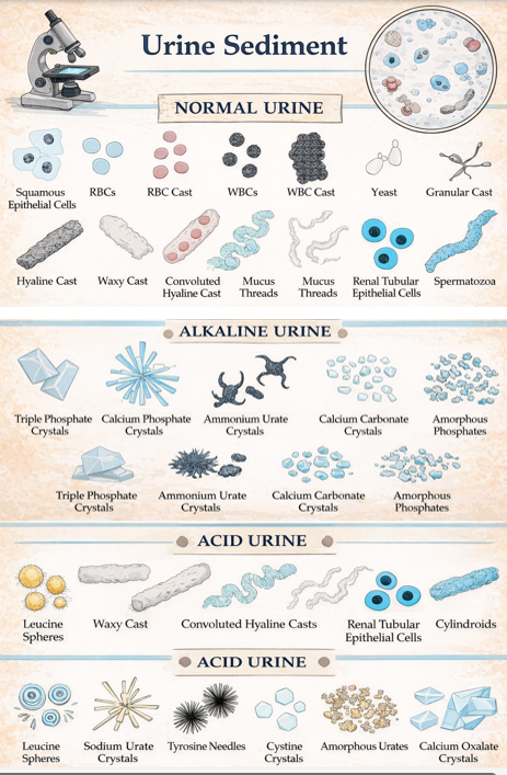
Observe urine under a microscope to examine cells and crystals.
§Sedimentation will produce better finding.
§Potentially able to identify type of bladder calculi based on type of crystal.
§Staining can be used to assist with making things clearer.
summary - urinalysis
Urinalysis is a fundamental diagnostic tool used to assess renal and urinary tract health. It consists of four main components: physical examination, chemical analysis, measurement of urine concentration, and microscopic examination.
Physical examination includes assessment of urine colour and turbidity. Normal appearance varies between species; for example, horses and rabbits naturally produce cloudy urine, which is not necessarily pathological. Diet, stress, and hydration can also affect urine colour.
Chemical analysis is commonly performed using dipsticks to assess parameters such as pH and the presence of biochemical components. Results are interpreted by comparing colour changes on the strip to reference scales.
Urine concentration is assessed by measuring urine specific gravity using a refractometer, providing information on renal concentrating ability.
Finally, microscopic examination of urine sediment allows identification of cells, casts, crystals, and microorganisms. Sedimentation improves diagnostic yield and can help identify the type of urinary crystals present, which may indicate bladder calculi.]
![<p><span>Urinalysis is a fundamental diagnostic tool used to assess renal and urinary tract health. It consists of four main components: physical examination, chemical analysis, measurement of urine concentration, and microscopic examination.</span></p><p style="text-align: justify;"><span><strong><u>Physical examination</u> </strong>includes assessment of urine colour and turbidity. Normal appearance varies between species; for example, horses and rabbits naturally produce cloudy urine, which is not necessarily pathological. Diet, stress, and hydration can also affect urine colour.</span></p><p style="text-align: justify;"><span><strong><u>Chemical analysis </u></strong>is commonly performed using dipsticks to assess parameters such as pH and the presence of biochemical components. Results are interpreted by comparing colour changes on the strip to reference scales.</span></p><p style="text-align: justify;"><span><strong><u>Urine concentration </u></strong>is assessed by measuring urine specific gravity using a refractometer, providing information on renal concentrating ability.</span></p><p style="text-align: justify;"><span>Finally, <strong><u>microscopic examination </u></strong>of urine sediment allows identification of cells, casts, crystals, and microorganisms. Sedimentation improves diagnostic yield and can help identify the type of urinary crystals present, which may indicate bladder calculi.]</span></p>](https://assets.knowt.com/user-attachments/8d6d8c91-0c85-4606-a9a3-d6f9a9935ce4.png)
If an animal cannot empty its bladder properly, what is the most likely long-term effect on kidney function?
the renal tract system is a connected pathway and pressure can travel backwards
that means bladder dysfunction can eventually affect the ureters, kidneys, glomerular filtration