Vision, Optic Tract, Optic Tract Injuries and Implications
1/130
There's no tags or description
Looks like no tags are added yet.
Name | Mastery | Learn | Test | Matching | Spaced |
|---|
No study sessions yet.
131 Terms
corrugator supercilii
muscle of the eye
depresses and pulls the eyebrow medially
corrugator supercilii expresses
expresses concern or confusion
orbicularis oculi
muscle of the eyes
involuntarily closes the eyelid such as when blinking or sleeping (palpebral fibers)
forcefully closes the eye such as when squinting or expressing pain (orbital fibers)
orbicularis oculi innervated by
CN7 (facial nerve)
extraocular muscles
7 muscles that act to move the eye and one muscle that elevates the eyelid (levator palpebrae)
levator palpebrae
elevates the eyelid
fast-twitch extra ocular muscles provide
linear and rotary motion of the eyeball, allowing for scanning the visual field
optokinetic reflex
stabilizes the visual field as the head moves through the surrounding environment
______ _______ of the eye muscles is necessary for coordinated, simultaneous movements, producing complementary images from the right and left visual fields
symmetrical cooperation
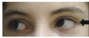
unilateral weakness or paralysis can lead to
diplopia
diplopia
double-vision; limits the ability of the eyes to scan the visual field
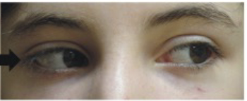
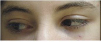
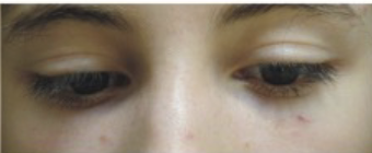
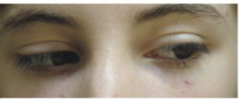
ptosis
refers to drooping of the eyelid and may result from weakness of the levator palpebrae
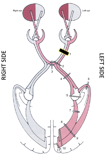
a complete lesion of the optic nerve anterior to the optic chiasm leads to
complete ipsilateral field loss
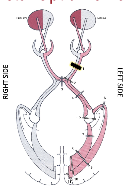
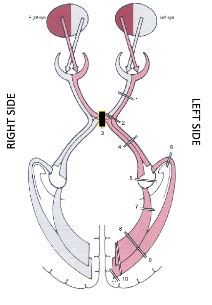
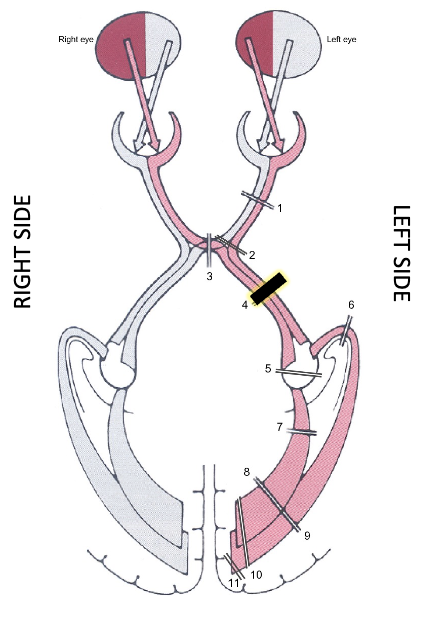
lesions at the optic chiasm cause
bitemporal deficits, or loss of peripheral vision on both sides
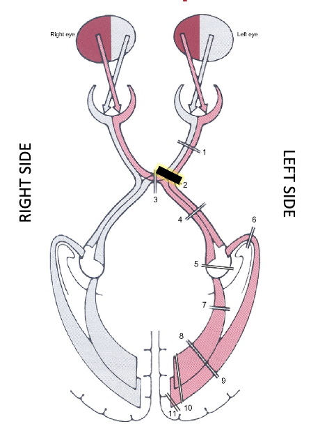
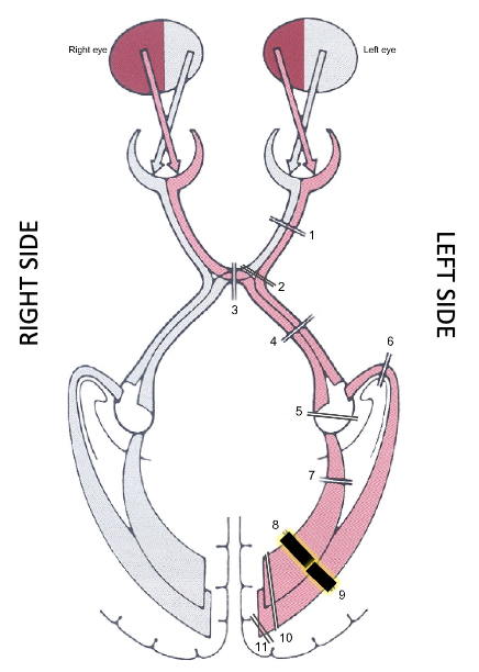
lesions posterior to the optic chiasm or in the occipital lobe of the brain contribute to
partial or full homonymous hemianopsia
homonymous hemianopsia
loss of vision on the same side of each visual field
individuals with visual field loss may benefit from
cueing to scan the environment or from specialized prism glasses to “fill in” the missing visual field
sclera and cornea make up the
outer shell
sclera
thick fibrous capsule around the eye, helps give shapecon
cornea
right at the front of the eye, attaches to the sclera, lets light in
anterior cavity has both an
anterior and posterior chamber
anterior chamber of anterior cavity is filled with
aqueous humor
anterior chamber of anterior cavity is between
iris and cornea
aqueous humor
watery fluid substance that provides pressure so that the eye doesn’t collapse in on itself
posterior chamber of anterior cavity is between
lens and iris
posterior chamber of anterior cavity is filled with
aqueous humor
lens
refracts light onto the retina at fovea centralis
ciliary body and suspensory ligaments help the lens to
change shape to refract light
posterior cavity is where?
behind the lens
posterior cavity is filled with
vitreous humor
vitreous humor
gelatinous, helps keep shape in the posterior cavity
choroid
vascular layer
extrinsic muscles of the eye (6 + 1)
4 rectus muscles, 2 oblique muscles +1
superior rectus
inferior rectus
medial rectus
lateral rectus
superior oblique
inferior oblique
levator palpebrae (+1)
superior rectus
pulls eye up
inferior rectus
pulls eye down
medial rectus
pulls eye medially
lateral rectus
pulls eye laterally
superior oblique
pulls eye down and out
inferior oblique
pulls eye up and out
optic nerve innervates the
retina
facial nerve innervates
orbicularis oculli
Cranial nerve 3 innervates
superior rectus muscle, medial rectus muscle, inferior oblique, inferior rectus muscle
cranial nerve 4 innervates
superior oblique muscle
CN6 innervates
lateral rectus muscle
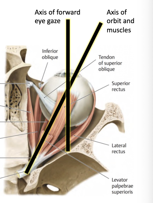
all muscles of eye attach at
common tendinous ring
true or false: axis of forward gaze and axis of orbit and muscles are aligned.
false

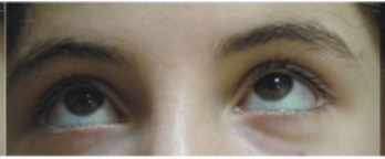
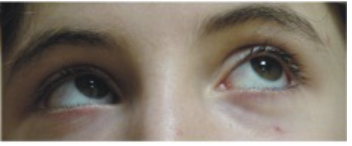
what muscles are involved?
right superior rectus
left inferior oblique
right superior rectus
left superior rectus
right inferior oblique
left superior rectus
right lateral rectus
left medial rectus
right medial rectus
left lateral rectus
right inferior rectus
left superior oblique
right and left inferior rectus
right superior oblique
left inferior rectus
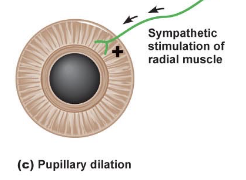
iris has 2 muslces
outer radial muscle, inner circular muscle
parasympathetic (r&d) stimulates the circular muscle resulting in
pupillary constriction from sphincter pupillae
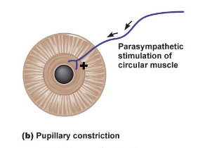
sympathetic (fof) stimulates the radial muscle causing
pupillary dilation from dilator pupillae
the optical apparatus mechanistically collects and focuses light at ___:____ on the retina
280:1
optic disc
where the optic nerve enters the eye and collects info from the retina; blindspot - there are no rods or cones
central 5 degress (radial) contains ____% of all RGCs (retinal ganglion cells)
12
central 10 degrees contains __% of RGCs
34
central 20 degrees contains __% of RGS
54
_____ input is a primary component of ADLs and IADLs
visual
two pathways from primary visual cortex (PVC)
dorsal stream and ventral stream
dorsal stream
the “where” pathway (to parietal lobe)
location, movement, spatial relations
ventral stream
the “what” pathway (to temporal lobe)
color, texture, shape, size, detail
memory
pre-chiasmatic lesions affect
one eye only
optical apparatus lesion
damage to lens, cornea, or sclera
post/retro-chiasmatic lesions usually affect
both eyes and require extensive screening
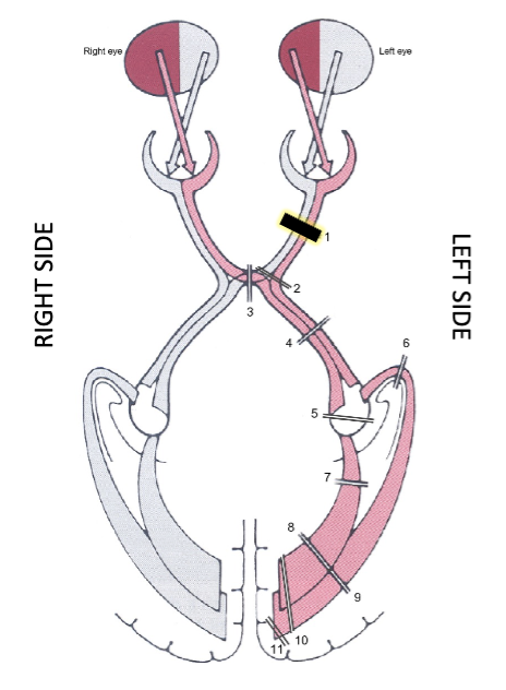
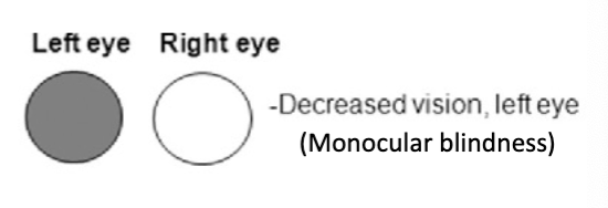
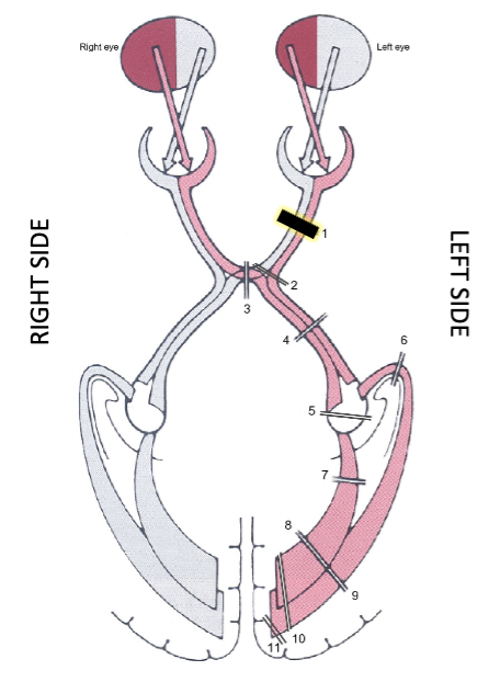
what is it? what does it result in?
distal optic nerve lesion
results in monocular blindness
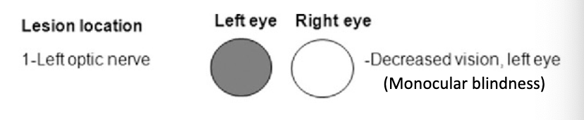
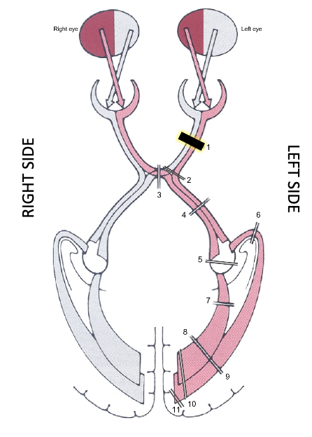
distal optic nerve lesion; progressive lesion of the optic nerve typically causes
tunnel vision
distal optic nerve lesion; acute lesion or untreated progression typically causes
unilateral blindness (anopia)

distal optic nerve lesion; partial acute lesion can cause
unilateral scotoma/anopia
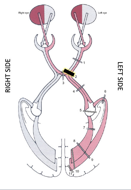
proximal optic nerve lesion
scotoma
partial loss of vision in a visual field
proximal optic nerve lesion
junctional scotoma

what is it? what does it result in?
midsagittal optic chiasm lesion
bitemporal hemianopia, ipsilateral fibers remain intact but decussation fibers are lesioned
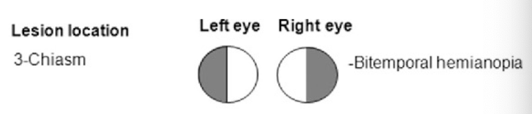
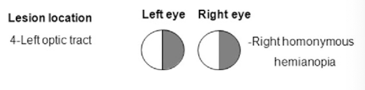
what is it? what does it result in?
optic tract lesion
complete right or left visual field deficit, right homonymous hemianopia
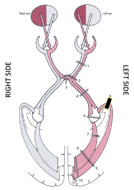
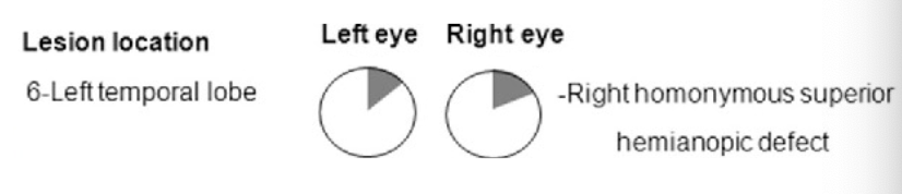
what is it? what does it result in"?
temporal lope optic radiations lesion
right homonymous superior hemianopic defect (“pie in the sky”)
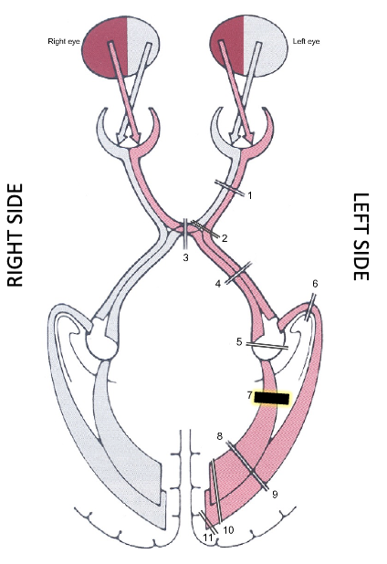
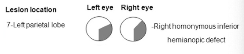
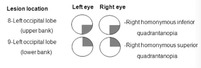
what is it? what does it result in?
parietal lobe optic radiations lesion
right homonymous inferior hemianopic defect (“pizza on the floor”)
what is it? what does it result in?
occipital lobe optic radiations lesion
“pizza on the floor” anopia, “pie in the sky” anopia, put together: homonymous hemianopia
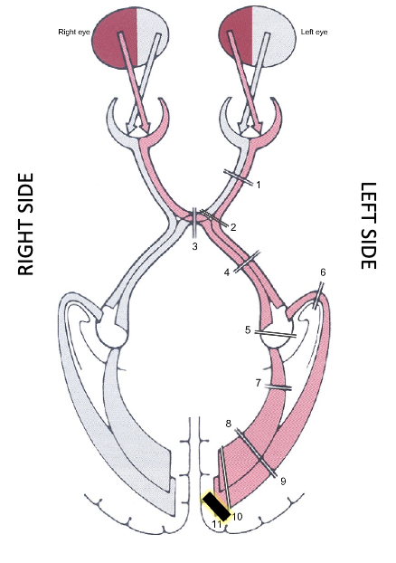
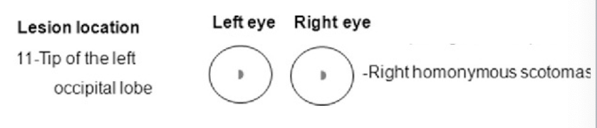
what is it? what does it result in?
occipital lobe optic radiations lesion
right homonymous scotomas
CVA lesions: Middle Cerebral Artery supplies
lateral surfaces of cerebral cortex (frontal, parietal, and temporal lobes)
CVA lesions: Middle Cerebral Artery complications:
contralateral hemianopsia, visuospatial deficits
CVA lesions: anterior cerebral artery supplies
medial aspects of cerebral cortex (frontal and parietal lobes)
CVA lesions: anterior cerebral artery complication:
homonymous hemianopsia
CVA lesions: posterior cerebral artery supplies
occipital lobe, inferior aspects of temporal lobe, parts of brainstem
CVA lesions: posterior cerebral artery complications
CN3 paralysis, homonymous hemianopsia, visual agnosia
CVA lesions: vertebral basilar artery supplies
medulla, pons, CNs, cerebellum
CVA lesions: vertebral/basilar artery complications
horner’s syndrome (pupil constriction, ptosis, anhidrosis), nystagmus, CN paralyses, paralysis of conjugate gaze (eyes don’t move together)
aphasia
loss of ability to understand or express speech
Broca’s Area
frontal lobe
motor speech
language production
expressive aphasia (Broca’s aphasia) — cannot express what they are trying to say
word salad
Wernicke’s Area
temporal lobe
associative auditory
language comprehension
receptive aphasia — can’t understand what someone is saying
Wernicke’s and Broca’s area would be impacted by CVA lesions to
middle cerebral artery and some of anterior cerebral artery and PCA
pre-chiasmatic lesions affect
one eye only
post-/retro-chiasmatic lesions usually affect
both eyes

acute lesion or untreated progression typically causes
unilateral blindness (anopia)
partial acute lesions can cause
unilateral scotoma/anopia