Lecture 17 Cell Attachment
1/126
There's no tags or description
Looks like no tags are added yet.
Name | Mastery | Learn | Test | Matching | Spaced |
|---|
No study sessions yet.
127 Terms
List what is necessary for cell viability
Cells need exposure to blood, extracellular fluid, or lymph
Majority of cells require attachment to a solid surface for viability,
growth, migration, and differentiation (create and/or secrete
proteins)
Cell type, shape, and behavior is dependent on the type of tissue it
forms
Discuss implications of cells growing on biomaterial surfaces
Biomaterial environment
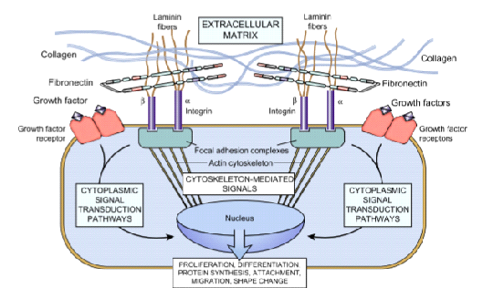
Cell adhesion is mediated by integrins and can trigger
biochemical signaling pathways in the cell
List and define methods for studying cell behavior
In vitro studies
Cell type, shape, and behavior is dependent on the type of tissue it
forms
ECM has different compositions for different tissues
Fibrillar proteins
Amorphous matrix
Adhesive molecules
Fibrillar proteins
Collagen, elastin
Amorphous matrix
glycosaminoglycans (GAGs), proteoglycans, hyaluronic acid
Adhesive molecules
Fibronectin, Laminin
Cell adhesion is mediated by
integrins and can trigger
biochemical signaling pathways in the cel
Cell adhesion is mediated by integrins and can trigger
biochemical signaling pathways in the cel
Lead to cell spread, growth, migration, differentiate
Cell behavior dependent of substrate characteristics like
adhesiveness and mechanical properties
Especially bind to fibronectin and vitronecti
In vitro studies
Helpful for medical science, growing tissues, test biomaterials
Microscopic examination
stains enhance tissue contrast
Histology
Pathology
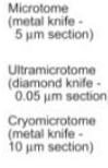
Steps
-ex vivo
-fixation
-dehydration and embedding
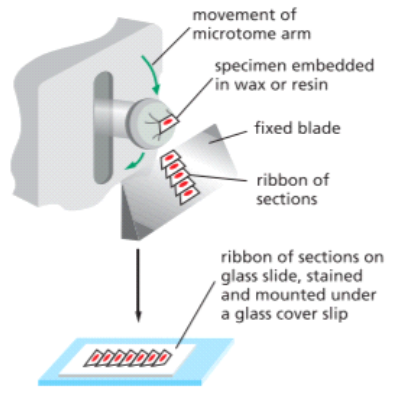
-sectioning
-staining and contrast
-microscopy
Fixation
Formalin, glutaraldehyde, frozen
cells need blood or some kind of fluid to be alive this provides
oxygen remove carbon dioxide provides nutrient and removes waste
where cells are have capillaries throughout
tissue all provide
oxygen and nutrients for cells do job need blood
want cells grown in or on devices so need way for blood to get
on the device
happy cells are attached cells most cells in body attached to either
each other ot ECM (proteins outside of cell)
If not attach the cell actually dies via
integrin, adherence ECM
ONLY TISSUE CELLS ALL NEED TO BE
ATTACHED
once attached would do its job:
proliferation
diffrerenitaie
migrate
all major things need to be attached first
proliferation
(mitosis) (tissue growth is mulplication of cell number not enlargement) (more cells to form tissue
differentiae
(do there function to create ECM and make proteins so DNA TO rna TO PROTEIN (makes cells specialized and proteins develop for the cell want to differentiate and form ECM which means for tissue(more ECM TO FORM TISSUE
migrate
(integrins on ECM proteins)
each type of tissue in the body
has different cells
bone cells
-osteoblasts
fibroblasts
general structural support cells usually in connective tissue are in all kinds of tissues in body and there job is to make ECM (structural component of the tissue)
Each tissue has
varying composition of ECM
ECM
is structural, not cells (think how if ECM must be to make a difference in mechanical
diff connective tissues-
bones lining deep connective tissue seen throughout
attach to the materials
want to attach
bone has
a ton of collagen-dispersed cells
3 ECM
fibrillar proteins
Amorphous proteins
adhesive proteins
fibrillar proteins
collagen + elastin create a structural framework for our ECM there is not a tissue in the body that does not have collagen many different types (collagen is most prevlent ECM prtein and is very stroong)
elastin-
elastic common in stretchy materials like blood vessels /linings and skin, ligaments
Amorphous proteins
filler material , gelatinous (jello-like), lubricants (common in plac with lots of friction want ot decrease, GAGS, proteoglycans , hyaluronic acid (material makes skin plump succulent and resilient)
wont be discussed as much is sugared and proteins keep hydrated
adhesive proteins
- connection points
-fibronectin
-laminin
fibronectin
(if figure how to adsorb in all materials inmplant generates good tissue integration connects cells too ECM (connects to the collagen)
laminin
conencts cells to ECM (sheets of ECM)
collagen
is a rebar structure support binds to
cells connected to
integrins, then laminin, and the red line is ECM, then
The job of another cell fibroblast cells job is to make ECM and be surrounded by ECM
cell attachment and tissue integration important for:
in vitro
in vivo implants
in vitro
- stem cells, regenerative medicine 3D tissues
in vivo implants
body to integrate recognize not attack an implant
have a material coated with proteins coming from tissue with the area and adsorb and have certain proteins attach to integrins that have diff shapes and bind on certain types of protein so for the cell to attach needs certain types of integrins
once cell attach would get alot of integrins attaching (integrins are like velcro the strength is from alot crowd together (aggregate together)) and create a strong attachment and spread out
have a material coated with proteins coming from
tissue with the area and adsorb and have certain proteins attach to integrins that have diff shapes and bind on certain types of protein so for the cell to attach needs certain types of integrins
once cell attach would get alot of
integrins attaching (integrins are like velcro the strength is from alot crowd together (aggregate together)) and create a strong attachment and spread out
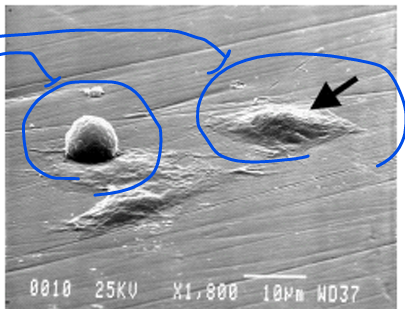
shows cell attaching this is a happy cell
that migrates, proliferate (/multiply) and for ECM which is differentiation DNA to mRNA to protein thus would form tissue
multiply number of cells and migrate to form tissue well
in vitro
this is the starting point to grow tissues and organs
pattern tissue with different cell types
control tissue growth
(can control how and direction of cells spreading and migrating)
poteintial to grow tissue and organs in vitro if can grow
cell attatchment to get
good tissue integration
it is important to study cell behavior tells us about the biocompatibility of our device
can study using :
in vivo
in vitro
in vitro
- cheaper easier and looks how material interacts with cells on a lab bench so take the material place in an environment with blood serum cell types to look at attachment
work involves working with a cell line, which are hard to kill but easy to grow in vitro.
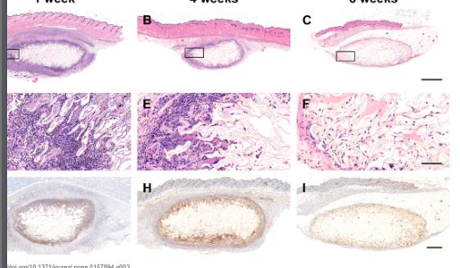
in vivo
- animal testing implant the device in the same location plant in people or the back of rats underneath the skin and wait six weeks and then explant (6 weeks is about the time most tissues take to heal) then cut out the implant and the tissue around it (really interstrested in the tissue around it to look for integration or signs of complications or cell types, proteins present, gives a sense of getting integration or complication)
genomics transcriptions
-DNA->mRNA
What diff ways mRNA was made in the tissue
tells what protein was formed
light microscopy
- tells about shape of cells
stains to enhance tissue contrast (/can be done in vivo and vitro work
histology
- microscopes to study tissue would need a thin slice of tissue and want it to be 1 cell thick (less then 100 microns thick tissue to see through tissue)
pathology
looking for disease tissue using a mechanism of disease (using techniques from the world of histopathology )
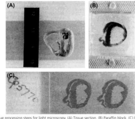
histopathology
take tissue in vitro or in vivo cut off body and do autopsy or biopsy and fixate the tissue (harden so when taken out the body, it would still keep the cells same place within the body as it preserves structural relationships gives the shape and orientation of cells, proteins, molecules and keep the same)
to fixate can freeze the tissue, use formolan or glutaraldehyde
after dehydrate it remove the water and embed it (replacing water with either parffin (stretchy waxy material) or epoxy (strong polymer glue) this creates stronger shape of tissue
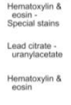
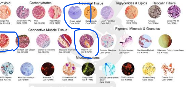
stain and contrast
most common is releveant for whole body which is the H+E stain tell which tissue
(hematoxylin -blue-shows all DNA and nuecli
eosin
-pink binds to cyto plasm ECM
shows CELL BODIES VS tissue structure when together looks purple
stains
highlighting specific proteins cell types
immunohistochemistry
- antibodies to bind and identify any protein then when add to the tissue would add a fluorophore to the antibody (fluoresce molecule that glows in the dark) can also add enzymes that change colors
how section tissue can matter for
ex section egg can see different form of cells so keep in mind
H+M white
is nothing can tell the cell type based on just the shapes
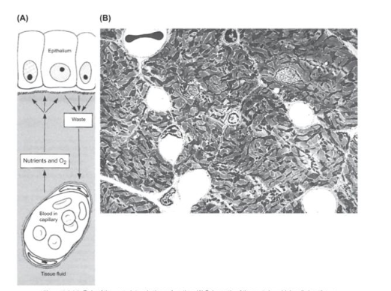
Figure 2.1.4.2 Role of the vasculature in tissue function (A) Schematic of the route by which oslls in a tissue obtain nutrition and oxygenation by diffusion from underlying capillaries. Metabolic waste products diffuse in the reverse direction and are rerrioved into the circulation, in each case, diffusion occum through the fluid that permeates the extracellular matrix. (B) Myocardum, a highly metabolically active tissue, has a rich capill lary network, as demonstrated by transmission electron microscopy. The six open round spaces are capi laries, with a red blood cell seen on cross-section in the capillary in the upper left comer ((A) Reproduced by permission from Cormack D.H. 1987. Ham's Histology, ninth ed. Lippincott, Philadelphia, PA)
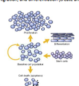
FIGURE 11.1.4.14 Acheving steady-state populations of cells. Total cell numbers in a baneline true population can be regulated by changes in stem cell input, loss of cells through programmed cell death apoptosis, cellular prolifwation or differentiation (Repro sduced by permission from Kumar V, Abbas, A.K. & fausto, N. (Eds (2005) Robbins and Cotran Pathologic Basis of Diane 7th edn Saunders Philidiripihu, PA)
ECM had different tissues
loose connective tissue
fibrous connective tissue
blood
bone
cartilage
adipose tissue
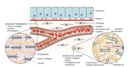
Figure 2.1.4.4 Main components of the extracellular matrix (ECM), including collagen, proteoglycans, and adhesive glycoproteins. Both epithelial cells and fibroblasts interact with ECM via integrins. Basement membranes and interstitial ECM have differing architecture and general composition, although there is some overlap between them. Some ECM components je.g., elastinį are not included for the sake of sim-plification
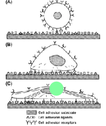
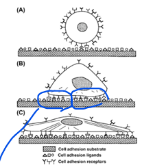
FIGURE II.1.5.16 Progression of anchorage-dependent cell adhe
sion (A) Initial contact of cell with solid substrate. (B) Formation of bonds between cell surface receptors and cell adhesion ligands. (C) Cytoskeletal reorganization with progressive spreading of the cell on the substrate for increased attachment strength. (Reproduced by permission from Massia, S. P. (1999). Cell-Extracellular Matrix Interactions Relevant to Vascular Tissue Engineering, In: Tissue Engi neering of Prosthetic Vascular Grafts, Zilla, P. & Greisler, H. Pieds RG Landes Co., 1999)

FIGURE II.1.3.1 Scanning electron microscopy image of MC3T3-E1 cells at various stages of adherence on polished titanium substrate demonstrating changes in cell morphology during attachment. Attached cells have a flattened morphology and form intimate con-tact with the substrate. Detached cells are round. SEM 1800X
MIGRATION, SHAPE CHANGE
FIGURE II.1.5.3 Integrin-ECM interaction. Schematic showing the mechanisms by which ECM (e.g., collagen, fibronectin, and laminin) and growth factors interact with cells, activate signaling pathways, and can influence gene expression, growth, motility, differentiation, and protein synthesis. Integrins bind ECM and interact with the cytoskeleton at focal adhesion complexes. This can initiate the production of intracel-lular messengers, or can directly mediate nuclear signals. Cell surface receptors for growth factors also initiate second signals. (Reproduced by permission from Kumar, V., Abbas, A. K., Fausto, N. & Aster, J. C. (eds.). (2010), Robbins and Cotran Pathologic Basis of Disease, 8th Edn. Saunders: Philadelphia, PA.)
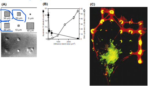
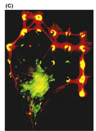
FIGURE II.1.5.17 Effect of spreading on cell growth and apoptosis (A) Schematic diagram showing the initial pattern design containing different-sized square adhesive islands and Nornarsku views of the final shapes of bovine adrenal capillary endothelial cells adherent to the fabri cated substrate. Distances indicate lengths of the square's sides. (B) Apoptotic index (percentage of cells exhibiting positive TUNEL staining) and DNA synthesis index (percentage of nudei labeled with 5-bromodeaxyuridine) after 24 hours, plotted as a function of the projected coll area. Data were obtained only from islands that contained single adherent cells, similar results were obtained with circular or square islands, and with human or bovine endothelial cells. (C) Ruorescence micrograph of an endothelial cell spread over a substrate containing a regular array of small (5 um diameter) circular ECM islands separated by nonadhesive regions created with a microcontact printing technique. Yellow rings and crescents indicate colocalization of vinculin igreen and F-actin (red) within focal adhesions that form only on the regulatory spaced circular ECM slands (A), (B) Reproduced by permission from Chen, C. S. et al. (1997) Geometric control of cell life and death Science, 276, 1425; (C) Reproduced by permission from Ingber, D, E. (2003). Mechanosensation through integrins: Cells act locally but think globally Proc Natl Acad Sci, 100, 1472)
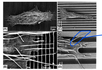
Figure 2.1.3.4 Effect of micro- and nano-patterns on comsal epithelial cell alignment. (A) Cells were aligried perpendicularly to ndges that were 70nm wide and 330 nm apart. (B) Expanded view of (A) show ing flopodia also aligned perpendicularly to the patterns (With increasing width and spacing (1900m ridge width, 2100nm spacing), cells were aligned with the ridges and (D) flopodia were guided by the topographic pattern. (Reprinted from Teixeira, A.1, McKie, GA, Foley J.D., Bertics, PJ, Nealey, PF.. Murphy, CJ, 2006. The effect of environmental factors on the response of human comeal epithelial cels to nanoscale substrate topography, Biomaterials 27 (21), 3945-3954, Copyright (2006), with permission from Elsevier)
Gross examination
Determines overall specimen configuration; many diseases and processes can be diagnosed
Light microscopy (LM)
Studies overall microscopic fissue architecture and cellular structure: different stains allow analysis of, e.g., calcium, fat, matrix, apoptotic cell death, etc.
Transmission electron microscopy (TEM)
Studies ultrastructure (fine structure) and identifies colls and their organelles and environment
Scanning electron microscopy
High-resolution 3D imaging
Genetic Analysis (Genomics)
Maintaining Tissue Architecture Relationships
Information on Total Tissue Without Architecture
protein analysis (proteomics)
maintaining tissue architecture relationships
know tissue to find out cell behavior
information on tota tissue without architecture
physicochemical analysis
maintaining tisue rchitexture relationships
information on total tissue withough architecture
microscopy: electron microscopy
electron microscopy
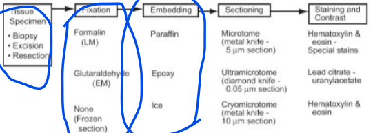
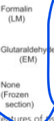
tissue specimen→fixation→embedding→sectioning→staining and contrast


stains diff type of protein and tissue wont ask but relevant to project
H+M white is nothing can tell the cell type based on just the shapes
Perfusion
delivery of blood to a tissue or an organ
Differentiation
is a fundamental biological process defined as the progressive specialization of cells, where they develop distinct structural and functional characteristics (phenotype). This process is driven by the differential expression of specific genes within a cell, leading to a distinct biological profile. As cells specialize, they typically lose the ability to interconvert into other cell types, and more of their "unnecessary" genes are usually irreversibly turned of
Proliferation
is a fundamental cellular process referring to the increase in cell number, achieved through cell division and replication. It is one of the core functional attributes shared by virtually all cells, alongside nutrient uptake, metabolism, and waste removal
Collagen
s the most abundant extracellular protein in the human body and is a ubiquitous and essential component of the native extracellular matrix (ECM). It is critically important for the biomechanical stability of tissues, and is intimately involved in processes such as cell adhesion, migration, growth, differentiation, morphogenesis, and wound healing.
Elastin
is a highly elastic, insoluble polymer that serves as the core structural protein of elastic fibers in the extracellular matrix (ECM). It is one of the principal fibrous structural proteins found in the body, alongside collagen and fibrinogen
Glycosaminoglycans (GAGs)
are a family of negatively charged linear polysaccharides that play crucial roles in the composition and function of the native extracellular matrix (ECM)
Proteoglycans
are a family of amorphous matrix macromolecules that are integral components of the native extracellular matrix (ECM) of soft tissues, along with glycosaminoglycans (GAGs) and hyaluronan
Hyaluronic acid
often referred to as hyaluronan, is a negatively charged linear polysaccharide that constitutes a major family of Glycosaminoglycans (GAGs
is distinguished among GAGs because it is the only GAG that is nonsulfated and generally does not attach to a core protein to form a proteoglycan. It is composed of repeating disaccharide units of N-acetylglucosamine and glucuronic acid
HA is found in the ECM of all body tissues. It plays an important structural role in articular cartilage and skin tissues
Fibronectin
is a key glycoprotein found in the extracellular matrix (ECM) and blood plasma, renowned for its strong cell-adhesive properties and its ability to bind numerous other ECM components, such as collagen, fibrin, and proteoglycans
Biochemical signaling pathways
are the molecular mechanisms by which cells receive and process cues from their environment—including those presented by biomaterials—to regulate essential cellular functions .
1. Mechanotransduction Pathways
2. Integrin and Adhesion Signaling
3. Soluble Factor and Cross-Talk Signaling
4. Immune/Inflammatory Signaling
Implanted biomaterials trigger pathways leading to inflammation:
Reception
The process begins when a signaling molecule, or ligand (such as a hormone, neurotransmitter, or growth factor), binds to a specific receptor. These receptors are typically located on the cell membrane (for hydrophilic ligands like peptides) or inside the cell (for hydrophobic ligands like steroid hormones). The ligand-receptor interaction is highly specific—much like a key fitting into a lock—and it triggers a conformational change in the receptor that activates it.
Signal Transduction
Once the receptor is activated, it initiates a cascade of intracellular signaling events. This often involves the activation of enzymes and the production of secondary messengers (such as cyclic AMP, inositol triphosphate, or calcium ions) that amplify the signal within the cell.
Many pathways rely on phosphorylation cascades, where a series of protein kinases activate one another in sequence, ultimately modifying target proteins that control cellular processes. G-proteins, tyrosine kinases, and ion channels are common players in these transduction systems. Through amplification and integration of signals, the cell ensures that small external changes can lead to significant and precise responses.