Lab cell division
1/11
There's no tags or description
Looks like no tags are added yet.
Name | Mastery | Learn | Test | Matching | Spaced | Call with Kai |
|---|
No analytics yet
Send a link to your students to track their progress
12 Terms
Mitosis
occurs in somatic cells
Somatic cells are all the cells in the body other than sex cells and sex cells form either sperm of oocytes
Somatic cell division: occurs when one cell divides, resulting in two identical cells. Somatic cell division is essential to produce the trillions of new cells for development and growth, as well as to replace old or dying cells and those destroyed from trauma or disease.
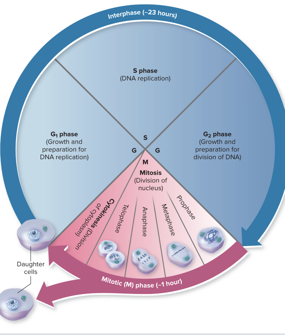
The cell cycle
depicts the steps in the replication of a somatic cell. It consists of all changes the cell undergoes as it divides into two identical cells called daughter cells.
There are two major phases in the cell cycle: interphase and the mitotic (M) phase.
Interphase
is the time the cell prepares for cellular division. This phase is distinctive when viewing cells with a light microscope because the DNA within the nucleus remains in the form of loosely coiled chromatin.
Interphase has three distinct phases: G1, S, and G2.
G phase
(also called the first gap stage) of the cell cycle, cells grow and produce new organelles and other structures needed for DNA replication.
Replication of the centrioles to produce two centriole pairs is also initiated in this phase.
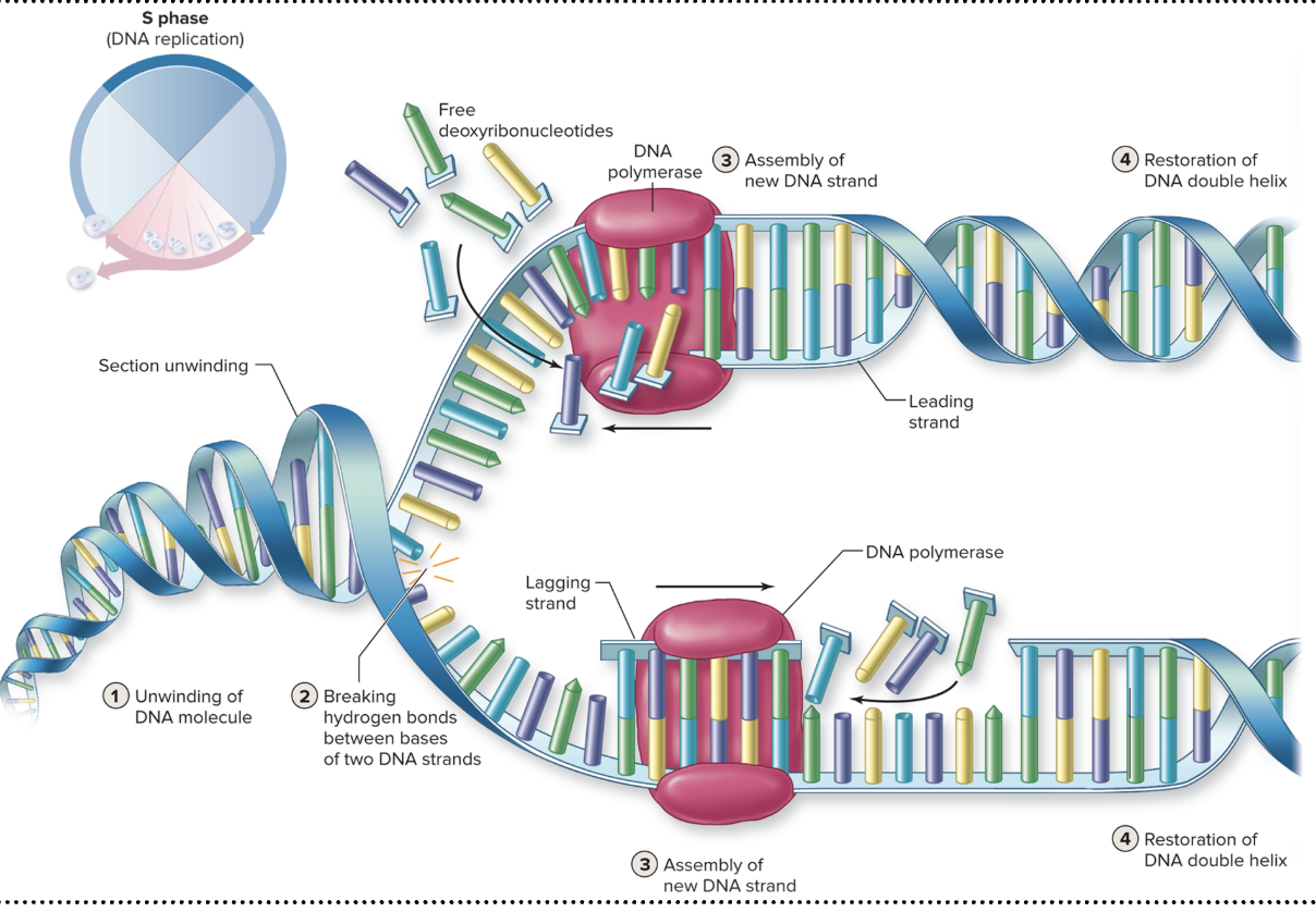
S phase
During the S phase (also called synthesis), the double helix strands of DNA are replicated. Forming DNA requires large numbers of deoxyribonucleotides and the enzyme DNA polymerase. All of these components are located within the nucleus.
The following steps in DNA replication involve unwinding, breaking, assembly, and restoration:
1: Unwinding of DNA molecule. The spiral, complementary DNA strands are unwound from each other by specific enzymes.
2: Breaking the parent strands apart. The hydrogen bonds holding the complementary bases together in the DNA strands are broken. Once the portions of strands are separated, binding proteins ensure that the strands remain separated.
3: Assembly of new DNA strands. Both strands of DNA are read as templates by DNA polymerase enzymes that move along both parental strands (one called the leading strand and the other the lagging strand because of how transcription takes place on each). DNA polymerase assembles new strands of DNA as complementary deoxyribonucleotides are paired. For example, if the base sequence of a small portion of a DNA strand is TTAGCTAGC, then the base sequence of the newly formed complementary DNA strand assembled by DNA polymerase would be AATCGATCG. Complementary base pairs are held together by hydrogen bonds. The bond between nucleotides in the DNA polymer is a phosphodiester bond.
4: Restoration of DNA double helix. The DNA double strands are returned to their coiled, helix structure.
The process continues until the entire lengths of both strands of DNA are replicated. The replicated DNA strands, now called sister chromatids, remain attached at a region called a centromere. The joined sister chromatids form a chromosome. The sister chromatids are separated at the centromeres during mitosis; following their separation, each is called a chromosome.
G2 Phase
The last part of interphase, called the G, phase (or the second gap phase), is brief
During this phase, centriole replication is completed (having produced two centriole pairs present within the cell) and enzymes and other structures needed for the mitotic phase of cell division are synthesized.

Mitotic Phase
Following interphase, cells enter the mitotic (M) phase. Two distinct division events occur in this phase to produce two new cells: mitosis, which is division of the nucleus, and cytokinesis, which is the division of the cytoplasm. Mitosis begins first.
Four consecutive phases take place during mitosis: prophase, metaphase, anaphase, and telophase, which can be remembered with the acronym P-MAT. Each phase merges smoothly into the next in a nonstop process.
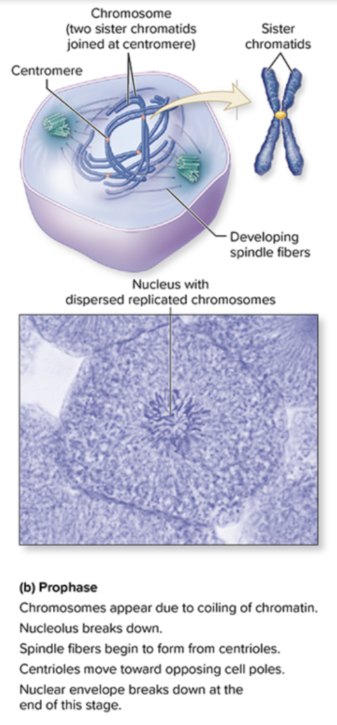
Prophase
is the first stage of mitosis
Chromatin becomes supercoiled into the chromoson that are more maneuverable and are less likely to become tangled during cell division. The DNA and protein within the chromatin coil, wrap, and twist, forming the chromosomes. Chromosomes are composed of the two sister chromatids, which resemble relatively short, thick rods. Chromosomes become noticeable during the prophase stage as dark-staining structures within the nucleus when are viewed with a light microscope.
In addition, the nucleolus breaks down and disappears. Elongated microtubules called spindle fibers begin to grow from the centrioles. The two centriole pairs are pushed apart by the elongating microtubules composing the spindle fibers; eventually, the centriole pairs come to lie at opposite poles (ends) of the cell.
The end of prophase is marked by the dissolution (disassembly) of the nuclear envelope. This permits the chromosomes to be moved by spindle fibers through the cytoplasm during the next stages of mitosis.
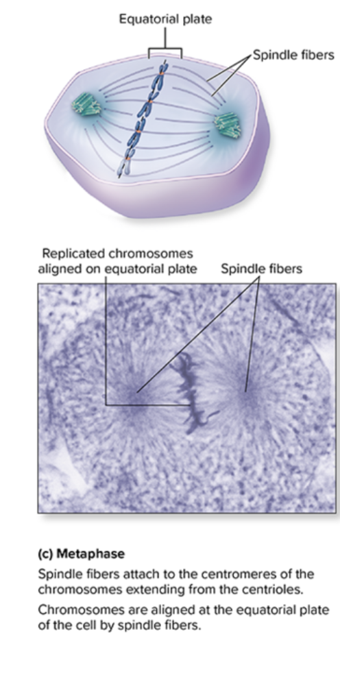
Metaphase
second stage of mitosis (step c), during which the chromosomes are aligned along an imaginary line in the middle of the cell (region called the equatorial plate).
This alignment occurs through the growth of spindle fibers from each centriole toward the chromosomes.
Some fibers attach to the centromere of each chromosome, directing their movement to the equatorial plate.
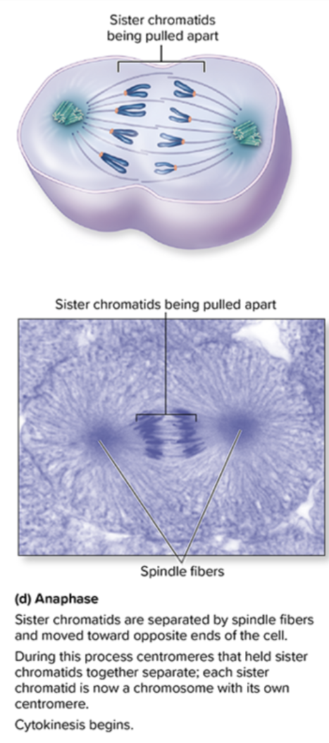
Anaphase
third stage of mitosis, initiates as the spindle fibers cause the sister chromatids to be moved apart toward the cell's poles; the centromere leads the way, and its "arms" trail behind (step d).
Each chromatid is now a chromosome composed of one DNA double helix with its own centromere
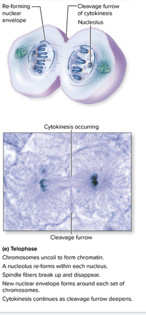
Telophase
begins with the arrival of a group of chromosomes at each cell pole.
Essentially, the processes of prophase are reversed in telophase.
The chromosomes begin to uncoil and return to the form of dispersed threads of chromatin, each new nucleus forms a nucleolus, the mitotic spindle breaks up and disappears, and a new nuclear envelope forms around each set of chromosomes.
Telophase signals the end of nuclear division.
Cytokinesis
is the other major event in the mitotic phase, and it is the division of the cytoplasm between the two newly forming cells.
The beginning of cytokinesis usually overlaps with anaphase and telophase of mitosis, and continues as mitosis ends.
A ring of microfilament proteins on the inner surface of the cell's plasma membrane contracts at the cell's equator. It pinches the mother cell into two separate cells in a manner analogous to the tightening of a belt.
The resulting cleavage furrow that appears indicates where the cytoplasm is dividing
Two new daughter cells are formed and cell division is complete.