Cardiovascular System 3.1
1/15
There's no tags or description
Looks like no tags are added yet.
Name | Mastery | Learn | Test | Matching | Spaced | Call with Kai |
|---|
No analytics yet
Send a link to your students to track their progress
16 Terms
Blood Vessel Layers
Tunica Externa - Connective Tissue
An External Elastic Membrane
Tunica Media - Smooth Muscle (Determines diameter of vessel)
An Internal Elastic Membrane
Tunica Intima/Internal - Single layer of Endotheluum
Arteries
Move blood away from heart
Thicker muscle layers
Are under higher pressure
Maintain shape
Have 2 different layers of Elastic Membranes: Internal & External
Veins
Move blood toward heart
Have valves to prevent backflow
Which Large Artery Carries Deoxygenated Blood?
Pulmonary Trunk & Pulmonary Arteries
Which Large Veins Carry Oxygenated Blood?
Pulmonary Veins
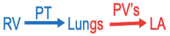
Pulmonary Circuit of Blood Flow
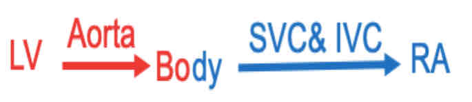
Systemic Circuit of Blood Flow
Heart Valves and their Purpose
AV Valves: Tricuspid and Bicuspid; prevent backflow into the Atria
Semilunar Valves: Aortic and Pulmonary; prevent backflow in the Ventricles
Role of Papillary Muscles and Chordae Tendinae
They anchor and control the movements of the Av Valve leaflets (cusps) so that they don’t swing into Atria.
SA Nodes
Located in superior wall of Right Atrium
Consists of nerves that fire eclectically impulses (go through heart)
Atria
Impulses from SA Nodes goes here first
Contracts Atrial muscles
AV Node
Located in inferior wall of Right Atrium
Gets electric impulse from Atria
AV Bundle
Get electrical current from AV Node
Splits, conducting the electrical impulses into the Right/Left Bundle branches
Purkinje Fibers
Branches from AV Bundles feed into these
Cause Ventricular Contraction
Conduction System of the Heart
SA Nodes → Internodal Pathways → AV Node → AV Bundle → RBB/LBB → Purkinje Fibers
What are the Septum’s?
Interatrial Septum (Between Aorta and PT)
Interventricular Spectrum (Between LV and RV)
Atrioventricular Spectrum (Between Atriums and Ventricles)