Spine and Skull Anatomy Review
1/60
Earn XP
Description and Tags
These flashcards cover key concepts from the spine and skull anatomy lecture notes, focusing on the various projections, structures, and techniques involved in imaging.
Name | Mastery | Learn | Test | Matching | Spaced | Call with Kai |
|---|
No analytics yet
Send a link to your students to track their progress
61 Terms
What are the main segments of the spine?
Cervical, Thoracic, Lumbar, Sacrum, Coccyx.
What is the purpose of the AP Axial projection for the Cervical spine?
To visualize the C3-T1 intervertebral disc spaces.
What position is needed for the Lateral projection of the Thoracic spine?
Upright, recumbent, or cross-table in true lateral position.
What angle is used for the AP Axial Coccyx projection?
10 degrees caudad.
What does the term 'Scottie Dogs' refer to in lumbar obliques?
It is an anatomical landmark used to identify lumbar vertebrae in oblique views.
In which projection of the sacrum are the sacral foramina visualized?
AP Axial Sacrum.
What anatomical structures are evaluated during the PA Axial projection of the skull?
Petrous ridges in the lower 1/3 of the orbits.
Which sinuses are visualized during the SMV projection?
Sphenoidal and maxillary sinuses.
What is the primary view for visualizing the SI joints?
AP Axial and Oblique projections.
In which projections for the Ribs are the ribs above and below the diaphragm visualized?
AP and PA projections for ribs above the diaphragm, and AP for ribs below the diaphragm.
Cervical Spine: AP Axial
Pt/Part Position: Upright or Supine, occlusal plane perp
CR: 15 - 20 cephalic, enters C4 (inferior to most prominent point of thyroid)
SID: 40”
What’s seen: C3 -T1, Intervertebral disc spaces open
Cervical Spine: Lateral (Grandy Method)
Pt/Part Position: Upright or X-table. Depress shoulders as much as possible. Elevate chin slightly to avoid superimposition of mandible rami and spine.
CR: Horizontal, enter C4
SID: 60 - 72”
What’s seen: Zygapophyseal joints, all 7 cervical vertebra and part of T1, no mandible and spine overlap
Cervical Spine: Odontoid (open mouth)
Pt/Part Position: Upright or Supine, occlusal plane perp
CR: Perp entering midpoint of mouth
SID: 30”
What’s seen: Entire Atlas, Axis & Dens, C1/C2, Zygapophseal joint (lateral masses)
Cervical Spine: Fuch (AP)
Pt/Part Position: Supine, lift chin until mental point and mastoid tips are aligned and perp
CR: Perp entering MSP, just distal to point of chin
SID: 40”
What’s seen: Entire dens in foramen magnum
Cervical Spine: AP/PA Axial Obliques
Pt/Part Position: Upright preferred, 45 degree oblique, extend chin if needed
CR:
AP: 15 - 20 degrees cephalic, enters C4
PA: 15-20 degrees caudad enters C4
SID: 60 - 72”
What’s seen: Interveterbal foramina, C3 - C7
Thoracic Spine: AP
Pt/Part Position: Upright or Supine
CR: Perp to T7 (3-4'“ below jugular notch or midpoint between jugular point and xiphoid) *Light 1.5 - 2" inches above shoulders
SID: 40”
What’s seen: All 12 thoracic vertebrae, spinous process in midline
*Use of anode heel affect
Thoracic Spine: Lateral
Pt/Part Position: Upright, recumbent, or x table, true lateral, arms at right angles to body
CR: Enters posterior half of thorax at T7 *Light 1.5 - 2'“ above shoulders
SID: 40”
What’s seen: Interveterbral foramina, ribs superimposed posteriorly, open intervertebral joint spaces
Thoracic Spine: Cervicothoracic Lateral (Swimmers)
Pt/Part Position: Upright or recumbent, extend arm closest to IR over head an depress other shoulder
CR: Pass through intervetrbal disc space of C7 - T1
SID: 60 - 72”
What’s seen: C4 - T3 clearly demonstrated, humeral heads separated
Thoracic Spine: AP/PA obliques
Pt/Part Position: Upright or recumbent, 70 degree oblique
CR: Enters level of T7
SID: 40”
What’s seen: Zygapophyseal Joints, all 12 thoracic vertebrae
Lumbar Spine: AP
Pt/Part Position: Upright or Supine, flex knees to reduce lumbar lordotic curve
CR: Perp to L4 (Iliac Crest) to see lumbosacral OR L3 (1.5” above crest) to see lumbar only
SID: 40”
What’s seen: All 5 lumbar vertebrae, open disc spaces, SI joints included
Lumbar Spine: Lateral
Pt/Part Position: Upright, recumbent, true lateral, if lumbar not horizontal angle caudad 5 degrees for men and 8 degrees for women
CR: Perp to coronal plane, L4 for lumbosacral, L3 for lumbar only
SID: 40”
What’s seen: Intervertebral foramina, lower thoracic to sacrum, superimposed iliac crests
Lumbar Spine: L5/S1 Lumbosacaral Junction “Spot”
Pt/Part Position: Recumbent, true lateral
CR: 2” posterior & 1.5” inferior to ASIS, runs parallel to inter iliac line (crests)
SID: 40”
What’s seen: Open lumbosacral joint
Lumbar Spine: AP/PA Obliques
Pt/Part Position: Upright or recumbent, 45 degree oblique
CR:
AP: 1” above iliac crest & 2” medial to elevated ASIS
PA: 1" above crest & 2” lateral from the spine on elevated side
SID: 40”
What’s seen: Zygapophyseal joints, “scotty dogs”, T12 to sacrum
Spine - SI Joints: AP Axial
Pt/Part Position: Supine
CR: 30 - 35 degrees cephalic midway between ASIS & pubic symphysis
SID: 40”
What’s seen: Sacroiliac joints open, L5 - S1 joint open as well
SI Joints: Oblique
Pt/Part Position: Recumbent RPO or LPO, 25 - 30 degree oblique. Elevate affected side
CR: Enters 1” medial to elevated ASIS
SID: 40”
What’s seen: SI joint of interest open
Sacrum: AP Axial Sacrum
Pt/Part Position: Upright or Supine
CR: 15 degrees cephalic, enters midway between ASIS & public symphysis
SID: 40”
What’s seen: Sacral foramina visualized, sacrum not foreshortened
Coccyx: AP Axial
Pt/Part Position: Upright or Supine
CR: 10 degrees caudad, enters 2” superior to pubic symphysis
SID: 40”
What’s seen: Coccyx free of superimposition
Sacrum and Coccyx: Lateral
Pt/Part Position: Recumbent or upright
CR:
Sacrum: enters 3-4” posterior to ASIS
Coccyx: 3-4” posterior & 2” inferior to ASIS
SID: 40”
What’s seen: Lateral sacrum or coccyx centered, coccyx segments all visualized
What positions show the intervertebral foramina?
Cervical - Oblique
Thoracic - Lateral
Lumbar - Lateral
What positions show the zygapophyseal joints?
Cervical - Lateral
Thoracic - Oblique
Lumbar - Oblique
AP Oblique projections for spines
Cervical (French): farthest from IR
Thoracic (Fried): farthest from IR
Lumbar (Chicken): closest to IR
SI Joints (Fingers): farthest from IR
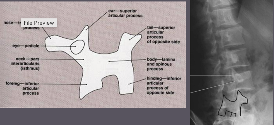
Name the parts of the scotty dog
***
Skull - Lateral
Pt/Part Position: Upright or recumbent, can oblique patient
Positioning Line: IOML parallel, Interpupillary line perp
CR: Enters 2” above EAM
Evaluation Criteria: Entire cranium, no rotation or tilt, superimposed orbital roofs and rami
Skull - PA Axial - Caldwell (can do PA also)
Pt/Part Position: Seated or Prone, forehead & nose on IR
Positioning Line: OML perp
CR: 15 degrees caudad, exit nation
Evaluation Criteria: Petrous ridges in lower 1/3 of orbits,
*If PA - (no axial): Petrous ridges fill orbits
Skull - SMV (submentovertical)
Pt/Part Position: seated, lean back so head vertex is on IR
Positioning Line: IOML parallel
CR: Enters between mandibular angles (gonion), passes through 3/4” anterior to EAM
Evaluation Criteria: Superimposed mental point & anterior frontal bone, symmetric petrous parts
Sinuses - Lateral
Pt/Part Position: Upright or recumbent, can oblique patient
Positioning Line: IOML parallel, interpulilary line perp
CR: Enters ½ - 1” posterior to outer canthus
Evaluation Criteria: Shows all 4 sinuses, no rotation
Sinuses - PA Axial Caldwell
Pt/Part Position: Seated or prone, forehead & nose on IR
Positioning Line: OML perp
CR: Horizontal, tilt IR 15 degrees, exit nasion
Evaluation Criteria: Petrous ridges in lower 1/3 of orbits, all 4 sinuses seen (sphenoid just between ethmoid)
Sinuses - SMV (submentovertical)
Pt/Part Position: Seated, lean back so head vertex on IR
Positioning Line: IOML parallel
CR: Enters between mandibular angles (gonion), passes through ¾ anterior to EAM
Evaluation Criteria: Superimposed mental point & anterior frontal bone, symmetric petrous parts
Skull - AP Axial - Towne
Pt/Part Position: Seated or supine
Positioning Line: OML perp is preferred, IOML as backup
CR:
If OML- 30 degrees caudad, enter 2.5” above glabella
if IOML - 37 degrees caudad, enter 2.5” above glabella
Evaluation Criteria: Foramen magnum with dorm sellae and posterior clinoids inside, entire occipital bone
Sinuses - AP Axial Towne
Not done
Skull - Parietoacanthial (Waters)
Not done for skull
Sinuses - Parietoacanthial (waters)
Pt/Part Position: Seated/upright, chin on bucky
Positioning Line: OML at 37 degrees
CR: Perp exiting the acanthion
Evaluation Criteria: Petrous ridges just below the maxillary sinus, frontal and ethmoid sinuses distorted. *Sphenoid sinus only seen of mouth opened
How much degree difference is there between the OML and OIML
7 degree
What should you do to your patient to get the skull as close to the IR as possible?
Oblique your patient
Sinuses should always be done _____ to visualize air/fluid levels
upright
What sinuses are the largest?
Maxillary
Facial Bones - Lateral
Pt/Part Position: upright or recumbent, true lateral
Positioning Line: IOML parallel, Interpupil perp
CR: Enters zygoma, halfway between outer cants and EAM
Evaluation Criteria: All facial bones included, zygoma centered. Superimposed orbital roofs and mandible.
Facia Bones - PA Axial Caldwell
Pt/Part Position: upright or prone, forehead and nose on IR
Positioning Line: OML perp
CR: 15 degrees caudad, exit nasion
Evaluation Criteria: Petrous ridges in lower 1/3 of orbits
Facial bones - parietoacanthial modified shallow waters
Pt/Part Position: Upright, chin on bucky
Positioning Line: OML at 55 degrees
CR: Perp exiting the acanthion
Evaluation Criteria: Petrous ridges below orbits (in maxillary sinuses)
Sternum - Oblique
Pt/Part Position: Upright, RAO, 15 - 20 degree oblique
CR: Midway between jugular notch and xiphoid (apron T7) and 1” lateral on elevated side
Evaluation Criteria: Sternum visualized over heart shadow
*use breathing technique to blur ribs
Sternum- Lateral
Pt/Part Position: Upright, true lateral, lock hands behind back and roll shoulders back
CR: Through mid sternum
Evaluation Criteria: Full inspiration for contrast, full sternum visualized
*Top of IR 1.5” above sternal notch
SC Joints - PA
Pt/Part Position: Upright or prone
CR: Enters T2 - T3
Evaluation Criteria: Both SC joints equally centered
SC Joints - Oblique - Bilateral
Pt/Part Position: Upright or prone, RAO or LAO, 10 - 15 degree oblique
CR: Enters T2 - T3
Evaluation Criteria: Joint of interest open & projected free of the spine
*RAO = right SC joint, LAO=left SC joint
Ribs - AP posterior ribs above diaphragm
Pt/Part Position: Upright or supine
CR: Enters apron. T7
Evaluation Criteria: Ribs 1-9 seen above diaghram
Ribs - PA anterior ribs above diaphragm
Pt/Part Position: Upright or supine
CR: Enters T7
Evaluation Criteria: Ribs 1 -10 seen above diaphragm
Ribs - AP posterior ribs below diaphragm
Pt/Part Position: Upright or supine
CR: Enters center of IR with bottom of plate 1.5” below costal margin (iliac crest)
Evaluation Criteria: Posterior ribs 8-12 seen below diaphragm
Ribs - AP oblique RPO or LPO
Pt/Part Position: Upright or supine, 45 degree oblique
CR:
Upper ribs: IR 1 ½ above shoulder
Lower ribs: IR 1 ½ below inferior costal margin or at crest
Upper: CR perpendicular, half way between MSP and lateral boarder of affected ribs
Lower: CR midway between crest and costal margin
Evaluation Criteria: Axillary portion of ribs seen for injured side, side of interest elongated
AP= Injured side closest to IR
PA = Injured side furthest from IR (PA away)
How does the heart, diaphragm and stomach lie in hypersthenic patients?
…
How does the heart, diaphragm and stomach lie in sthenic patients?
…
How does the heart, diaphragm and stomach lie in hyposthenic patients?
…
How does the heart, diaphragm and stomach lie in asthenic patient?
…….