Chapter 12 : Spinal Cord and Spinal nerves
1/28
Earn XP
Description and Tags
CHAPTER 12: Spinal Cord & Spinal Nerves • Spinal Cord (Structure, Function) • Spinal nerves • Meninges (Layers, Roles) • Meningitis (Causes, How ID, Treat, Complications (NOT treat Quickly) • Injections (Spinal & Epidural Anesthesia): Advantages of each • White vs Gray Matter • Reflex (Definition, 5 parts) • Reflex Classifications, Stretch, Polysynaptic • Spinal Nerve Structure / Layers • Spinal nerves #, where exit, MIXED nerves • Plexus (Definition, Advantage, 4 Plexus area, Why overlap?) • For Each Plexus: What nerve(s) did we discuss, what role do they perform, what happens with injury CHAPTER 12 pt 2 : Spinal Cord & Spinal Nerves {CONTINUED} • Thoracic Outlet Syndrome (ALL) • Dermatomes (Definition, Function?) • Spinal Stenosis
Name | Mastery | Learn | Test | Matching | Spaced | Call with Kai |
|---|
No analytics yet
Send a link to your students to track their progress
29 Terms
Describe the structure of the Spinal cord. (location,
Location: Extends from the foramen magnum to the 2nd lumbar vertebrae level
Forms the Conus medullaris (wider @ top w two areas of enlargement, Separates into Cauda equine (horse tail)
Describe the functions of the spinal cord
major communication with between the Brain and perineal nervous system
integration of info and produces a response through REFLEX MECHANISMS
What nervous system does the spinal cord belong to ?
The Central Nervous system (CNS)
How many layers do the Meninges have?
What are they called?
three.
Dura Mater
Arachnoid mater
Pia Mater
Meninges (1): Dura Mater
epidural space ( btwn bone & dura mater)
Extremely strong
Separates to enclose the Dural Venous Sinuses
2 layers
Superficial Periosteal Layer (not in spinal cord)
Deeper meningeal layer
Meninges (2): Archnoid Mater (location?
Location: under subdural space (btwn dura and arachnid mater)
Archnoid granulations protrude into superior sagittal sinus
Absorbs cerebral spinal fluid into Venous Blood of Sinus
Meninges (3): Pia Mater
Location: Subarachnoid space (btwn Arachnoid and Pia mater)
spider- web like
Blood vessels and cerebral spinal fluid here
“gentle mother” delicate with tiny BV’s
Clings to brain
What are the Meninges?
Def: 3 thin connective tissue membranes that cover and provide protection for the Central nervous system
they also enclose the venous sinuses
they also have Ceriveal spinal fluid (csf)
Meninges (1) : Dura Mater: What are the 3 types of separations that forms the Dural Septa (abbrev.- FFT)
Falx Cerebri
Falx Cerebelli
Tentorium Cerebelli
What is Meningitis?
what is it caused by?
Meningeal inflammation
Caused by bacteria or Virus
Injections: Spinal Anesthesia (block)
prevents pain in lower body regions
Advantage : stronger anesthesia into CSF with a faster effect
Injections: Epidural Anesthesia
needle not thru dura mater, drugs diffuse into CSF
Advantage: longer lasting
what is Gray matter?
consists of neuron cell bodies, dendrites and axons
What is white matter?
Consists of myelinated axons → nerve tracts
Define reflex
Automatic response to Stimulus, No conscious thought drives it
Protective (Somatic Reflexes)
Maintains homeostasis (Autonomic Reflexes) = Stable BP.
5 parts of reflex
stimulus→ sensory (afferent) neuron → Integration center (interneuron) → motor neuron (efferent) → effector
What are the 2 Reflex classifications?
Monosynaptic : only 1 synapse
Sensory receptor: muscle spindle
SIMPLEST REFLEX
Polysynaptic : more than 1 synapse, interneuron
prevents falling by shifting weight
withdrawal reflex: flexor response to pain stimuli
crossed extensor reflex: @ same time as withdrawal reflex, stimulates extensor muscles to support body
Spinal nerve structure
Axon
Surrounded by Endoneurium = delicate loose CT (areolar)
Fascicles
packaged
surrounded by Perineurium = dense irregular CT
Perineurium in concentric cell layers
up to 15 layers thick
Epineurium
binds fascicles together to form NERVE
dense irregular CT
CT coverings make periphearal nerves TOUGH
Spinal nerves are part of what NS?
Where do they emanate from?
Peripheral nervous system (31 pairs)
Emanate from the spinal cord
Spinal nerve
Short (1-2 cm)
Quickly branches into Dorsal, ventral ramus and meningeal branch
Where do the 5 spinal nerve areas exit?
Cervical = C1-C8 and C1 exits btwn skull and C1 vertebrae
Thoracic = T1-T12
Lumbar = L1-L5
Sacral = S1- S5 and Exits from sacrum thru sacral foramina
Coccygeal = Co (1)
What are mixed nerves?
made up of Sensory and motor
Define Plexus. What are the plexuses formed by?
Intermingling of nerves. Formed from Ventral Rami of different spinal nerves = Roots join together.
What are the advantages of the Plexus’s
Damage to 1 spinal segment/ root = NOT fully paralyzed limb muscle
name the 4 plexus areas. + associated nerves
Cervical Plexus
Spinal nerves: C1-C4
Brachial Plexus
Spinal nerves: C5- T1
Lumboscaral Plexus
spinal nerves: L1-L4
Coccygeal Plexus
spinal nerves: S5-Co
For Each Plexus: what role do they perform, what happens with injury.
CP:
Deep in the neck
intervals superficial neck skin (cutaneous nerves) & hyoid bone muscle
phrenic nerve: innervates the diaphragm
Irritation of Phrenic nerves = hiccups (diaphragm spasms)
Injury= paralyzed diaphragm and respiratory arrest
BP (brachial plexus)
5 roots that are deep to the sternocleidomastoid
Axillary
Radial
Musculocutaneus
Ulnar nerves
Median nerves
Injury: xs Arm Pull or Blow to Superior Shoulder
Severe = Weaken or Paralyze Upper Limb
LSP
Lies within Psoas Major Muscle
Femoral Nerve (under Inguinal ligament: Motor branches to Quads, Cutaneous to anterior thigh & Medial lower leg)
Obturator Nerve (Through Obturator foramen) Innervates Adductors
Sciatic Nerve = Tibial & Common Peroneal Nerves = L4-S3: LARGEST NERVE IN THE BODY
Coccygeal Plexus
Motor innervation to Pelvic Floor Muscles & Sensory Cutaneous over Coccyx skin area.
Thoracic Outlet Syndrome (Cause, S&S, Diagnosis, treatment, complications, preventions)
compression of Brachial plexus OR subclavian artery or vein.
Cause: car accident, Repetitive injury (Job, Sport), Pregnancy, Anatomical differences (Cervical rib)
Risk Factors: Women (3x) > Men, Ages 20-50.
Signs & symptoms : Numbness in arm/fingers, Shoulder & Neck Pain, Arm fatigue with activity, Weak grip.
Diagnosis: Ultrasound (1st = Vascular), Xray or MRI (Cervical rib),
Treatment: Physical Therapy, Antinflammatory meds, Injections (Steroid, Botox), Surgery
Complications: Repetition injury: Long term injury= Chronic pain /
Prevention: Avoid carrying heavy Backpack, Daily stretches, Exercises keep shoulder muscles strong
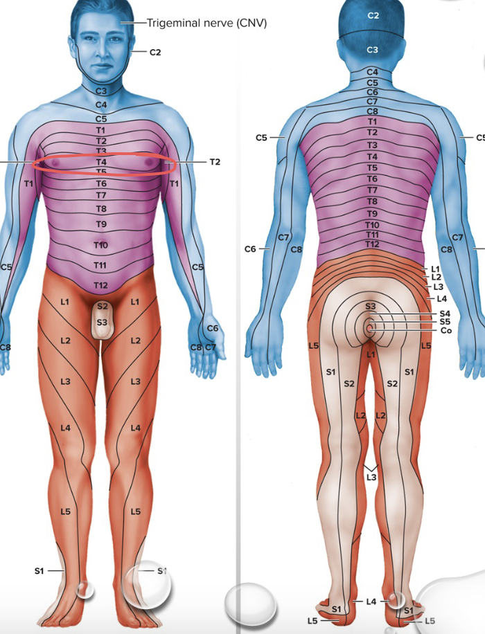
What are Dermatomes?
Area of Skin innervated by Cutaneous branches of single spinal nerve
All except C1 contribute
ID nerves damaged with Spinal cord injury
Spinal Stenosis
problem: spinal canal / nerve roots narrowing causing compression of nerves