CH 6 Bones and Tissues
 Call Kai
 Learn
Learn Practice Test
 Spaced Repetition
 Match
Match Flashcards
 Knowt Play
1/112

There's no tags or description
Looks like no tags are added yet.
Name | Mastery | Learn | Test | Matching | Spaced | Call with Kai | Chat |
|---|
No analytics yet
Send a link to your students to track their progress
113 Terms
What is cartilage?
supporting connective tissue
What are the characteristics of cartilage?
no blood vessels
flexible tissue
consist of water (60-80%)
formed by chondrocytes (from embryonic state)
What are the types of cartilage?
hyaline
elastic
fibrocartilage
Where can you find hyaline cartilage?
ends of bones in joints, connecting the ribs to the sternum, and supporting the respiratory tract.
every part of articulation
Where can you find elastic cartilage?
cartilage in external ear, epiglottis, eustachian tube, larynx
Where can you find fibrocartilage?
in intervertebral discs, pubic symphysis, and menisci of the knee.
What three things make up bones?
specialized cells
protein fibers
extracellular matrix (vascularized )
What are the functions of bone?
support
mineral storage
blood cell production
protection
movement
production of fat storage
Bone function: support
framework for attachment of tissues and organs
Bone function: mineral storage
calcium and phosphate
Bone function: protection
organ such as heart and brain are protected
Bone function: movement
bones are levers, bones change magnitude and direction of force generated by muscles
Bone function: production of fat storage
production of osteocalcin influences fat storage (reduction), insulin production (increasesand sensitivity, and glucose metabolism.
Low osteocalcin in bone →
reduced insulin sensitivity and increased fat mass.
High osteocalcin in bone →
increased insulin sensitivity and reduced fat mass.
Four specialized cell types of bones
osteocyte
osteoblast
osteoprogenitor
osteoclast
Origin of cells (bone)
mesenchym
Parts of bone: cells

Osteocyte
mature bone cell that maintain the bone matrix
main; round with extensions → pass chemical info

Osteoblast
immature bone cell that secretes osteoid, the organic bone matrix

Osteoprogenitor
stem cell that divides to produce osteoblasts

Osteoclast
multinucleate cell that secretes acids and enzymes to dissolve bone matrix

Osteogenesis
The process of bone formation, which involves the differentiation of osteoblasts and production of the bone matrix.
uses the bone cell type called osteoblasts to create new bone tissue.
Osteolysis
The process of bone resorption where osteoclasts break down bone tissue, releasing minerals and collagen into the bloodstream.
uses the bone cell type called osteoclasts to dissolve the bone matrix.
liberate mineral storage
What is the importance of fibers in bone?
tolerate stretching, twisting, and bending (spiral orientation)
provide an organic framework for the formation of mineral crystals

What is responsible for a majority of the bone weight?
collagen fibers represent 1/3 of bone weight
What is the matrix in bone?
calcium phosphate: hydroxyapatite
What combination of the matrix is found in the lamellae?
hydroxyapatite crystals + protein fibers = protein-crystal

Where are protein crystals?
Protein crystals are found in the bone's lamellae, where they contribute to its structural integrity.
Two types of bone
compact and spongy
Compact bone?
external wall
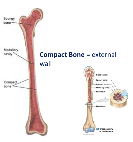
Spongy bone location
internal layer
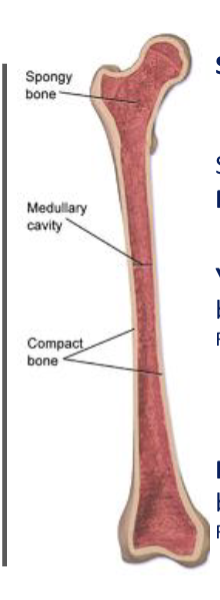
Function of spongy bone
protects the medullary cavity (bone marrow)
Two types of bone marrow in spongy bone
yellow and red
What is yellow marrow?
dominated by adipocytes
Where can you find yellow bone marrow?
found in shaft and end of long bones
What is red bone marrow?
tissue dominated by blood cells
Where can you find red marrow?
found in flat bones and long bones
What is the functional unit of compact bone?
osteon or Haversian system

What lamellae make up compact bone?
concentric
circumferential
interstitial

Concentric lamellae
of each osteon are parallel to the long axis of the bone (bull’s eye target)
Circumferential lamella
located at the internal and external surface of the bone
Interstitial lamellae
fill the space between osteons
Structure of spongy, trabecular, or cancellous bone
parallel, thick, branching plates or bars
Trabeculae
arrangement of spongy bone is into parallel thick branching plates or bars
Importance of trabeculae in spongy bone
reduce the weight of skeleton = easier movement of bones
How do trabeculae help with force?
force (ie body weight) is directed downward to large bones to distribute it
hip to the metaphysis
across the knee joint to the rest of the leg

Parts of a long bone (ie humerus and femur)
epiphysis, metaphysis, diaphysis (shaft), articular surface
Most superior portion/ tips of long bone
epiphysis
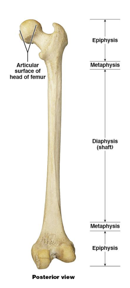
Metaphysis
the region between the epiphysis and diaphysis of a long bone, where growth occurs and the bone widens.

Diaphysis (shaft)
the long, tubular main section of a long bone that provides strength and support.

What is periosteum made from?
connective tissue → dense irregular
Function of periosteum
isolate and protect the bone
provides a route and place to attachment for insertion points to tendons and ligaments
Importance of periosteum
important in bone growth and repair
rich in blood vessels and nerve

Diagram: periosteum
periosteum: fibrous layer (bone repair), osteogenic layer (produce new cells)
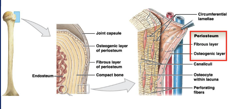
Endosteum locations
lines the medullary cavity (marrow)
lines inner surfaces of central canals and perforating canals
covers the trabecular of spongy bone
Importance and diagram of endosteum
bone growth and repair

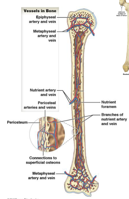
How is blood supplied to bones?
blood vessels and artery
What is the nutrient artery and nutrient vein?
enter through nutrient foramen
to supply blood to the bone's medullary cavity and trabecular bone.

Types of vessels for bone blood supply
epiphyseal
metaphyseal
periosteal
Epiphyseal vessels
supply medullary cavities of the epiphyses (through foramina)
Metaphyseal vessels
supply to diaphyseal surface
bone is replacing cartilage
ossification of bones
Periosteal vessels
supply superficial osteons (compact bones)
Diagram of blood supply to bone
Nerve supply in the bones
periosteum
sensory nerves

Periosteum role in nerve supply (bone)
contains a network of lymphatic vessels and sensory nerves
What do sensory nerves in bone innervate?
endosteum
medullary cavity
epiphyses
Why are bone injuries painful?
Bone injuries are painful due to the stimulation of sensory nerves in the periosteum and surrounding tissues, which transmit pain signals to the brain.
lots of nerves!
What are the seven bone shapes for classification?
sutural (Wormian)
Pneumatized
short
irregular
flat
long
sesamoid
Some Pirates Sneak Some Interesting Flat Logs
Sutural bones
small, flat, oddly shaped bones found between the flat bones of the skull along the sutures
they develop from separate centers of ossification

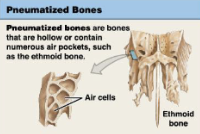
Pneumatized bones
bones that are hollow or contain numerous air pockets, such as the ethmoid bone
Short bones
boxlike in appearance
external surfaces are covered by compact bone, but the interior contains spongy bone
carpal bones and tarsal bones

Irregular bones
have complex shapes with short, flat, notched, or ridged surfaces
internal structure is equally varied
vertebrae that form the spinal column and several bones in the skull

flat bones
thin, roughly parallel surfaces of compact bone
spongy bone sandwich
strong but relatively light
form the roof of the skull, sternum, ribs, and scapulae
protect underlying soft tissues and have an extensive SA for the attachment of skeletal muscles
aka parietal bones
thick layers of compact bone aka internal and external tables
diploe: spongy bone between tables

Long bones
long and slender
diaphysis, 2 metaphyses, 2 epiphyses, medullary cavity
upper and lower limbs
humerus, radius, ulna, femur, tibia, fibula

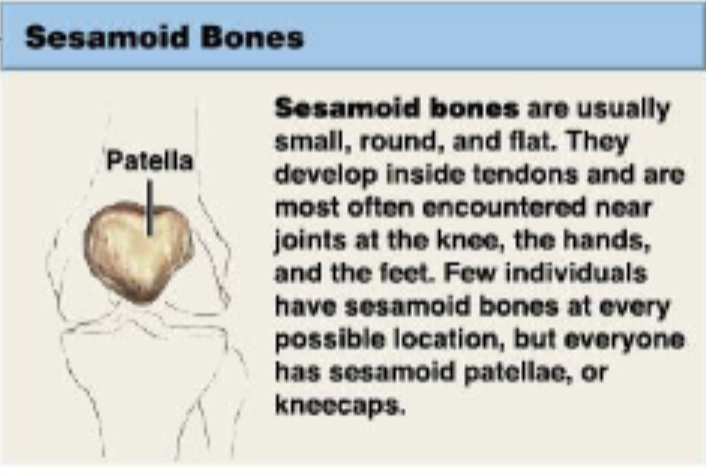
Sesamoid bone
small, round, flat
develop inside tendons and are most often encountered near joints at the knee, hands, and feet
kneecaps
Bone markings or surface features function: depression and openings
passageway of blood vessels and nerves
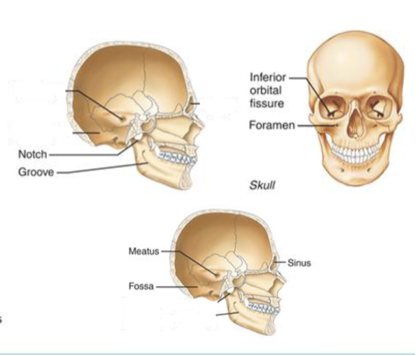
Bone markings or surface features TYPES: depression and openings
foramen
groove
fissure
notch
fossa
meatus
sinus
foramen
round or oval opening through a bone
groove
furrow
fissure
narrow, slitlike opening
notch
indentation at the edge of a structure
fossa
shallow basinlike depression in a bone, often serving as an articular surface
meatus
canal-like passageway
sinus
cavity within a bone, filled with air and lined with mucous membrane
Bone markings or surface features TYPES: surfaces that form joints
head
facet
condyle

head
bony expansion carried on a narrow neck
facet
smooth, nearly flat articular surface
condyle
rounded articular projection, often articulates with a corresponding fossa
Bone markings or surface features TYPES: projections that are sites of muscle or ligament attachment
tuberosity
crest
trochanter
line
tubercle
epicondyle
spine

tuberosity
large rounded projection; may be roughened
crest
narrow ridge of bone; usually prominent
trochanter
very large, blunt, irregularly shaped process (only examples are on the femur)
line
narrow ridge of bones; less prominent than a crest
tubercle
small rounded projection or process
epicondyle
raised area on or above a condyle
spine
sharp, slender, often pointed projection
Bone markings or surface features: process and ramus

Process
any projection or bump
Ramus
an extension of a bone that forms an angle with the rest of the structure
L shape