Lab 12 Diagrams

0.0(0)
Studied by 0 people
call kaiCall Kai
Locked
learnLearn
examPractice Test
spaced repetitionSpaced Repetition
heart puzzleMatch
flashcardsFlashcards
GameKnowt Play
Card Sorting

1/4

encourage image

There's no tags or description

Looks like no tags are added yet.

Last updated 6:57 PM on 7/25/26
Name
Mastery
Learn
Test
Matching
Spaced
Call with Kai
Chat

No analytics yet

Send a link to your students to track their progress

5 Terms
1
New cards
<p>Simple Synovial Joint Anatomy Diagram</p>

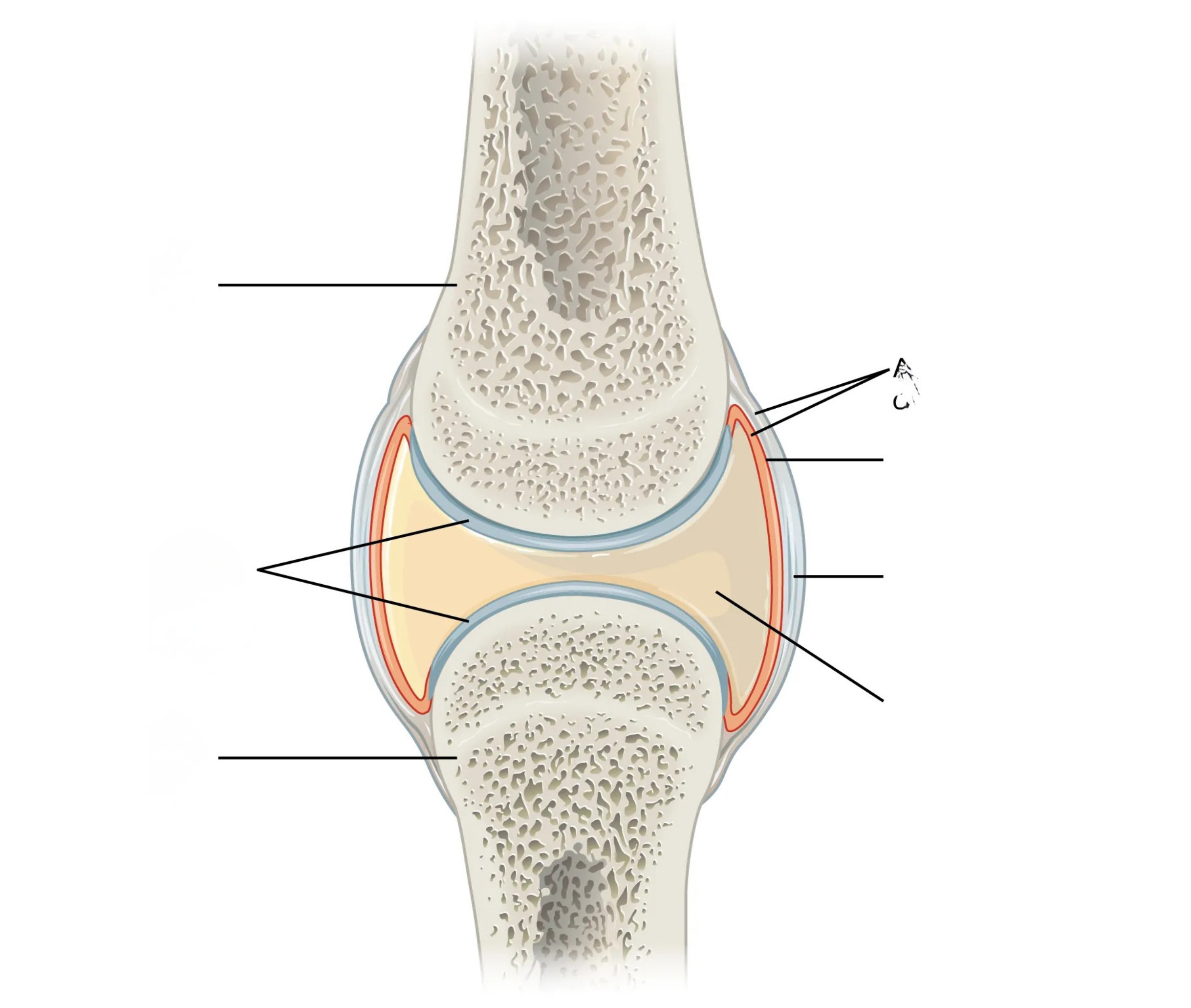
Simple Synovial Joint Anatomy Diagram

Ligament: band connecting bone to bone, holds the joint together
Articular cartilage: smooth cushion covering the bone ends, reduces friction
Joint (articular) capsule: the whole covering that encloses the joint — made of the fibrous capsule + synovial membrane
Fibrous capsule: tough outer layer of the capsule
Synovial membrane: inner layer of the capsule, makes the synovial fluid
Cavity with synovial fluid: fluid-filled space inside the joint that lubricates and nourishes it
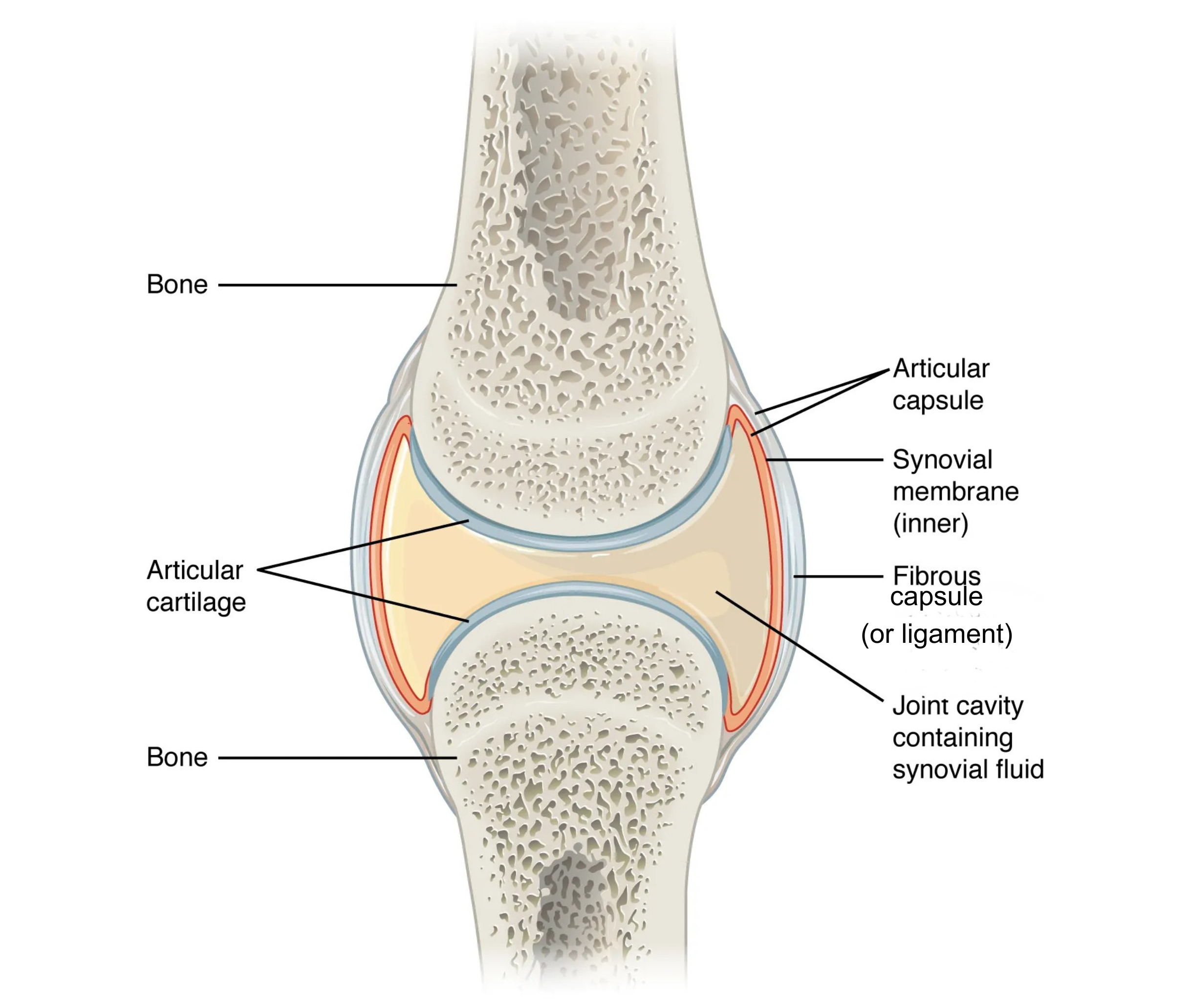
<p>Ligament: band connecting bone to bone, holds the joint together<br>Articular cartilage: smooth cushion covering the bone ends, reduces friction<br>Joint (articular) capsule: the whole covering that encloses the joint — made of the fibrous capsule + synovial membrane<br>Fibrous capsule: tough outer layer of the capsule<br>Synovial membrane: inner layer of the capsule, makes the synovial fluid<br>Cavity with synovial fluid: fluid-filled space inside the joint that lubricates and nourishes it</p>
2
New cards
<p>Knee (Tibiofemoral) Joint Diagram (Front &amp; Back Views)</p>

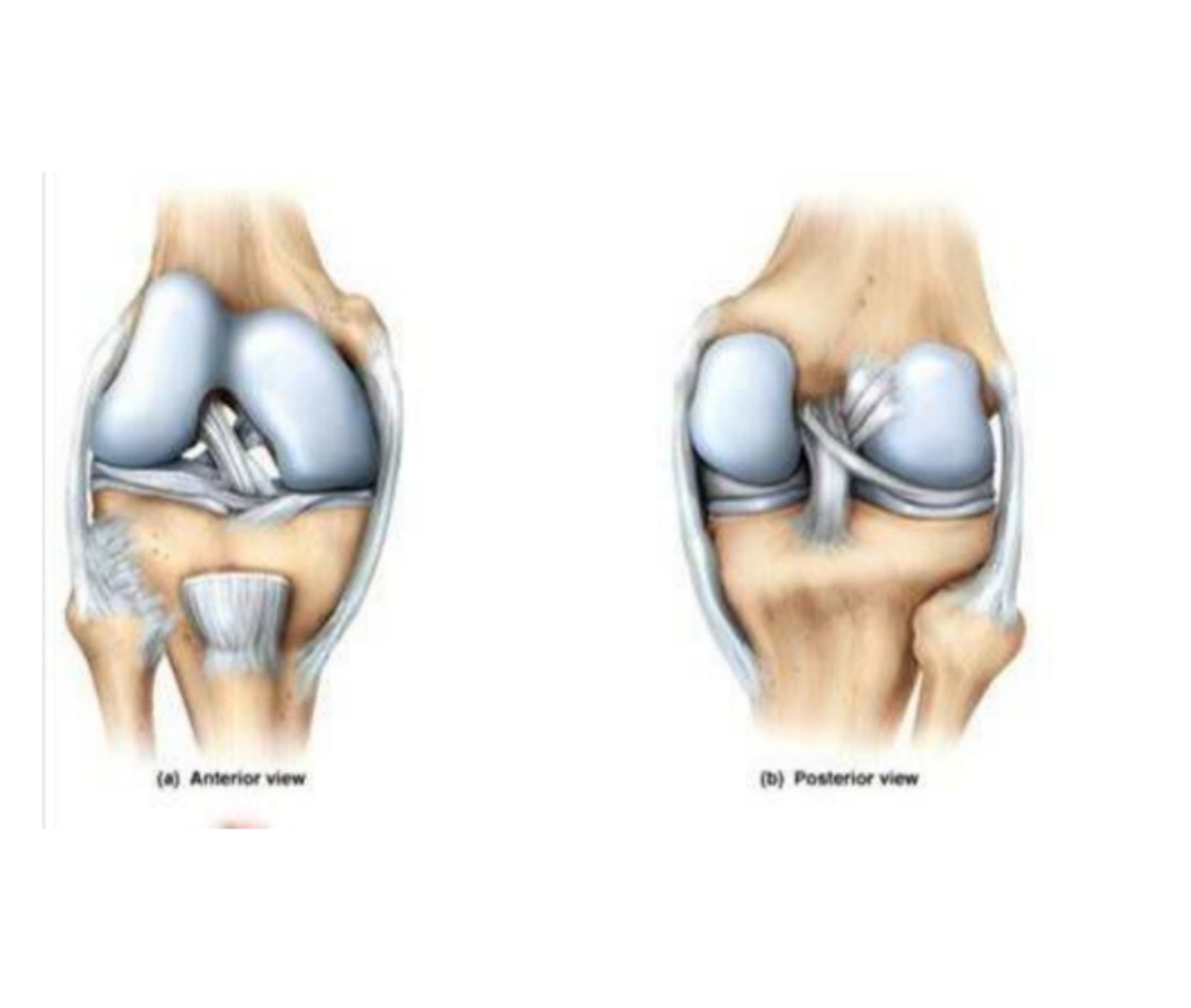
Knee (Tibiofemoral) Joint Diagram (Front & Back Views)

Femur: thigh bone
Tibia: shin bone
Fibula: thin outer lower-leg bone (doesn't bear much weight)
Lateral condyle of femur & medial condyle of femur: the two rounded knobs at the bottom of the femur that rest on the tibia
Articular cartilages: smooth cushions covering the bone ends
Medial meniscus & lateral meniscus: cartilage pads that act as shock absorbers
ACL (anterior cruciate ligament): stops the shin bone from sliding too far forward
PCL (posterior cruciate ligament): stops the shin bone from sliding too far backward
Medial collateral ligament & lateral collateral ligament: stop the knee from bending side to side
Patellar ligament (cut): connects the kneecap to the shin bone
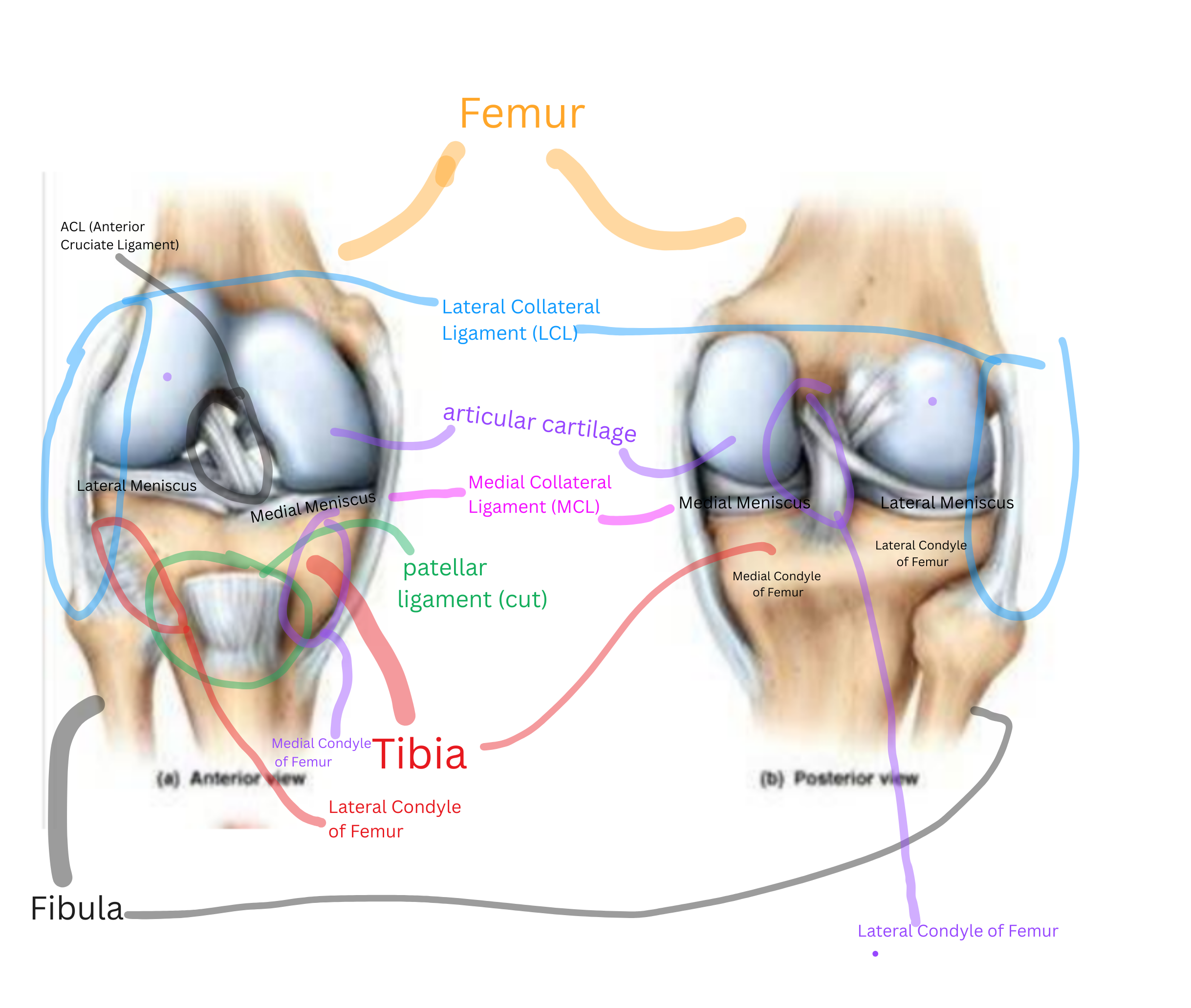
<p>Femur: thigh bone<br>Tibia: shin bone<br>Fibula: thin outer lower-leg bone (doesn't bear much weight)<br>Lateral condyle of femur &amp; medial condyle of femur: the two rounded knobs at the bottom of the femur that rest on the tibia<br>Articular cartilages: smooth cushions covering the bone ends<br>Medial meniscus &amp; lateral meniscus: cartilage pads that act as shock absorbers<br>ACL (anterior cruciate ligament): stops the shin bone from sliding too far forward<br>PCL (posterior cruciate ligament): stops the shin bone from sliding too far backward<br>Medial collateral ligament &amp; lateral collateral ligament: stop the knee from bending side to side<br>Patellar ligament (cut): connects the kneecap to the shin bone</p>
3
New cards
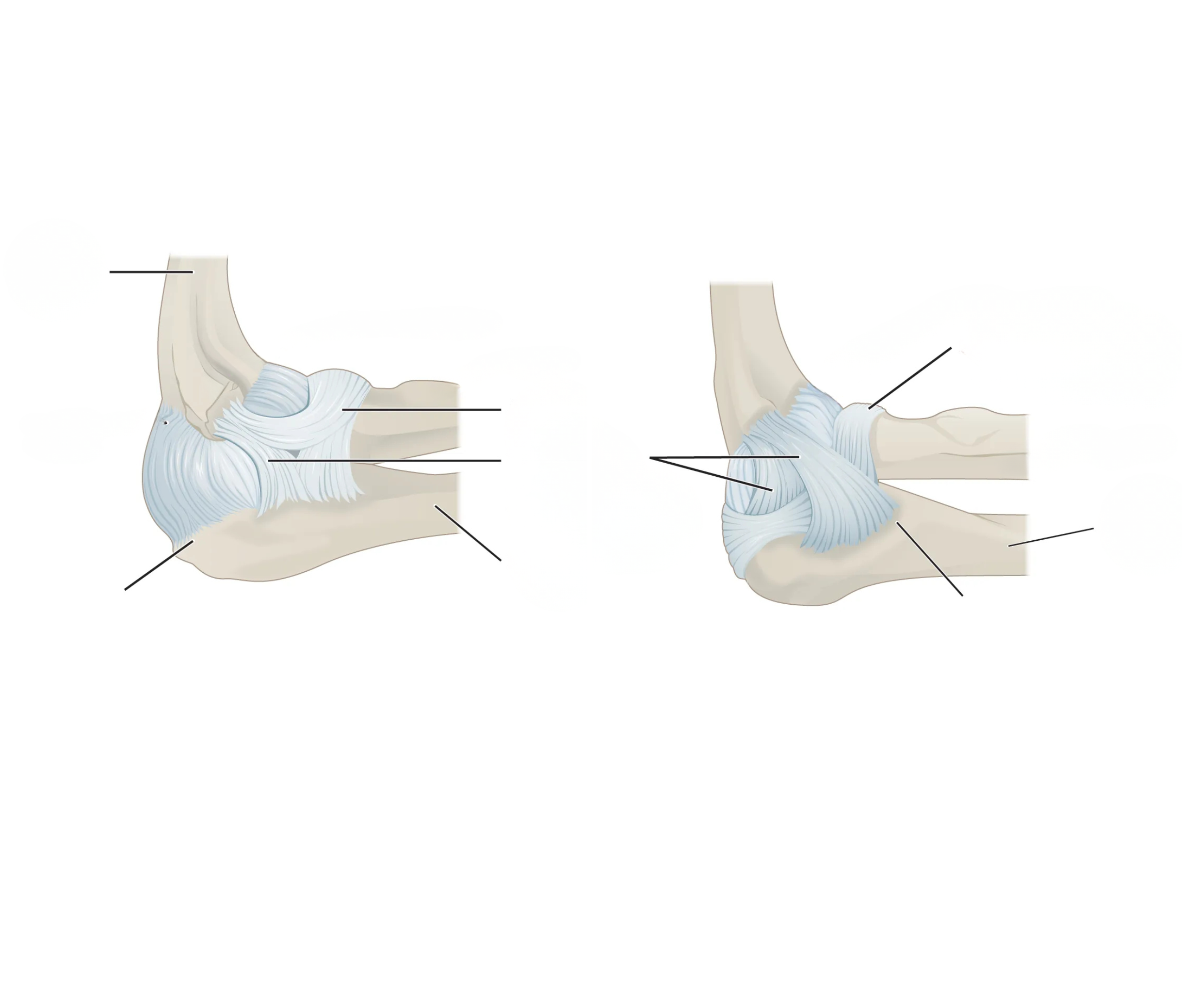
<p>Elbow Joint Model/Diagram</p>

Elbow Joint Model/Diagram

Humerus: upper arm bone
Ulna: forearm bone that forms the hinge with the humerus
Olecranon process: bony bump at the back of the elbow (part of the ulna)
Coronoid process: bony point at the front of the ulna that locks into the humerus when the elbow bends
Ligaments: hold the joint together
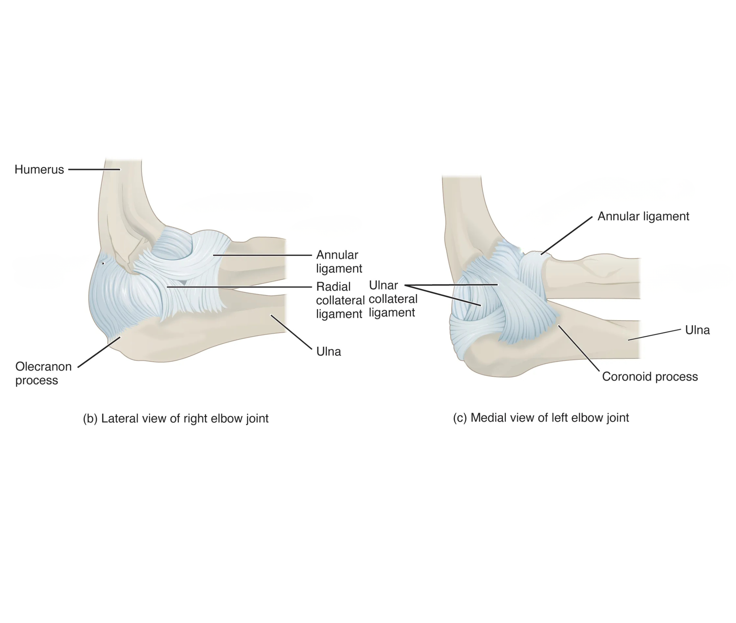
<p>Humerus: upper arm bone<br>Ulna: forearm bone that forms the hinge with the humerus<br>Olecranon process: bony bump at the back of the elbow (part of the ulna)<br>Coronoid process: bony point at the front of the ulna that locks into the humerus when the elbow bends<br>Ligaments: hold the joint together</p>
4
New cards

Shoulder Joint Model/Diagram

Humerus: upper arm bone

Scapula: shoulder blade

Glenoid cavity: the shallow socket on the scapula that the humerus head sits in

Coracoid process: hook-shaped bony bump on the scapula, for muscle attachment

Acromion process: bony tip of the scapula forming the point of the shoulder

Ligaments: loose and few — allows lots of movement but gives little stability 
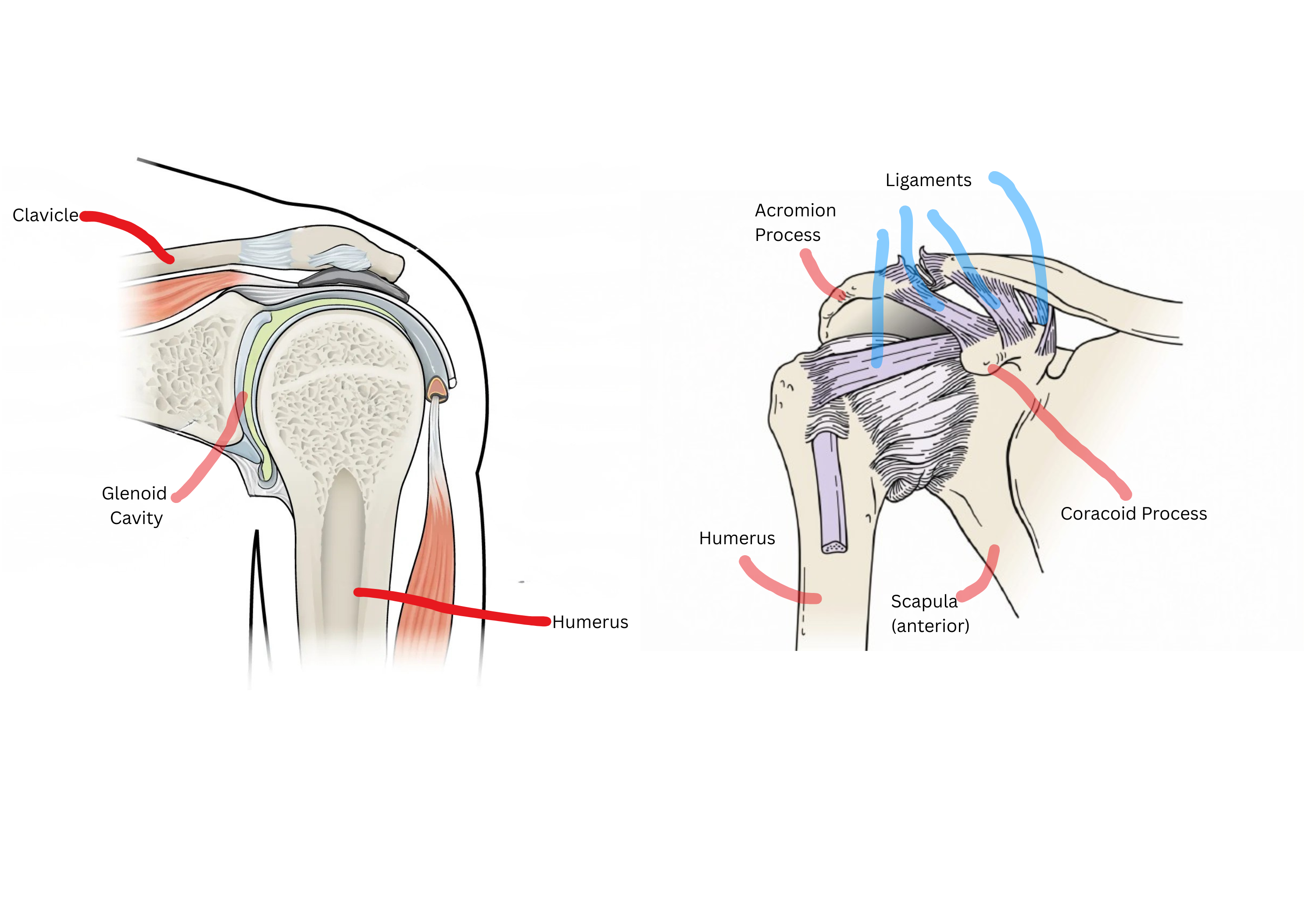
<p><span style="background-color: transparent;">Humerus: upper arm bone</span></p><p><span style="background-color: transparent;">Scapula: shoulder blade</span></p><p><span style="background-color: transparent;">Glenoid cavity: the shallow socket on the scapula that the humerus head sits in</span></p><p><span style="background-color: transparent;">Coracoid process: hook-shaped bony bump on the scapula, for muscle attachment</span></p><p><span style="background-color: transparent;">Acromion process: bony tip of the scapula forming the point of the shoulder</span></p><p><span style="background-color: transparent;">Ligaments: loose and few — allows lots of movement but gives little stability&nbsp;</span></p>
5
New cards
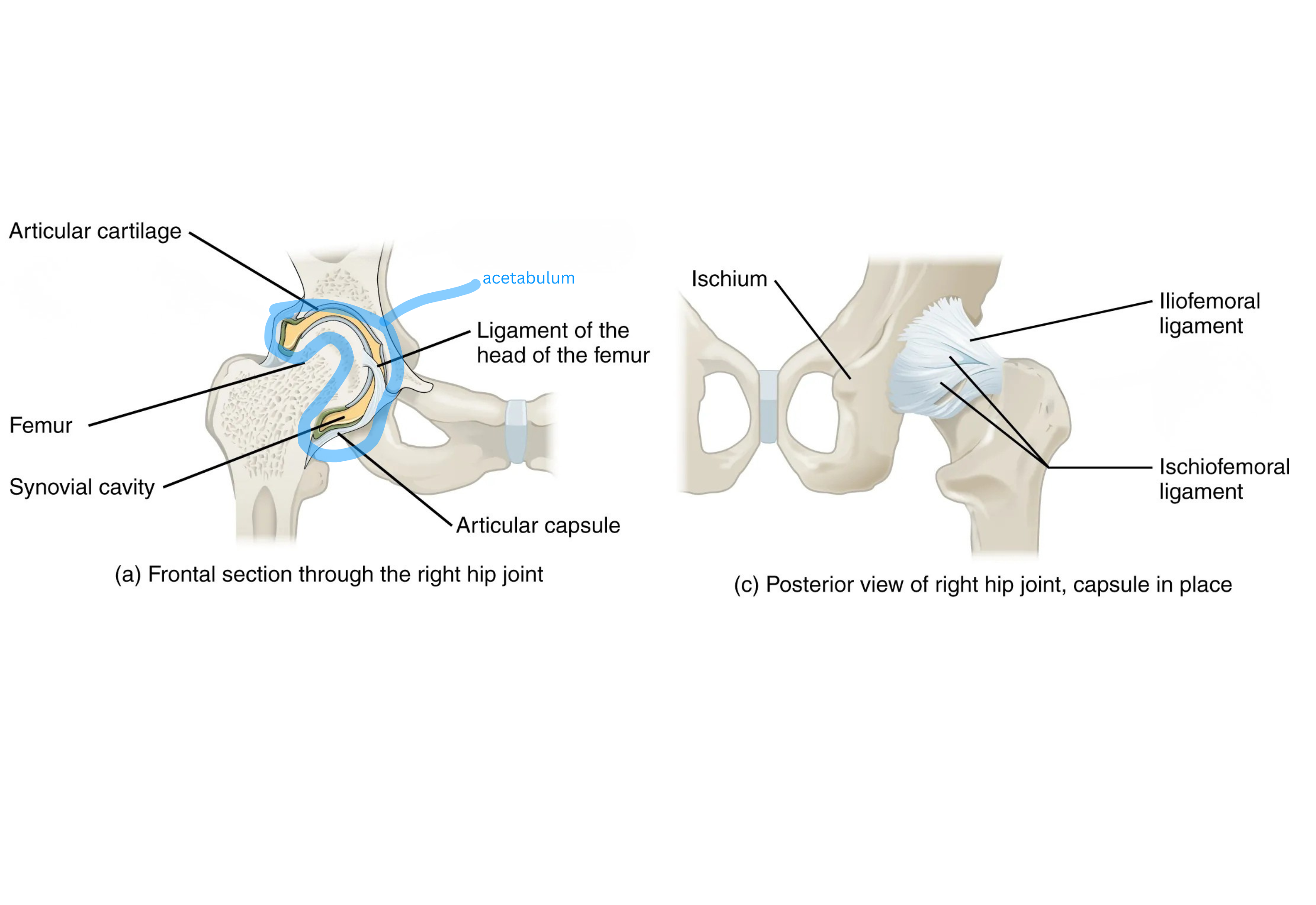
<p><span style="background-color: transparent;">Hip Joint Model/Diagram</span></p>

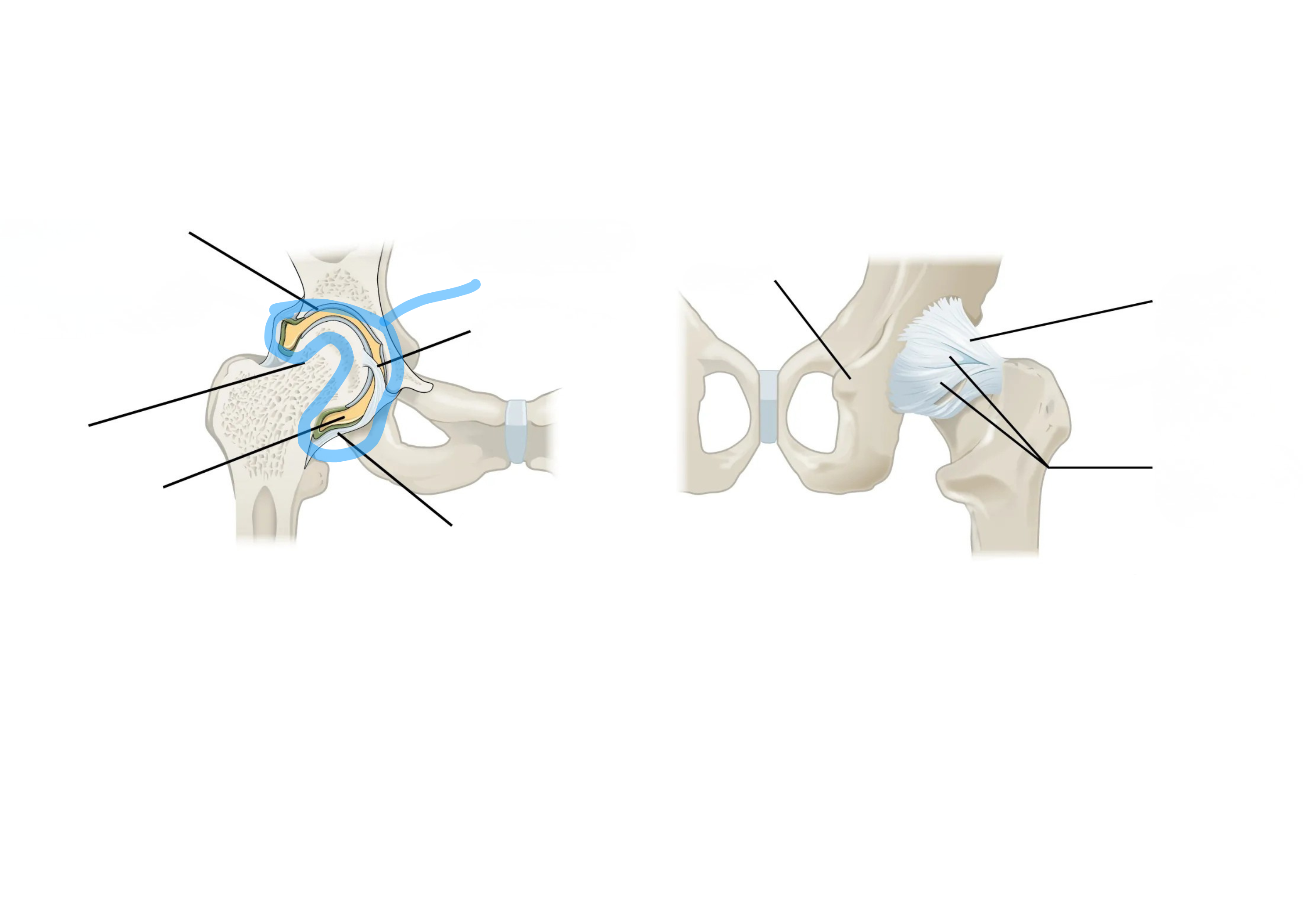
Hip Joint Model/Diagram

Acetabulum: the deep hip socket

Head of femur: ball-shaped top of the thigh bone that sits in the acetabulum

Iliofemoral ligament: strong ligament on the front of the joint, limits how far back the leg can extend
<p><span style="background-color: transparent;">Acetabulum: the deep hip socket</span></p><p><span style="background-color: transparent;">Head of femur: ball-shaped top of the thigh bone that sits in the acetabulum</span></p><p><span style="background-color: transparent;">Iliofemoral ligament: strong ligament on the front of the joint, limits how far back the leg can extend</span></p>