Lab 1 anat and phys
 Call Kai
 Learn
Learn Practice Test
 Spaced Repetition
 Match
Match Flashcards
 Knowt Play
1/77

There's no tags or description
Looks like no tags are added yet.
Name | Mastery | Learn | Test | Matching | Spaced | Call with Kai | Chat |
|---|
No analytics yet
Send a link to your students to track their progress
78 Terms
Planes
Transverse, Horizontal, Median (midsaggital plane), Sagittal Plane

Directional Terms
Cranial, caudal, rostral, dorsal, ventral, lateral, medial superficial, deep, palmar plantar, proximal, distal

Thorax
upper body cavity
Abdomen
Dorsal body cavity
Set of tail
Base of tail
Pinna
External ear (ossicle)
Crown
Top of head
Orbit
Eye socket
Stop
Divit between eyes
Muzzle
Nose
Throatlatch
Adam’s apple
Carpus
Wrist
Metacaprus
Palm
Digits
fingers
Elbow
You should know what a f*cking elbow is
Tarsus
Ankle
Metatarsus
Sole
Crest
Back of neck
Withers
Between shoulder blade
Hindfoot and forefoot
Rear and front feet
Dewclaw
Thumb
Leg/thigh
it’s a leg, it’s a thigh. What are u stupid
Forearm
Between hand and elbow
Flank
Side of animal behind ribs, holds up internal organs and supports weight

Stifle
Knee equivalent, L

Hock
Ankle/heal equivalent, M
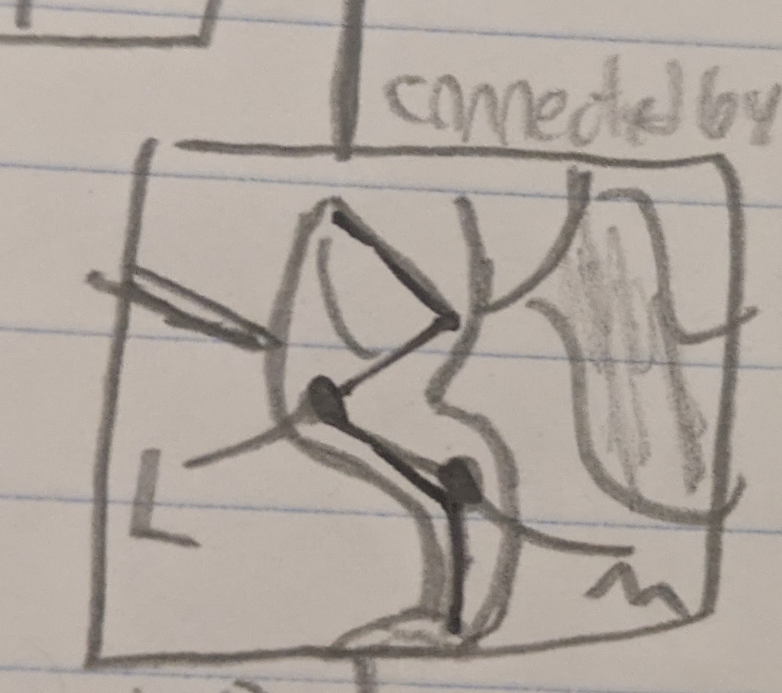
Palpation
Technique where use hands to feel body
4 stomachs of ruminent
Rumen, reticulum, omasum, abomasum
Rumen
1st stomach, microbes ferment plant fiber like cellulose
Reticulum
2nd stomach, forms cud, sends to mouth for rechewing
Omasum
3rd stomach, absorbs water/salts from food
Abomansum
4th stomach, true stomach, acids/enzyme finish digestion
Mustelid
Mink/otter fam
Tissues (epithelial)
Squamous, cuboidal, pseudostratified, columnar, and transitional
4 main tissue types
Epithelial, connective, muscular, neural
Simple vs stratified
1 layer vs 2 layer
Connective tissue types
Skeletal, Cardiac, Smooth
4 types of primary tissue
Epithelial, Connective, Muscle, Neural/nerve
Epithelial types
Lung, epidermis/skin, kidney tubules, small intestine, pseudostratified columnar
Lung (epithelial)
1 layer, squamous, can type of tissue can also be seen in heart

Epidermis/skin (epithelial)
Stratified squamous

Kidney Tubules (epithelial)
1 layer, cuboidal, also found in glandular ducts

Small Intestine (epithelial)
Simple columnar, not ciliated, has GLOBET CELLS (circular) that release mucous, also in stomach
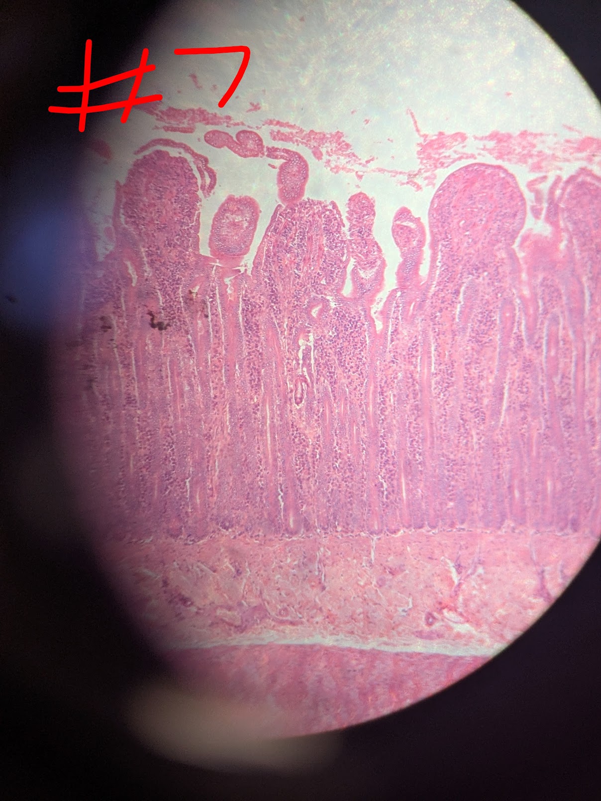
Pseudostratified Tissue
1 layer, ciliated at tip, also in trachea, cilia moves trapped particles across body

Connective Tissue
Dense regular, hyaline cartilage, blood
Dense regular (CT)
In tendon

Hyaline cartilage (CT)
In trachea, has empty spaces, fibrocartilage in intervertebral disk, elastic cartilage in auricle

White blood cells
Here

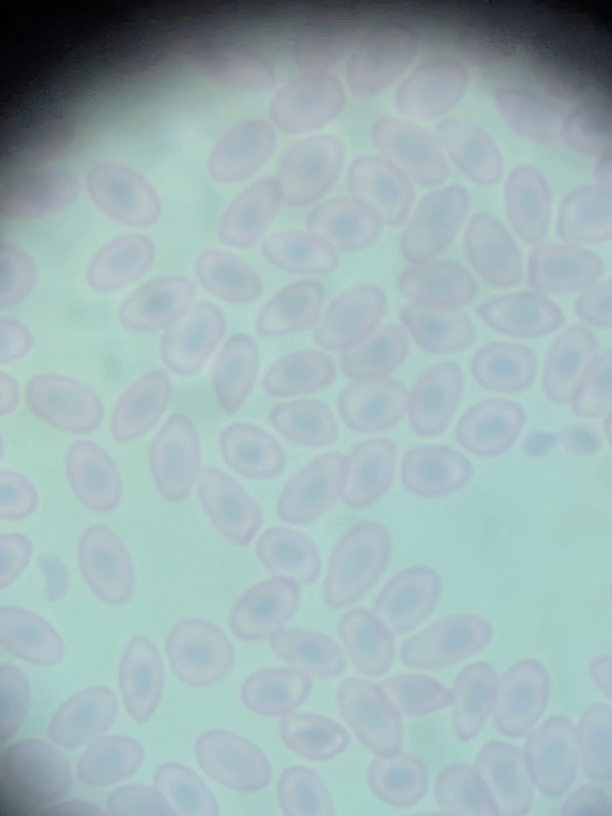
Red blood cells
Here
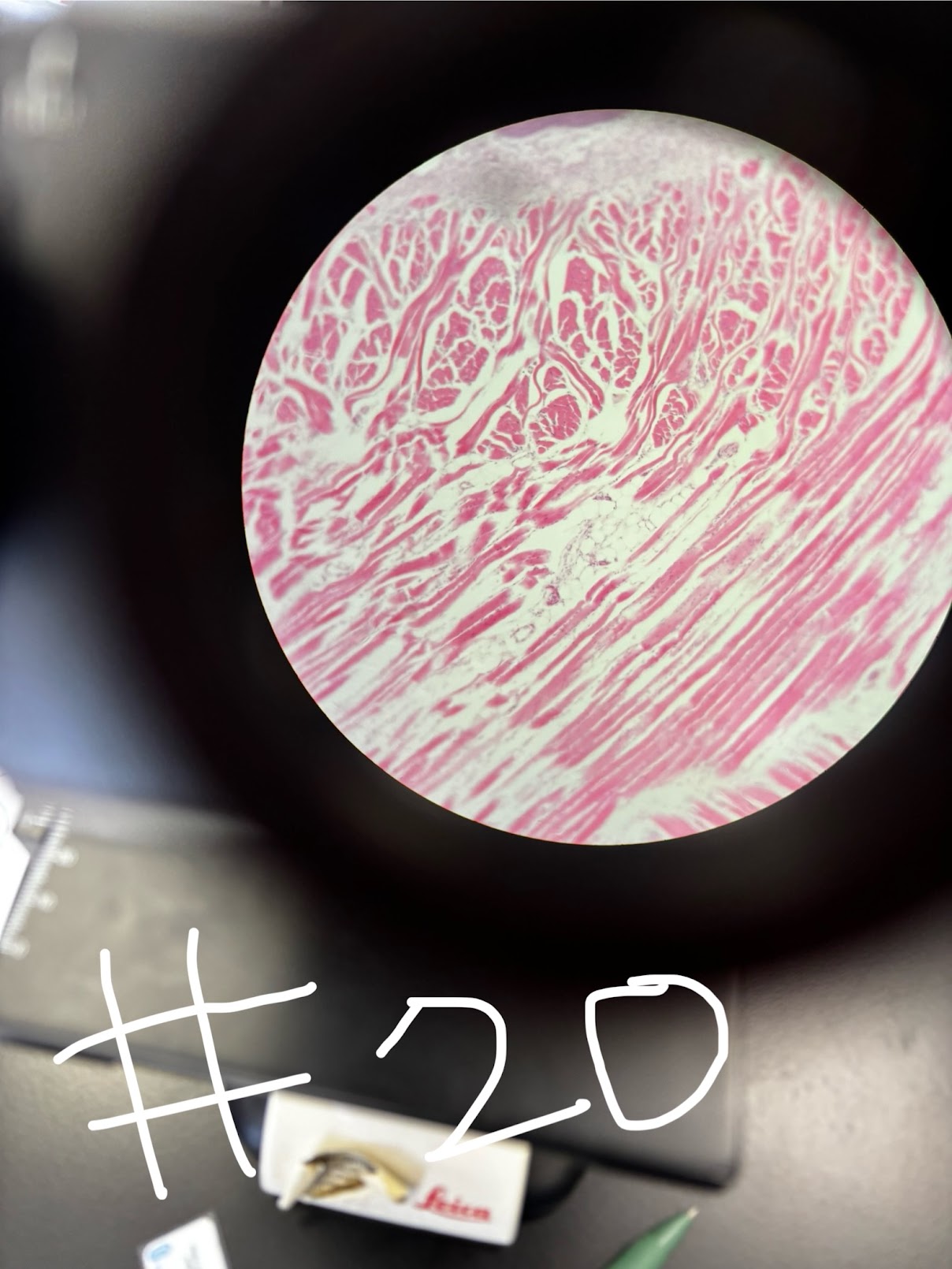
Skeletal Muscle
Voluntary, striated, long and cylindrical, mutlinuclei
Cardiac muscle
Short, branched, uninucleate, involuntary, striated, INTERCALCATED DISKS (coordinate heart beat)
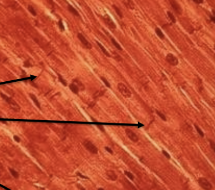
Smooth muscle
Involuntary, not striated, spindle

Nervous tissue
Has neurons (main part), dendrites (lines), neuroglia is white bubble with neuron (supporting)

Loose vs dense CT location
(beneath epithelia, hypodermis, blood vessels, organs, nerves) vs (tendons and ligaments)
Pectoralis Major
In red

Pectoralis Minor
In blue

Latissmus dorsi
In yellow

External Oblique
In green

Rectoral abdominis
in black

Triceps (long)
In green, looking right, lateral (outside), forelimb

Triceps (lateral)
In red, looking right, lateral (outside), forelimb

Dorsoepitrochlearis
In blue, looking right, lateral (outside), forelimb
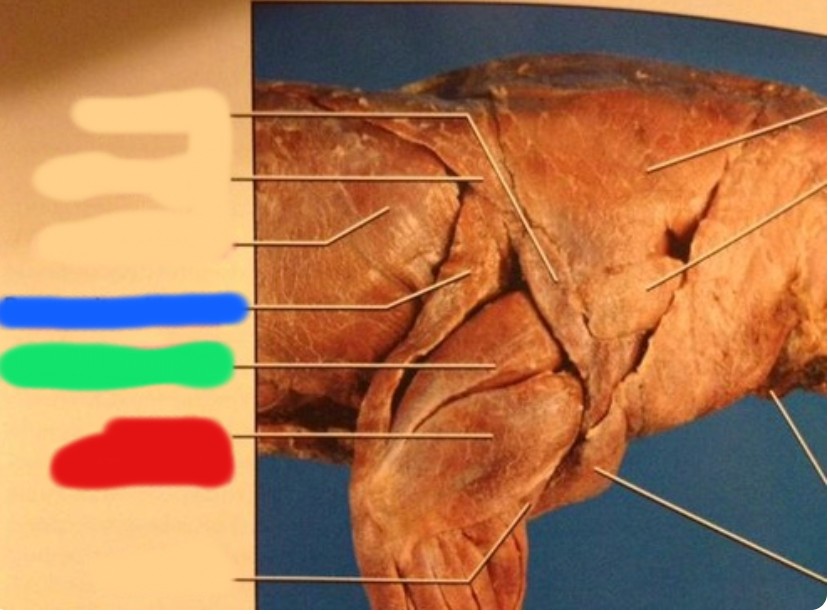
Biceps Brachii
In red, looking left, Medial (underside), forelimbs
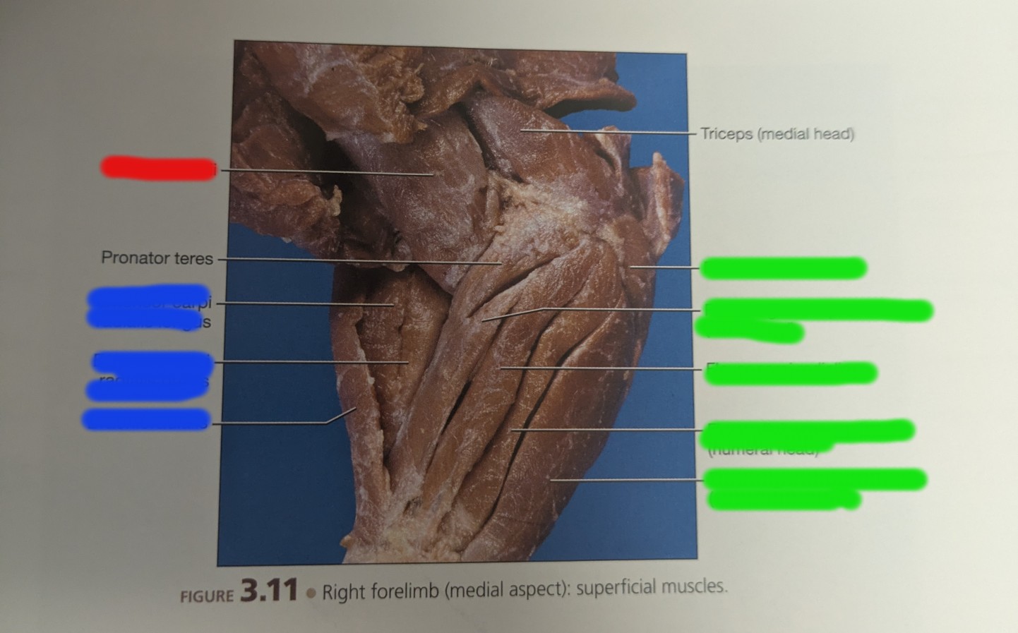
Carpal extensor muscle
Moves palm up, blue, medial (underside), forelimb, looking left

Carpal flexor muscles
Moves palm down, green, medial (underside), forelimb, looking left

Gluteus Maximus
Blue, outside (lateral), looking right, hindlimb

Tensor Fascia Latae
Red, outside (lateral), looking right, hindlimb

Vastus Lateralis
Purple, under, temporal fascia latae, outside (lateral), looking right, hindlimb

Biceps Femoris
Yellow, outside (lateral), looking right, hindlimb, SEMITENDINOSUS above it, CAUDOFEMORALIS next to semi

Sartorius
Green, outside (lateral), looking right, hindlimb, wraps around

Gracilis
Green, underside of thigh, above gastrocnemuis

Gastrocnemius
Green, shin bone, below gastrocnemuis

Extensor muscles
Increases angle, cranial and dorsal
Flexor muscles
Decrease angles, caudal and ventral (palmar)
Origin
Proximal to trunk, stiff
Insertion
Distal to trunk, movable
Adduction
Brings limb closer to medial plane
Abduction
Brings limbs further from from medial plane