(BIO 201) Lab Exam 1
 Call Kai
 Learn
Learn Practice Test
 Spaced Repetition
 Match
Match Flashcards
 Knowt Play
1/27

There's no tags or description
Looks like no tags are added yet.
Name | Mastery | Learn | Test | Matching | Spaced | Call with Kai | Chat |
|---|
No analytics yet
Send a link to your students to track their progress
28 Terms
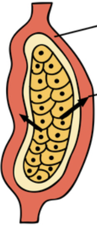
Name the type of gland.
Endocrine gland — hormones are secreted from cells (yellow) directly into bloodstream
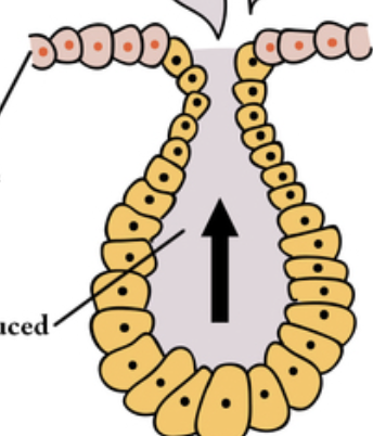
Name the type of gland.
Exocrine gland — duct/tubule releases hormone onto skin surface
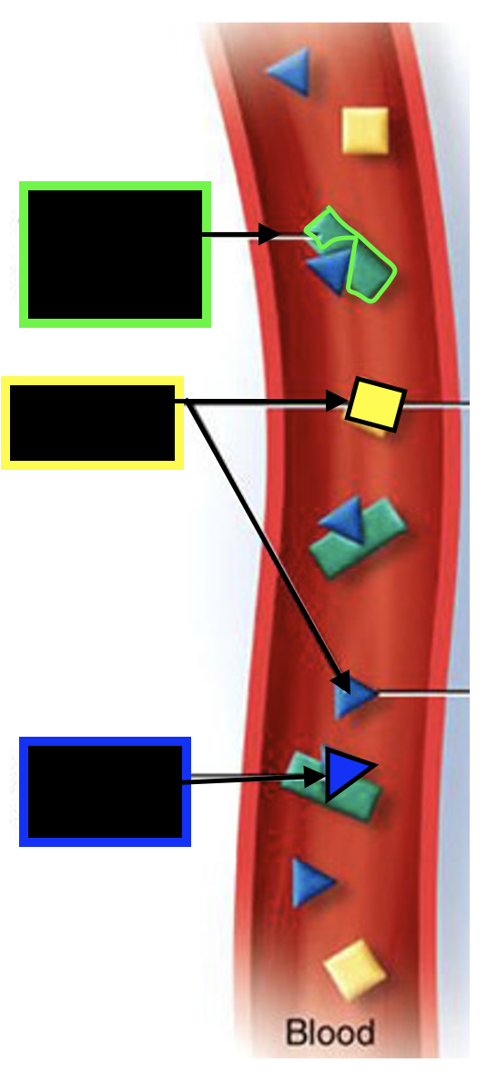
Identify the structures.
Green — transport protein
Yellow — free hormone
Blue — bound hormone
Which process is this diagram showcasing?
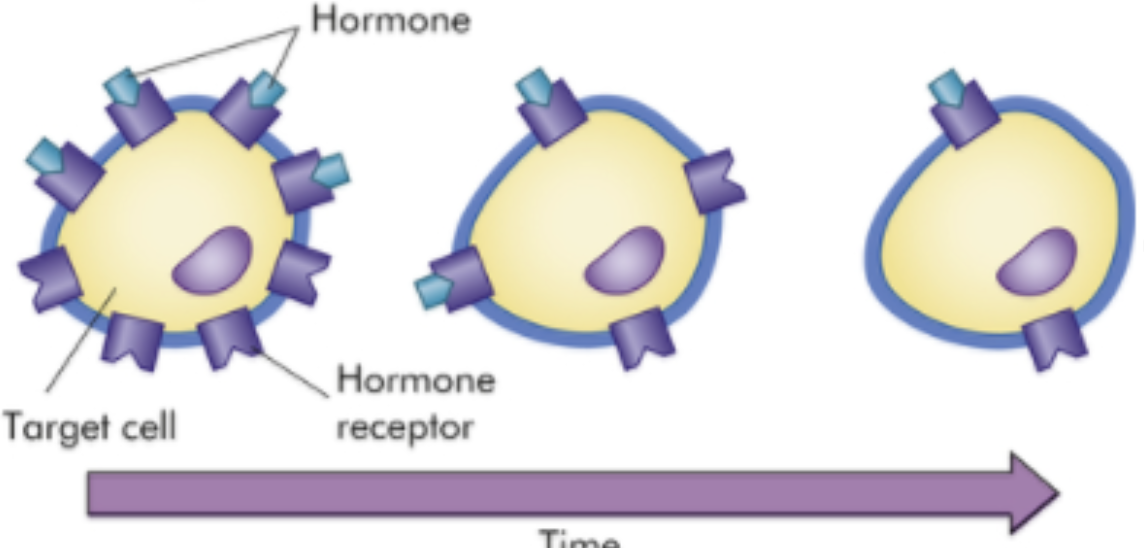
Briefly explain what occurs during this process.
Down-regulation
process by which target cells decrease number of receptors in response to prolonged exposure to a high level of hormone in blood
Which process is this diagram showcasing?
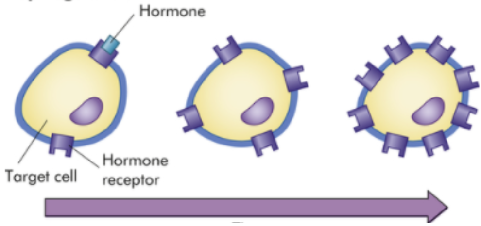
Briefly explain what occurs during this process.
Up-regulation
process by which target cells produce a greater number of receptors in response to a temporary increase in hormone level in blood
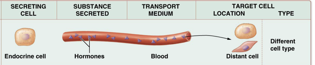
Type of cell signaling?
Endocrine cell signaling

Type of cell signaling?
Paracine cell signaling

Type of cell signaling?
Autocrine cell signaling
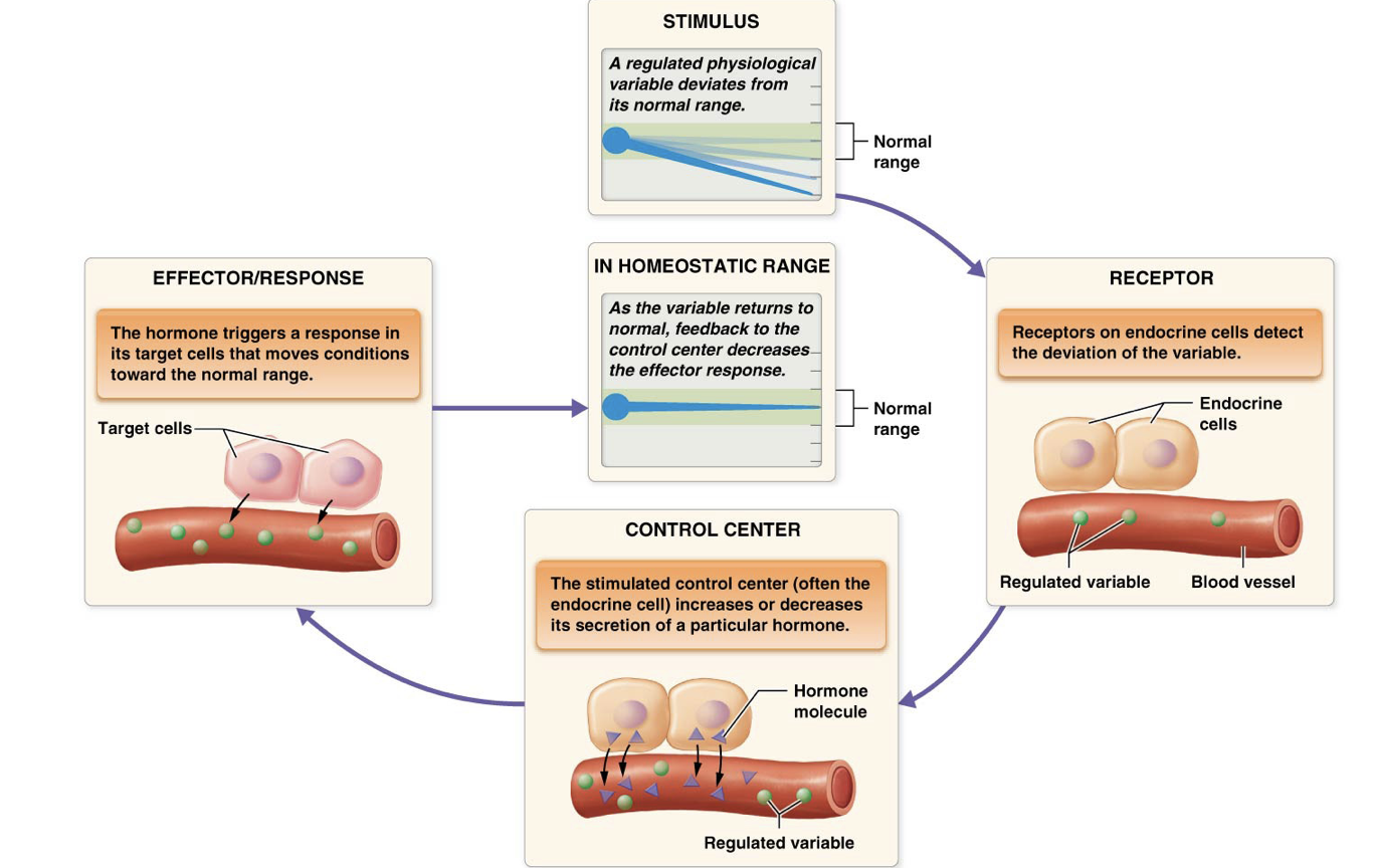
Is this feedback loop negative or positive?
Negative
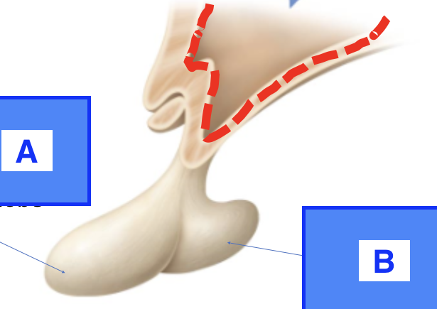
Name lobes A & B
A — anterior lobe
B — posterior lobe
(i) What system is displayed in this image? Function?
(ii) Identify structures A, B, and C
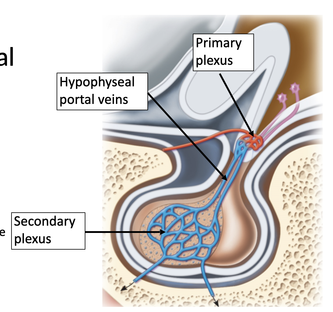
Hypothalamo-hypophyseal portal system
BVs connecting hypothalamus to ant.pit.
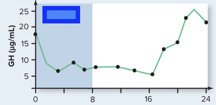
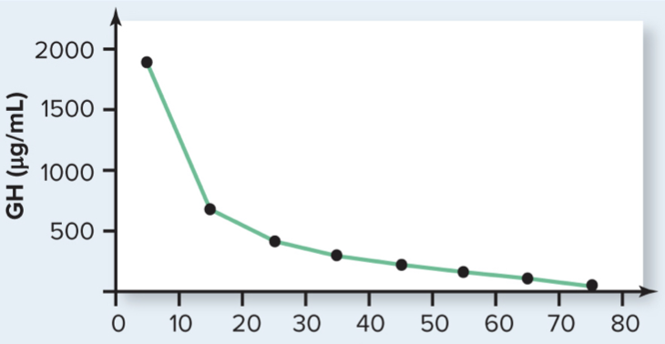
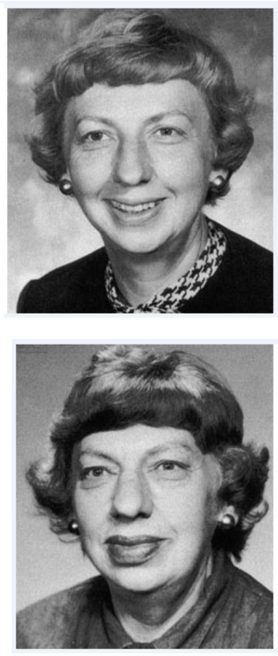
Thesee images are showcasing individuals with which condition?
Acromegaly (overprudction of a hormone)
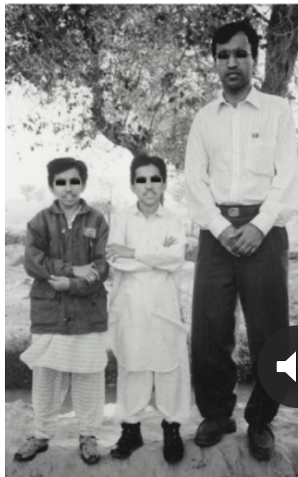
In this image, the poeple on the left side have which condition?
Pituitary dwwarfism
Growth Hormone — Name its 4 target tissues.
liver
adipose tissue
muscle tissue
bone & cartilage
ADH — Name its 2 target tissues.
1) kidneys
2) brain
Oxytocin — Name its 2 target tissues.
Mammary gland
Uterus
Thyroid Gland
Location
Anatomical structure
Histology / Cellular structure
Location
Anatomical Structure
R & L lobes connect at midline by isthmus
Histology/cellular structure
Highly vascularized
Composed of thyroid follicles (function units of gland)
Each follicle has 2 main parts:
Follicular cells
single layer of cuboidal epithelial cells forming follicle's border
secretes thyroglobulin (precursor to thyroid hormone)
Colloid
Iodine‑rich, gel‑like material in follicle lumen
stores thyroglobulin
Parafollicular cells
small clusters in b/w follicles
secretes calcitonin
Describe the chemical structure of thyroid hormones (T3 and T4).
How do T3/T4 differ from other monamine hormones?
T3: amino acid core bound to 3 iodine atoms
T4: amino acid core bound to 4 iodine atoms
T3/T4 are more hydrophobic than other monoamine hormones.
T or F: Both T3 and T4 are physiologically active.
True.
Parathyroid Glands
Location
Anatomical structure
Histology / Cellular structure
Location
3-5 separate glands on posterior surface of thyroid (sits on either side of pharynx/throat)
Anatomical Structure
Histology/cellular structure
Chief cells (AKA principal or parathyroid cells) → secrete PTH
Oxyphil cells → eoisionophilic cells; visibly larger & less numerous than chief cells, have lots of dots
Adrenal Glands
Location
Anatomical structure
Histology / Cellular structure
Location
Sits directly on superior aspect of each kidney
Anatomical Structure
Roughly pyramid-shaped
Histology/cellular structure
Medulla
Medulla
Chromaffin cells
secrete Epinephrine & Norepinephrine
Cortex
Zona glomerulos
secrete mineralcorticoid (aldosterone)
Zona fasciculata
secrete glucocorticoid (cortisol)
Zona reticularis
secrete androgen (DHEA)
Which part of adrenals has typical endocrine function?
Cortex
What part of adrenals are neuroendocrine tissue?
Medulla — secretes catecholamines
Describe the anatomy/histology of thyroid gland.
Location
Anatomical Structure
Histology/cellular structure
Describe the anatomy/histology of thyroid gland.
Location
Anatomical Structure
Histology/cellular structure
Describe 3 major functions of thyroid gland.
(1) Thermoregulation + Regulation of metabolic rate
sets basal metabolic rate (amount of E required by body at rest)
increases rate of catabolic reactions (breaking down macromolecules)
increasing rate of anabolic reactions (building up macromolecules)
generates heat
(2) Promotion of growth & development
bone & muscle growth
NS development
(3) Synergism w/ SNS
increases (up-regulates) production of receptors for sympathetic neurotransmitters
consequently helps in regulation of BP & HR