Back 1
 Call Kai
 Learn
Learn Practice Test
 Spaced Repetition
 Match
Match Flashcards
 Knowt Play
1/19

There's no tags or description
Looks like no tags are added yet.
Name | Mastery | Learn | Test | Matching | Spaced | Call with Kai | Chat |
|---|
No analytics yet
Send a link to your students to track their progress
20 Terms
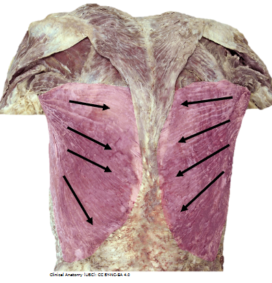
What’s the OIAN of this muscle
O: inferior 6 thoracic vertebrae, sacrum, crest of the ilium, ribs 10-12
I: floor of the intertubercular sulcus of the humerus
A: extends, adducts, and medially rotates humerus
N: thoracodorsal n. (C6, C7, C8)
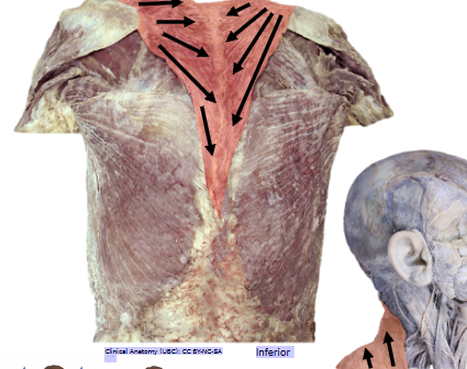
What’s the name of this muscle, and its OIAN? Anatomy (UBC): CC BY-NC-SA 4
Trapezius
O: external occipital protuberance, nuchal ligament, spinous processes of C7 to T12
I: clavicle; scapula; acromion
A: extends head; elevates and pulls shoulder posteriorly; stabilization and movement of the scapula
N: motor innervation CN XI (spinal accessory nerve); sensory cervical plexus (C3;C4)
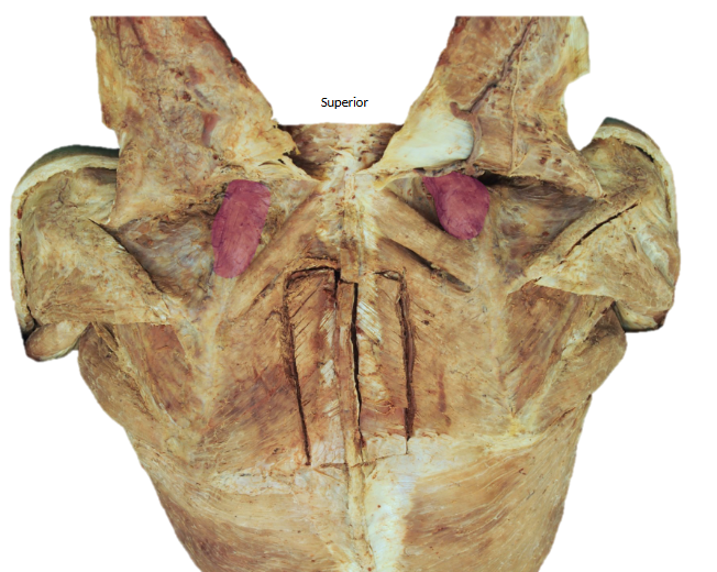
What’s the name of this muscle, and its OIAN
Levator Scapulae
O: Transverse processes of C1 to C6
I: Superior portion of the medial scapula border
A: Scapula Elevation
N: (C3, C4) Cervical Nerves & Dorsal Scapular Nerve (C4,C5)
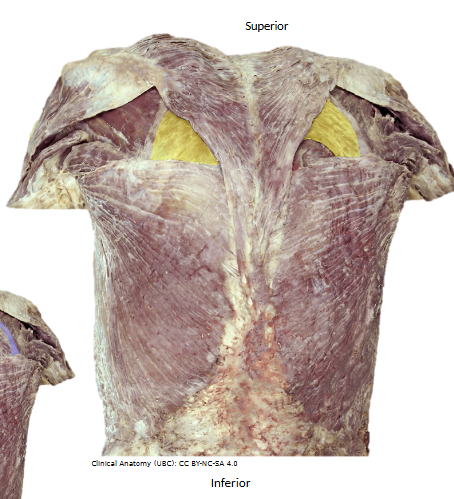
What’s the name of this muscle, and its OIAN
Rhomboid Major
O: Spinous processes of T2-T5
I: Medial border of the scapula
A: Superior-medial rotation of the scapula; scapula support; inferior rotation of the glenoid cavity
N: Dorsal scapular n. (C4, C5)
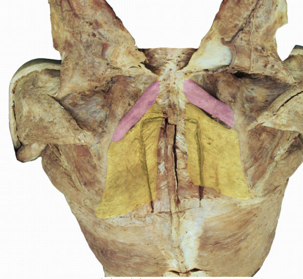
What’s the name of the muscle in pink, and its OIAN
Rhomboid Minor
O: Ligamentum Nuchae (Inferior portion); CVII to T1 spinous processes
I: Spine of the scapula (medial)
A: Adduction (retraction) and elevation of the scapula
N: Dorsal Scapular Nerve (C4,C5)
What muscles border the point of auscultation
inferiorly by the latissimus dorsi
medially by the inferior border of the trapezius
laterally by the medial border of the scapula formed by the teres major and infraspinatus
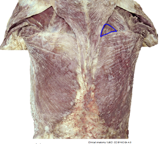
What is the structure outlined in blue called an what is its purpose
Point of auscultation
This is an important anatomical landmark that allows for better auscultation and fewer impedances during the pulmonary examination.
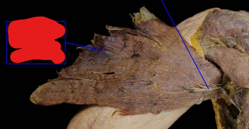
What are the two blue lines pointing to
The thoracodoral neurovasculature
The latissimus dorsi
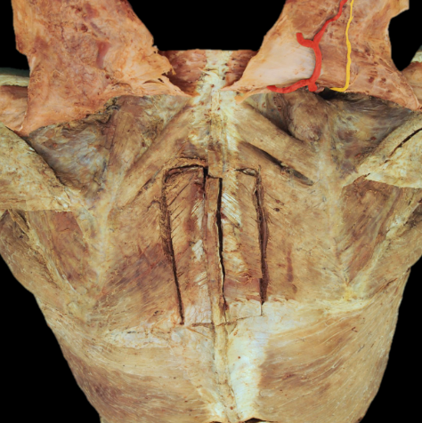
what are the highlight nerve and artery
Accessory nerve and transverse cervical artery
What’s the name of the muscle, and its OIAN
Serratus Posterior Superior:
O: T11-L
I: Ribs 2-5 superiorly
A: Elevates the ribs (respiration facilitation)
N: 2nd through 5th intercostal nerves
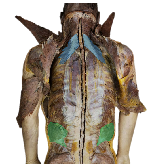
What’s the name of the muscle in green, and its OIAN
Serratus Posterior Inferior
O: Spino
I: Ribs 2-5 superiorly
A: Elevates the ribs (respiration facilitation)
N: 2nd through 5th intercostal nerves
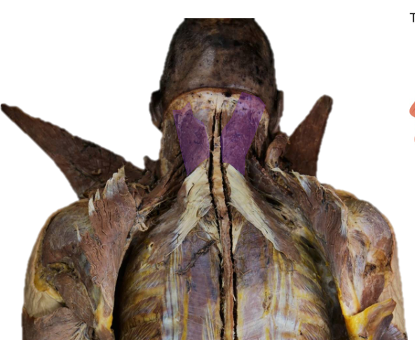
What’s the name of the muscle, and its OIAN
Splenius Capitis
O: Nuchal ligament; C7-T6 spinous processes
I: Mastoid process of temporal bone and superior nuchal line on occipital bone
A: Unilateral: flexion and rotation of the neck laterally
Bilateral: head and neck extension
N:Posterior rami of spinal nerves
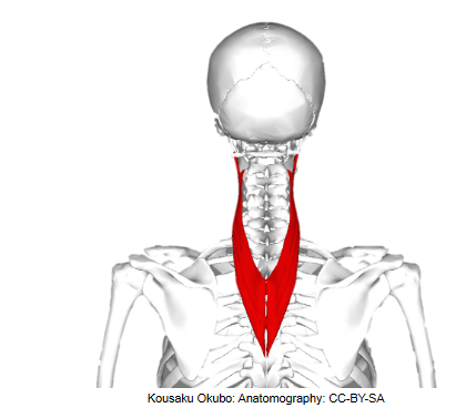
What’s the name of the muscle, and its OIAN
Splenius cervicis
O: Nuchal ligament; C7-T6 spinous processes
I: C1 to C3/C4 vertebral transverse processes
A: Unilateral: flexion and rotation of the neck laterally
Bilateral: head and neck extension
N: Posterior rami of spinal nerves
What the names of the erector spinae muscles and their functions and innervations
A: Primary extensor of the back; contributes to stabilization of
lateral rotation, and flexion of the vertebral column
N: Posterior rami of spinal nerves
what are the name of the gutter muscles and their OIANs
Rotatores, Multifidus, and Semispinalis
O: Transverse Processes of adjacent vertebrae
I: Spinous processes of more superior vertebrae
A: Extension, Rotation, and Stabilization of the vertebral column
N: Posterior Rami of Spinal Nerves
what is the lumbar triangle and where is it located
The lumbar triangle is a thin area of the abdominal wall, and as such it is a site of potential weakness that can lead to a lumbar hernia, also known as Petit’s hernia
what structures make up the lumbar triangle / inferior lumbar triangle
The boundaries are:
• the iliac crest inferiorly
• the latissimus dorsi muscle anteromedially
• the external oblique muscle posterolaterally
• the internal oblique muscle as the floor
what common type of hernia can be found a the inferior and superior lumbar triangle
Grynfelt hernia, also called the superior lumbar triangle, is defined by the quadratus lumborum muscle, twelfth rib, and internal oblique muscle and is also a back herniation
lumbar hernia, also known as Petit’s hernia (inferior in position to a grynfelt hernia
what are muscles attach to the nuchal ligament
Traps, Rhomboid minor, Serratus Posterior Superior, Splenius capitis and cervicis