Week 1 - Epithelium
1/74
There's no tags or description
Looks like no tags are added yet.
Name | Mastery | Learn | Test | Matching | Spaced | Call with Kai |
|---|
No analytics yet
Send a link to your students to track their progress
75 Terms
What are the 4 basic tissues?
1) Epithelium
2) Connective tissue
3) Muscle
4) Nervous tissue
Describe the cellularity and vasculature of epithelium.
- Very cellular; little intercellular material
- Avascular
What are the two functions of epithelium?
1) Lines body surfaces, body cavities, and organ surfaces
2) Forms glands for secretion
What are the 3 borders of epithelium?
A) Apical (luminal)
B) Lateral
C) Basal
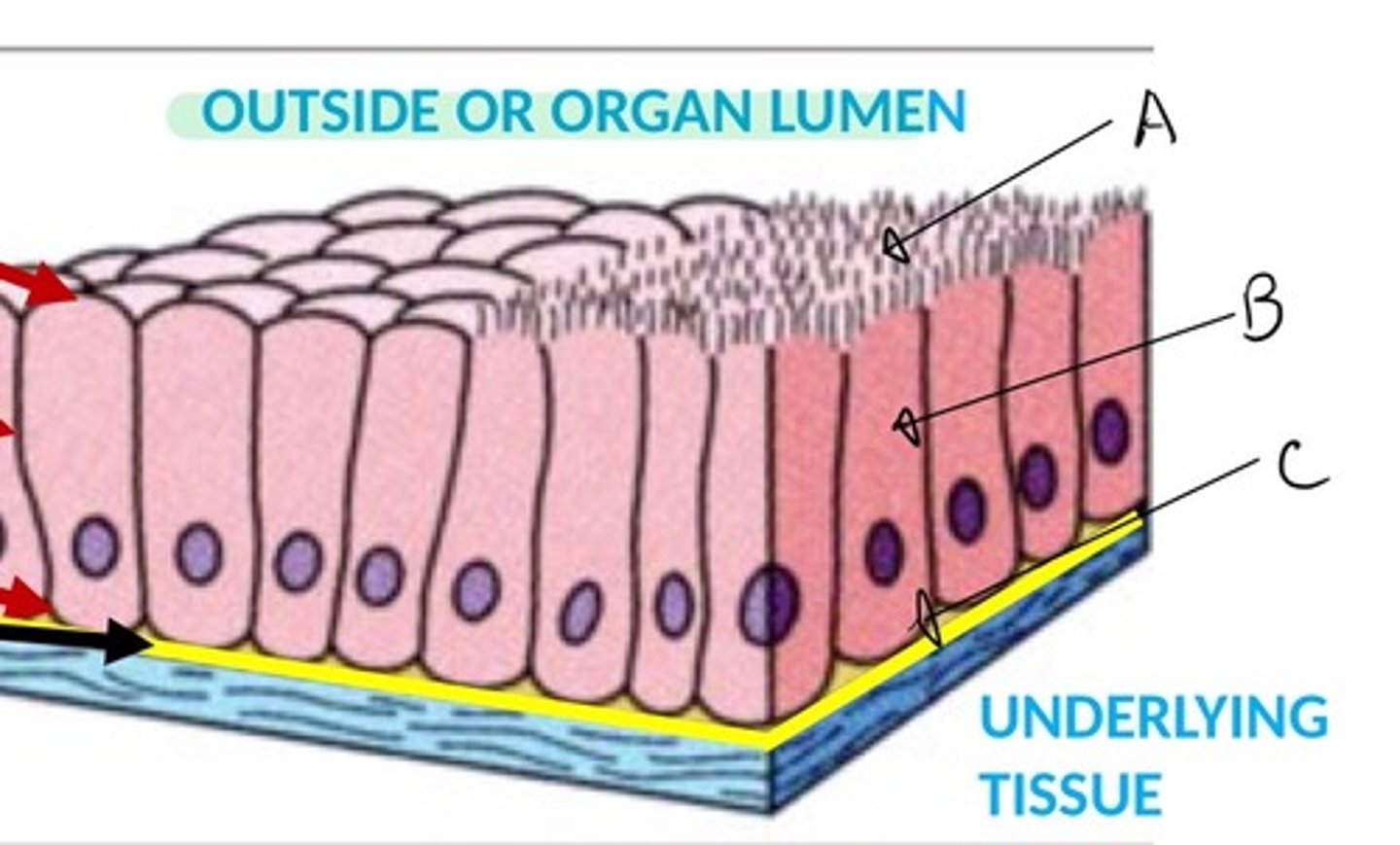
Basal border of epithelium is attached to the what?
- Basement membrane
What is the basement membrane?
- Extracellular material between epithelial cells and underlying CT
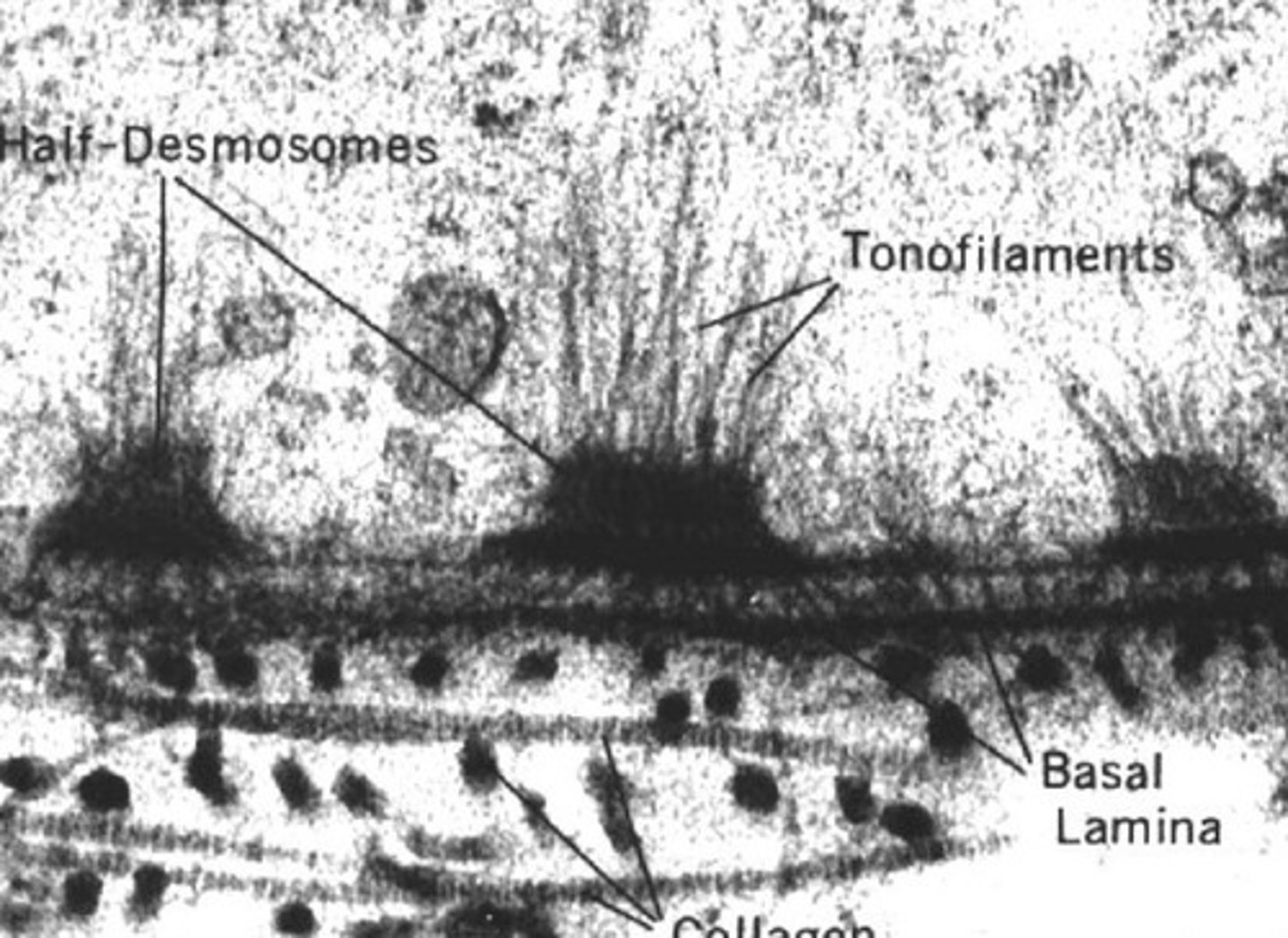
When using electron microscopy, what can one tell about the basement membrane?
- It is shown to have two layers: the basal lamina that is nearest to the epithelial cells and a deeper layer, the reticular lamina, that is more diffuse
What is the basal lamina of the basement membrane? The reticular lamina?
- Basal lamina: Noncellular, made from epithelium and is extracellular layer seen ultrastructurally
- Basement membrane: the entire structure seen with the light microscope
What is the basal lamina made of? What does it do?
- Collagen
- Provides structural support
What are two ways to classify epithelium?
1) Number of layers present
2) Cell shape at the apical (free) surface
What are the 3 ways to classify epithelium based on the number of layers present?
1) Simple epithelium = single layer of cells
2) Stratified epithelium = More than one cell layer
3) Specialized = Pseudostratified and transitional
What are the 3 cell shapes? Desribe them
1) Squamous = Cells are flat
2) Cuboidal = Cells are as tall as they are wide
3) Columnar = Cells are taller than they are wide
What are the two specialized terms for simple squamous epithelium we have learned? Describe them.
- Endothelium = Lines blood and lymph vessels
- Mesothelium = Lines body cavities and outer surface of many organs
What is the function of simple squamous epithelium?
- Protection; secretion which reduces friction
Where are simple cuboidal epithelium found?
- Found in organs that are specialized for secretion (salivary glands and thyroid follicles) and diffusion (kidney tubules)
What is the function of simple columnar epithelium? Where?
- Function in secretion and absorption - lining of stomach, small intestine, large intestine
- Function in secretion, transportation, and protection - Lining of uterus and uterine tubes
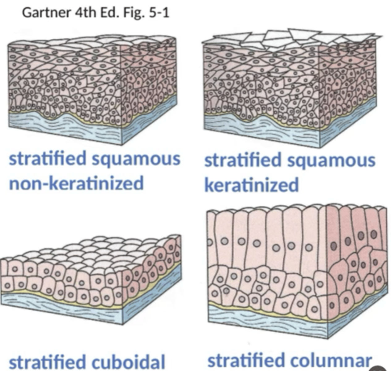
Which type of stratified epithelium can be keratinized?
- Stratified squamous epithelium
What are the 3 types of stratified epithelium?
1) Stratified squamous epithelium (keratinized or not)
2) Stratified cuboidal epithelium
3) Stratified columnar epithelium
What is another name for keratinized stratified squamous epithelium?
- Cornified stratified squamous
Where is keratinized stratified squamous epithelium found?
- Covering the general body surface, buccal cavity, anal region, ruminant forestomach
What is another name for nonkeratinized stratified squamous epithelium?
- Noncornified stratified squamous epithelium
Where is nonkeratinized stratified squamous epithelium found?
- Found in nasal vestibule, oral, esophageal and anal portions of digestive system, cornea of eye, conjunctiva, portions of the male and female urogenital systems
Where is stratified cuboidal epithelium found?
- Ducts in sweat glands, mammary glands, and salivary glands
In stratified columnar epithelium, what is the shape of the intermediate cells (if present)?
- Polygonal
Where is stratified columnar epithelium found?
- Portions of the upper respiratory tract, ducts of some glands
Describe the appearance of pseudostratified ciliated columnar epithelium.
- Looks stratified but it is not; All cells contact the basement membrane (basal lamina). Some cells do NOT reach the apical surface of the epithelium.
What are the functions of pseudostratified ciliated columnar epithelium? Where is it found?
- Functions include secretion, absorption, lubrication, protection, and transportation.
- Found in the lining of the upper respiratory tract, parts of the urogenital systems and ducts of some glands
Transitional epithelium is ____________. Where is it found?
- Distensible
- Lines regions of the urinary tract
What are the 3 plasma membrane specializations of the apical surface of epithelium we have learned?
1) Microvilli
2) Cilia
3) Stereocilia
What are microvilli?
- Cytoplasmic projections from apical surface of cell
With a light microscope, one can see microvilli as a _________________ ___________________.
- Brush border
Microvilli are made with _______________ covered by __________ ______________. What is their function?
- Microfilaments
- Cell membrane
Are microvilli motile?
- No
Where are microvilli located?
- Mainly in intestinal tract; proximal renal tubules; some other sites
What is the terminal web?
- Network of actin filaments, intermediate filaments and spectrin; helps support cell structure
What is the glycocalyx?
- Extracellular material on outside of microvilli
What does the glycocalyx contain?
- Disaccharidases and peptidases
Where are cilia located and what do they contain?
- Located on surface of specialized epithelial cells
- Contain microtubules
Are cilia motile?
- Yes
What is the function of cilia?
- Move in specific direction to move fluid or particles
Where can one find cilia in the body?
- Pseudostratified columnar epithelium of respiratory system; parts of reproductive tract
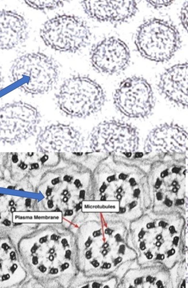
What is the "ultrastructure" of cilia?
- Microbtules ("9+2"); nine sets of double microtubules on periphery and two microtubules centrally
- Attached to cell by basal body
What are three differences between cilia and villi?
- Cilia are motile while villi are not
- Cilia are composed of microtubules while villi are composed of microfilaments
- Cilia are responsible for movement while villi are responsible for absorption
Explain how cilia are related to basal bodies.
- Cilia are inserted (and held to the cell) by basal bodies which are composed of microtubules in a nine triplet pin-wheel arrangement and located just underneath the cell membrane.
Compare the appearance of microfilaments and microtubules.
- When using an electron microscope, microfilaments (villi) appear as dots which are randomly organized, microtubules (cilia) appear as circles that are organized.
Are stereocilia true cilia?
- Not true cilia
Stereocilia are more like cilia or villi?
- Villi
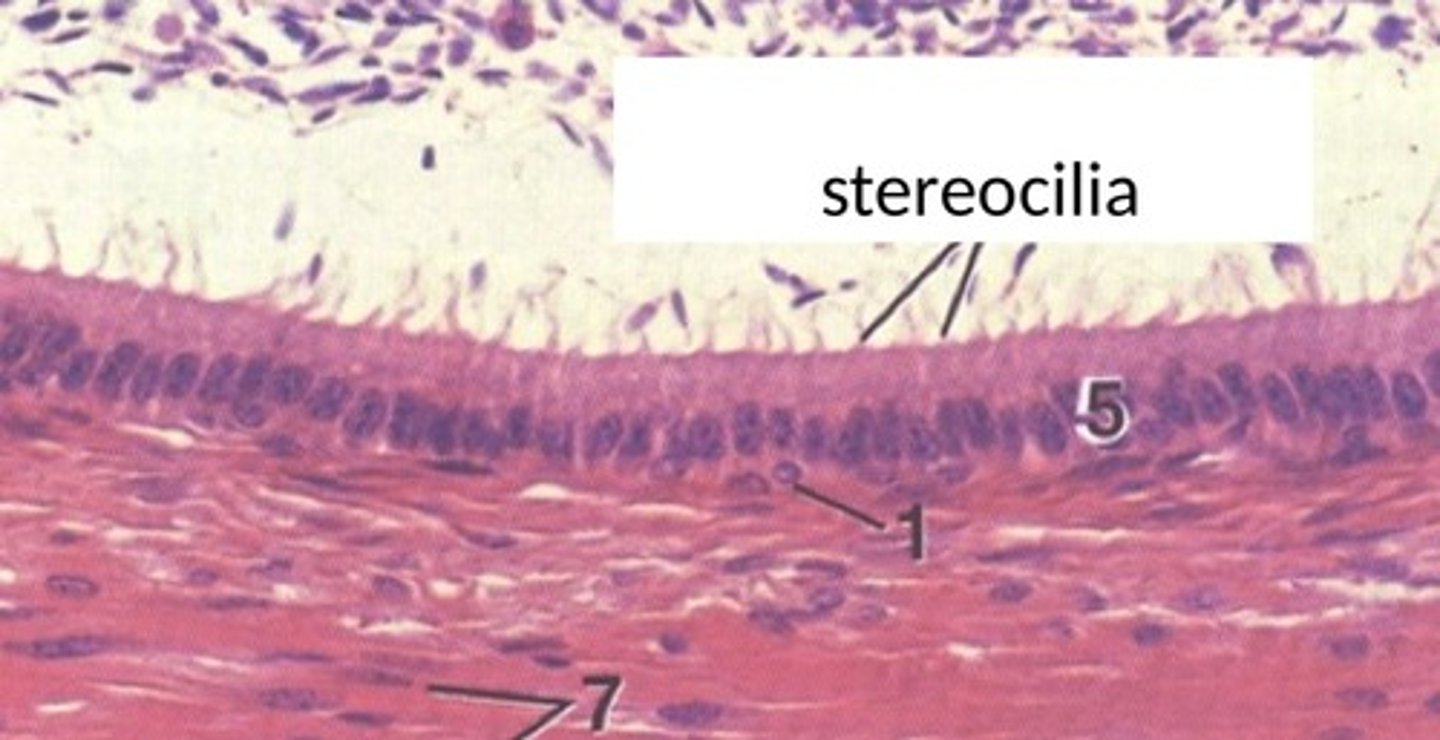
Describe the structure of stereocilia.
- Long microvilli composed of microfilaments
Are stereocilia motile?
- No
Where are stereocilia found?
- Epididymis; sensory hair cells of cochlea
What is the function of stereocilia?
- Increase SA for absorption in epididymis in epididymis; signal generation in cochlea
What are the junctional complexes discussed in this course?
1) Tight junctions (Zonula occludens)
2) Anchoring junctions: Ahderens junctions, desmosomes, hemidesmosomes
3) Communicating junctions - gap junctions
What is the function of tight junctions (zonula occludens)?
- Seals neighboring cells together in an epithelial sheet to prevent leakage of molecules between them.
What is the location of tight junctions?
- Blood brain barrier, blood air barrier (alveoli of lungs), bladder
What are examples of tight epithelia and leaky epithelia?
- Tight epithelia: Kidney = distal convolute tubule; collecting duct; Liver = bile ducts
- Leaky epithelia: Kidney proximal tubule
Describe the function of adherens junctions.
- Joints an actin bundle in one cell to a similar bundle in a neighboring cell to bind cells together
Describe the function of the desmosome.
- Reduce rotation of the cell via joining intermediate filaments between adjacent cells
Describe the function of the hemidesmosome.
- Anchors intermediate filaments in a cell to basal lamina
Describe the function of gap junctions.
- Form pores between cells for ions and molecules
What are the two major groups of glands? Describe them.
1) Exocrine: Secrete product via a duct to the surface of their epithelial origin
2) Endocrine: Ductless; Secrete into the blood or lymphatic vessels for distribution
What is the difference between simple and compound multicellular exocrine glands?
- Simple glands: Do not branch
- Compounds glands: Ducts branch
What are the five shapes of simple glands?
1) Tubular
2) Coiled tubular
3) Branched tubular
4) Acinar
5) Branched acinar
What are the types of compound glands?
1) Compound tubular
2) Compound acinar
3) Compound tubuloascinar
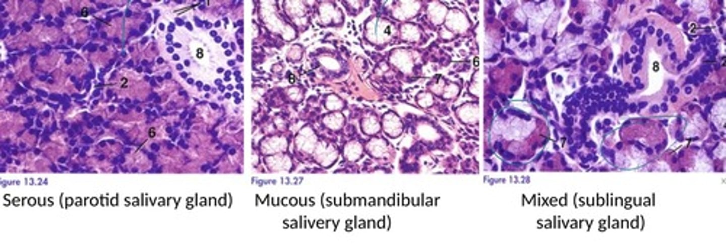
What are the three types of exocrine glands as classified by type of secretion?
1) Serous
2) Mucous
3) Mixed
How can you tell the difference between a serous gland or mucous gland?
- Serous glands tend to appear very basophilic (purple) while mucous glands tend to appear "white and foamy".
What are the three types of exocrine glands based on the mode of secretion? Describe them.
1) Holocrine: Secretory cell dies and becomes the secretory product
2) Merocrine: Secretion occurs via exocytosis; secretory cell stays intact
3) Apocrine: A small part of the apex of the cell is lost with the secretory product
What is the primary example of the unicellular exocrine gland?
- Goblet cell
What are unicellular exocrine glands?
- Isolated secretory glands in an epithelium
Describe goblet cells.
- Arranged individually in the epithelium lining the digestive tract and parts of the respiratory tract; Secretions protect the tract lining
What is the embryonic origin of external surface epithelium?
- Ectoderm
What is the embryonic origin of glands?
- Ectoderm and Endoderm
What is the embryonic origin of ependymal cells lining the neural canal?
- Ectoderm
What is the embryonic origin of mesothelium?
- Mesoderm
What is the embryonic origin of endothelium?
- Mesoderm
What is the embryonic origin of internal surface epithelium?
- Endoderm