PATH 3810 Acute and Chronic Inflammation
 Call Kai
 Learn
Learn Practice Test
 Spaced Repetition
 Match
Match Flashcards
 Knowt Play
1/80

There's no tags or description
Looks like no tags are added yet.
Name | Mastery | Learn | Test | Matching | Spaced | Call with Kai | Chat |
|---|
No analytics yet
Send a link to your students to track their progress
81 Terms
inflammation pros
beneficial host response to foreign invaders and necrotic tissue, helping to eliminate pathogens and initiate healing processes.
inflammation cons
inflammatory reaction and subsequent repair process can cause tissue damage
main components of inflammation are
vascular reaction
cellular response
activated by mediators derived from plasma proteins and cells
the 5 R’s of inflammation
Recognition of injurious agent
Recruitment of leukocytes
Removal of agent
Regulation (control) of response
Resolution (repair)
the outcome of acute inflammation is either…
…elimination of the insult, followed by reduced inflammation and tissue repair, OR persistent cell injury leading to chronic inflammation
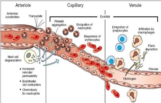
i looked at this diagram regarding arterioles, capillaries, and venules and inflammation and i understand
acute inflammation
rapid response to cell injury or invading microbes and other foreign substances that is designed to deliver leukocytes and plasma proteins to the site of injury

i looked at this diagram regarding normal vs abnormal capillary beds and i understand
stimuli for acute inflammation
infections
trauma: physical, chemical
necrosis
foreign bodies
hypersensitivity reactions
vasodilation of arterioles results in…
…locally increased blood flow, which causes engorgement of downstream microcirculation, which leads to erythema and warmth of tissue

vasodilation cross-section

vasodilation longitudinal section
neutrophils
most abundant leukocyte
segmented nucleus (up to 5 lobes), heterochromatic chromatin, membrane-bound cytoplasmic granules, scarce organelles
phagocytosis
phagocytosis involves…
…phagosome-lysosome fusion to form phagolysosome, generating H2O2 and superoxide radicals by respiratory burst
neutrophil degranulation
secrete granule contents (myeloperoxidase) into extracellular environment

neutrophils <3
eosinophils
respond to parasitic infections and allergies
migration stimulated by chemotactic factors produced by mast cells and basophils
common in type 1 hypersensitivity reactions due to IgE receptors
nucleus has 2-3 lobes

eosinophils!
lymphocytes
smallest leukocyte (so petite)
second most abundant
circulate via blood and lymphatics between lymphoid tissues and all other tissues
round, densely stained nucleus and small amount of agranular cytoplasm
amount of cytoplasm depends on state of activity
B lymphocytes
produce antibodies, differentiate into plasma cells in tissues
T lymphocytes
secrete cytokines that modulate activities of other lymphocytes
natural killer cells
a type of large lymphocyte
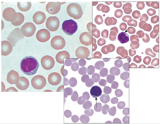
lymphocytes !
monocytes
largest leukocyte
precursors of tissue macrophages (highly motile phagocytes)
large eccentric, indented nucleus with nucleoli
extensive cytoplasm with frequent pseudopodia on surface
granules include myeloperoxidase, acid hydrolases, membrane adhesion proteins, TNF-a
capable of continuous lysosomal activity using aerobic and anaerobic pathways
antigen-presenting cell (APC)

monocyte
monocyte-macrophage system / mononuclear phagocyte system
consists of circulating monocytes, monocytic bone marrow precursors, and tissue macrophages both free and fixed
includes Kupffer cells, microglia, Langerhans cells, alveolar macrophages, APCs of lymphoid tissues, and osteoclasts
basophils
least abundant
very similar to mast cells, but the exact relationship is unknown
bilobed nucleus
large basophilic cytoplasmic granules that are metachromatic when stained by toluidine/methylene blue
granules are membrane-bound, round, and contain densely packed electron-dense material (contain proteoglycans and histamine)
unclear function, but involved in allergic reactions
very mysterious apparently
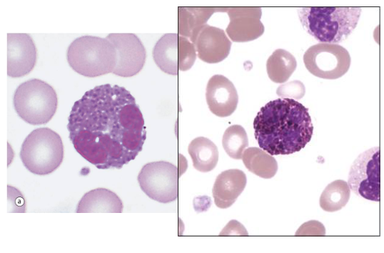
basophils
vascular changes of inflammation
increased vascular permeability
transudate and exudate (edema)
endothelial cells contraction
transudate
ultrafiltrate of blood plasma which moves from capillaries into tissues
exudate
protein-rich fluid and cells move into interstitium
edema
fluid accumulation in extravascular spaces which may be transudate or exudate

margination of neutrophils along endothelial surface as stasis
stasis
occurs as protein-rich fluid moves into extravascular tissue, erythrocytes become more concentrated, blood viscosity increases, circulation slows

i looked at this poor resolution diagram of transudate vs exudate and i understand

electron micrograph of a capillary
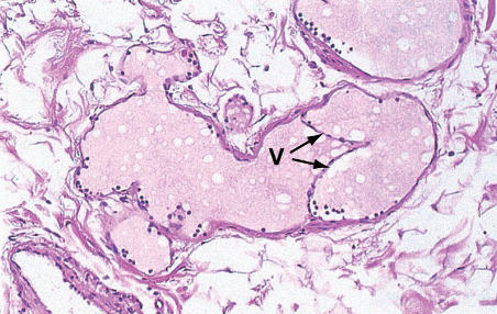
lymphatic vessel
V = valve
lymphatic vessels
lymph flow is increased during inflammation to help drain edema from extravascular space
diapedesis / transmigration
special characteristic of leukocytes that allows then to migrate through vessel walls
they squeeze through intercellular junctions between endothelial cells
leukocytes degrade basement membranes with collagenases
chemotaxis
migrate toward sites of injury in extracellular tissues along a chemical gradient
mediators include bacterial products, cytokines, complement, and leukotrienes
endothelial injury
results in vascular leakage due to endothelial cell necrosis and detachment due to direct injury to endothelial cells or leukocyte-mediated endothelial injury
irreversible
begins immediately after injury and persists for hours or days until repair: immediate sustained response
may induce delayed prolonged leakage

electron micrograph of cell migration from microcirculation
leukocyte emigration
type of emigrating leukocyte varies with age of inflammatory response
neutrophils predominate during the first 6 to 24 hours and are replaces with monocytes in 24-48 hours
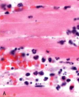
early (neutrophilic) infiltrates and congested blood vessels
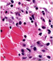
later (mononuclear) cellular infiltrates

kinetics of edema and cellular infiltration
outcomes of acute inflammation
resolution
progression to chronic inflammation
scarring/fibrosis
abscesses (which leads to fibrosis)
fibrosis means…
…scarring
morphologic patterns of acute inflammation
serous inflammation
fibrinous inflammation
suppurative/purulent inflammation
ulcer: a local defect or excavation of the surface of an organ or tissue that is produced by necrosis of cells and sloughing of necrotic inflammatory tissue

serous inflammation
serous inflammation
characterized by outpouring of watery, protein-poor fluid, derived from serum or secretions of mesothelial cells lining peritoneal, pleural, or pericardial cavities

fibrinous pericarditis (gross)

fibrinous pericarditis (microscopic)
F = exudate
P = pericardial surface
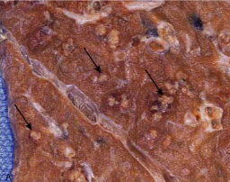
bacterial abscess of the lung (gross)

bacterial abscess of the lung (microscopic)
abscess
focal collections of pus caused by seeding of pyogenic organisms into a tissue or by secondary infections of necrotic tissue
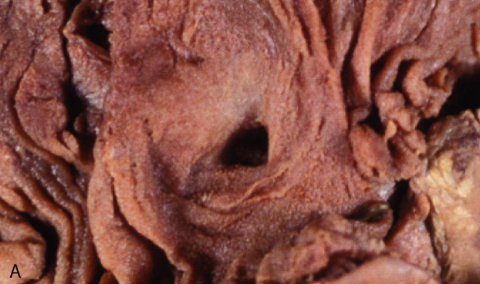
duodenal ulcer (gross)

duodenal ulcer (microscopic)
ulcers occur when…
…tissue necrosis and resultant inflammation exist on or near a mucosal surface.
features of the acute phase of ulcers
neutrophils and vascular dilations
features of the chronic phase of ulcers
scarring at lateral margins and along the base
chronic inflammation
inflammation of prolonged duration in which active inflammation, tissue injury, and healing proceed simultaneously
chronic inflammation is characterized by…
…infiltration with mononuclear cells, tissue destruction, and repair with angiogenesis and fibrosis
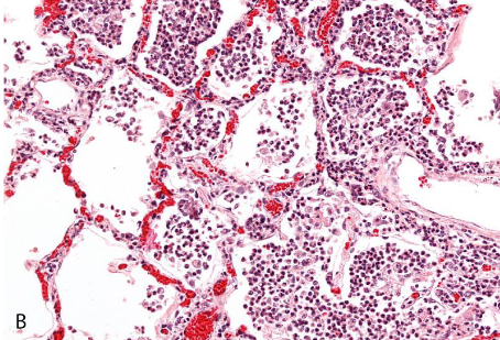
acute inflammation of the lung

chronic inflammation of the lung
granulomatous inflammation
characterized by patterned aggregation of activated macrophages that assume epithelioid appearance
granulomatous inflammation causes
persistent T cell responses to certain microbes
M. tuberculosis
T. pallidum
Fungal infection
foreign bodies

granuloma
keys to granuloma formation
chronic macrophage activation by T cell derived cytokines
morphologically typical and characteristic arrangement of epithelioid macrophages and multinucleated giant cells
systemic effects of inflammation / acute phase reaction
consist of cytokine-mediated effects on a variety of organ systems and manifests as
fever
elevated acute phase proteins in plasma
leukocytosis
tachycardia/hypertension
septic shock
What are the two main components of inflammation?
Vascular reaction and cellular response
Which of the following best describes acute inflammation?
A rapid response to cell injury or invading microbes that delivers leukocytes and plasma proteins to the site of injury
What can occur when an injurious stimulus persists following acute inflammation?
Persistent cell injury leading to chronic inflammation
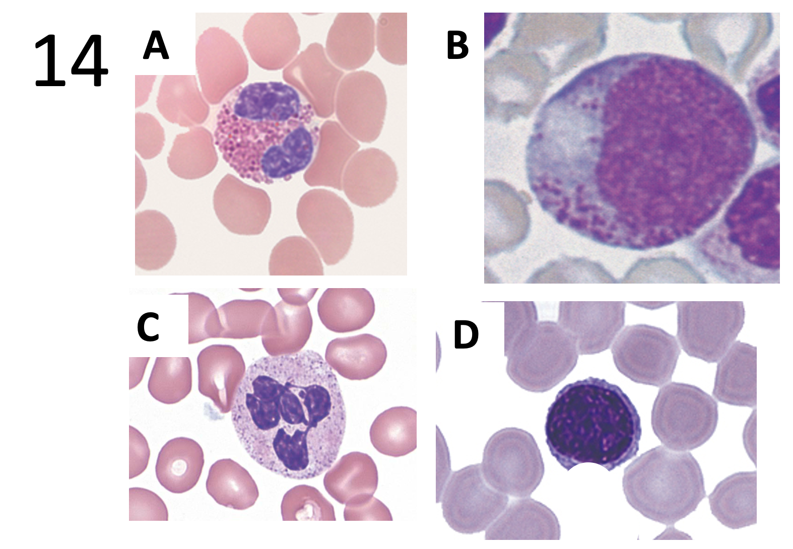
Choose from the pictures: which leukocyte is the precursors of tissue macrophages?
Picture B
Which statement is FALSE regarding the White blood cells?
Eosinophils are decreased in numbers in parasitic infections and allergies
Which of the following is increased in order to remove edema from extravascular space?
lymph flow

Which of the following types of inflammation is characterized by the accumulation of a watery, protein-poor fluid?
Serous

Which of the following best describes the process shown in the images provided?
Fibrinous inflammation

Which of the following is NOT observed in a typical granuloma?
central core of lymphocytes

An abscess is a localized form of which type of inflammation
Suppurative

Regarding the image, which of the following best describes the distribution of the pathologic process shown (HINT: look at arrows)?
Multifocal