Bio 315: Lab Exam 1
 Call Kai
 Learn
Learn Practice Test
 Spaced Repetition
 Match
Match Flashcards
 Knowt Play
1/356

There's no tags or description
Looks like no tags are added yet.
Name | Mastery | Learn | Test | Matching | Spaced | Call with Kai | Chat |
|---|
No analytics yet
Send a link to your students to track their progress
357 Terms
Cranial cavity
Where brain is

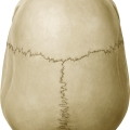
Calvaria
The "skull cap" (structure)
Cranial Base
The floor of the cranial cavity (structure)
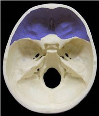
Anterior Cranial Fossa
Top (depression)

Middle Cranial Fossa
Middle (depression)

Posterior Cranial Fossa
Bottom, back of head (depression)

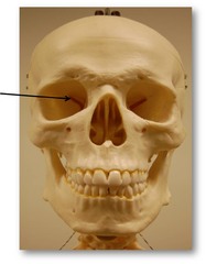
Orbits
"Eye sockets"
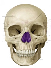
Nasal cavities
Space where nose is

Bony nasal septum
Line down center, divides

Piriform aperture
pear shaped, Outside/outline of nose
Hard palate
Roof of mouth, "brain freeze"

Zygomatic arch
Bone in cheek

Infratemporal fossa
Space

Coronal Suture
side to side

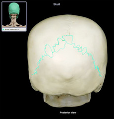
Lambdoidal Suture
Shaped like the Greek letter lambda, curved back
Sagittal Suture
Sagittarius is the archer--think of an arrow, straight down center, cuts in half

Squamous Suture
Scaly (like fish), side of skull, behind where the ear would be

Supraorbital Margin
Above eye (feature)

Parietal Bones
Top of head

Occipital Bone
Back of head

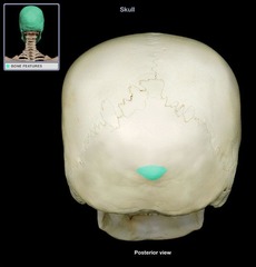
External Occipital Protuberance
bump on back of head (feature)
Occipital Condyles
Articulate with the first cervical vertebra, smooth bumps (feature)

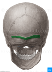
Nuchal lines
(feature)
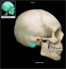
Mastoid Process of the Temporal Bones
(projection)
Styloid Process of the Temporal bones
Pointy, medial anterior (projection)

Zygomatic Process of the Temporal bones
Back of cheek (projection)

Petrous Portion
Pinch it, contains ear organs of hearing and balance (structure)

Mandibular Fossa
Depression

Crista Galli of the ethmoid bone
ridge on top (structure)

Cribriform Plate of the ethmoid bone
holes (structure)

Perpendicular Plate of the ethmoid bone
Helps form the bony nasal septum, hook (structure)

Orbital Plate of the ethmoid bone
Medial wall , flat surfaces, smooth (structure)
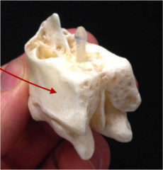
Superior and Middle Conchae of the ethmoid bone
(structures)

Greater Wing of the sphenoid bone
Curved structures, lateral

Lesser wing of the sphenoid bone
Pointy, smaller, medial

Sella Turcica
Saddle in center (depression)

Pterygoid Processes
Legs of the butterfly; 2 below wings

Medial and Lateral Plates of the pterygoid processes
M= plates on inside, smaller
L= wings that are further out

Body of Mandible
Bottom of teeth

Dental Alveoli of the mandible bone
"Tooth sockets" (depression)

Angle of the Mandible
Curve up, where it turns (feature)

Ramus of the Mandible bone
Straight up (feature)

Coronoid Process of the mandible bone
Dolphin fin closer to teeth (projection)
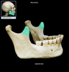
Condylar process of the mandible bone
Further back, blob on back

Head of Mandible
Part that sits in a depression in the maxilla only

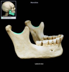
Mandibular Notch
"U" shape in center
Body of Maxilla
mustache area (structure)

Infraorbital Margin of the maxillae bone
lower part of eye socket (feature)

Palatine Process
Part of the hard palate, (structure)

Temporal Process (of the zygomatic bone)
Cheek bone (projection)

Horizontal Plate of Palatine Bones
Mid roof of mouth

Perpendicular Plate of Palatine Bones
lateral wall of nasal cavity, up and down, sides of "H", red part going backwards (structure)

Opening for nasolacrimal duct of the lacrimal bones
Looks like fat on the side of nasal cavity (space)

Inferior Nasal Conchae
Bumps, inside of eyes, small, yellow flaps
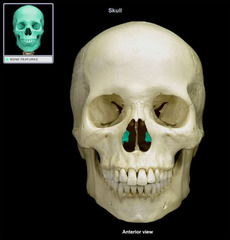
Vomer
Makes up part of the bony nasal septum

Supraorbital Foramen
Indent above eye, small groove in frontal bone

Infraorbital Foramen
Opening under eye (hole)

Mental Foramen
2 openings/holes on chin

Optic Canal
Most medial hole in orbit, next to boomerang lookin hole

Superior Orbital Fissure
Top corner of the boomerang;

Inferior Orbital Fissure
Bottom lateral corner of boomerang

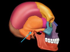
External Acoustic Meatus
Ear hole
Cribriform Foramina
(Collection of holes)

Foramen Rotundum
hole (most anterior)

Foramen Ovale
biggest hole out of the closest 3 (middle)

Foramen Spinosum
Lateral to biggest hole, smallest (lateral)

Foramen Lacerum
Jagged edge, most posterior, extends

Internal Acoustic Meatus
divot above melted jelly bean
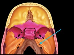
Jugular Foramen
throat, looks like a "J", melted jelly bean

Hypoglossal Canal
Under jugular foramen

Foramen Magnum
space in large bottom hole

Carotid Canal
Internal carotid artery (slightly anterior to foramen lacerum)

Incisive Fossa
hole Behind upper incisor teeth

Greater Palatine Foramen
Near upper molar teeth; back of mouth

Mandibular Foramen
Internal surface of mandible; where spring is attached

Lesser Palatine foramen
two smaller openings located behind the greater Palatine foramen

Frontal Sinuses
Paired; rarely equal in size, forehead

Maxillary Sinuses
Largest, smooth area

Ethmoid Air Cells
Above sinus, back of eye sockets
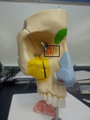
Hyoid Bone
(structure) anterior neck
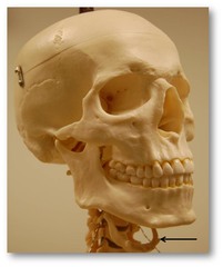
Greater horn of the hyoid bone
the larger, posterior projection from the body of the hyoid bone

Lesser horn of the hyoid bone
sticks up in the anterior portion

Cervical Vertebrae (7)
Upper neck/back - first 7 vertebrae (region)

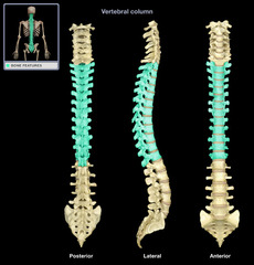
Thoracic Vertebrae (12)
Middle back, make up rib section (region)
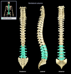
Lumbar Vertebrae (5)
Lower back (region)
Sacrum
almost bottom 5 connected

Coccyx
point at bottom of spine, tailbone (region)

Thoracic Kyphosis
Hump back, hint: c shape (curvature)

Sacral Kyphosis
Bottom c shape (think about where this is facing in the body) (curvature)

Cervical Lordosis
What is this curvature?
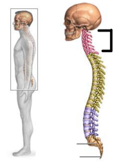
Lumbar Lordosis
backwards c shape (curvature)

Vertebral Canal
where spinal cord would sit (space)

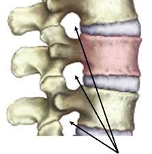
Intervertebral Foramina
between two vertebrae (opening)
body of vertebra
solid anterior portion of a vertebra

vertebral arch
surrounds vertebral foramen (structure)

Laminae of the vertebral arch
"roof" of the vertebral arch

Pedicles of the vertebral arch
"walls" of vertebral arch

Spinous Process of the vertebrae
The portion of the vertebrae that sticks out posteriorly. (projection)

Transverse Processes of the vertebrae
two lateral projections from the vertebral arch (projection)

Superior Articular Processes of the vertebrae
Smooth, "giraffe horns" or "antlers" on top(projection)
