Topic 6
1/37
There's no tags or description
Looks like no tags are added yet.
Name | Mastery | Learn | Test | Matching | Spaced |
|---|
No study sessions yet.
38 Terms
what is the role of microorganisms in decomposition of organic matter?
Microorganisms such as bacteria and fungi are an important part of the carbon cycle. when plants and animals die microorganisms secrete enzymes that decompose dead organic matter into small molecules so they can respire. when microorganisms respire these small molecules methane (CH4) and CO2 are released and this recycles carbon into the atmosphere.
How do scientists estimate time of death of a body?
-Rigor Mortis (stiffening of muscles )
-Body temperature
-Forensic entomology (colonisation of a variety of different insects).
-extent of decomposition.
-stage of succession.
how does body temperature allow us to estimate TOD?
all mammals produce heat from metabolic reactions like respiration. human body temp 37degrees
from TOD body metabolic reactions slow down and eventually stops causing body temp to decrease until it equals temp of surroundings process known as algor mortis.
conditions such as clothing, body weight and air temp can affect cooling rate.
how does degree of muscle contraction allow us to estimate TOD?
about 4-6 hours after death muscles in dead body starts to contract and become stiff ( rigor mortis).
Rigor mortis is when the muscle cells become deprived of oxygen.
Respiration still takes place in muscle cells but its anaerobic respiration. which causes build up of lactic acid.
Ph of cells decrease due to lactic acid inhibiting enzymes that produce ATP.
No ATP means bonds between myosin and actin in muscles cells become fixed and body stiffens.
how does forensic entomology allow us to estimate TOD?
when the body dies its quickly colonised by a variety of different insects.
TOD can be estimated by identifying the type of insect present on the body- e.g flies often the first insects to appear a few hours after death. other insects like beetles colonise the body at a later stage.
TOD can also be estimated by identifying the stage of life cycle the insect is in. blowfly larvae hatch from eggs about 24 hours after they are laid so if only bowfly eggs are found on a body you could estimate TOD was no more than 24 hours ago.
different conditions affect insects life cycle such as drugs humidity oxygen and temp.
What’s a DNA profile ?
is a fingerprint of an organisms DNA
everyone’s DNA is different except identical twins so DNA profile is unique to you
DNA profiling can be used to identify people and to determine genetic relationships between humans between animals and between plants
How is a DNA sample obtained?
obtained from the organism the DNA profile is being made from blood and saliva
What’s PCR?
PCR is used to make millions of copies of specific regions of the DNA in just a few hours.
DNA needs to be amplified so there enough to make a DNA profile PCR has lots of stages and is repeated over and over to make lots of copies
How is PCR used to amplify DNA?
a reaction mixture is set up containing DNA sample, DNA polymerase, primers and free nucleotides.
Primers are short pieces of DNA that are complementary to the bases at the start of the fragment you want.
DNA polymerase is an enzyme that creates new DNA strands
The DNA mixture is heated to 95 degrees to break the hydrogen bonds between two strands of DNA
The mixture is cooled to between 50 and 65 degrees so that the primers can bind (anneal) to the strands.
72 degrees mixture is heated so DNA polymerase can work.
DNA polymerase lines up free DNA nucleotides alongside each template strand. complementary base pairing means new complementary strands are formed.
two new copies of fragment of DNA are formed and one cycle of PCR complete.
each PCR cycle doubles the amount of DNA.
how to calculate number of DNA fragments or strands after PCR?
N x 2^n
N= staring number of fragments
n= cycles
Why is a fluorescent tag added?
view under UV light to visualise bands
what’s gel electrophoresis used for?
Gel electrophoresis is used to separate out the DNA fragments according to length
DNA is placed into a well in a slab of agrose gel and covered in buffer solution that conducts electricity
an electrical current is passed through move to the opposite charges
shorter DNA fragments move faster and trvael further through the gel
what can DNA profiling be used for?
paternity tests.
prevent inbreeding of animals by seeing how closely related they are
what’s a virus?
• Acellular → not cells, do not have a cell-surface membrane, do not have organelles or cytoplasm
• Non-living → unable to replicate independently, do not carry out any metabolic reactions e.g. respiration
Whats a bacteria?
• Prokaryotic cells need certain conditions to grow and multiply
→ optimum temperature and pH for enzyme activity
→ glucose for respiration
→ oxygen for aerobic respiration (unless anaerobic respiration can be carried out)
→ source of other nutrients (e.g. amino acids) to allow growth
• Prokaryotic DNA
→ not associated with histone proteins
→ a circular DNA molecule found
in the cytoplasm
→ some bacteria have plasmid
What defence against pathogens do we have?
Phagocytosis
Lysozyme
inflammation
skin
What is a non specific response?
a response not towards any antigen in specific.
it occurs in the same way for all micro organisms regardless of the pathogen.
what happens in the non specific response ( not antigen specific attacks microorganisms )of inflammation?
Occurs when pathogens enter the body through a wound or infection site.
The site becomes red, warm, swollen, and painful.
The immune system recognises foreign antigens on the pathogen’s surface.
Part of the non-specific immune response.
Damaged white blood cells and mast cells release histamine.
Histamine causes:
Vasodilation of arterioles → increased blood flow → redness & heat.
Increased permeability of capillaries → plasma, white blood cells, and antibodies leak into tissue → oedema (swelling).
Cytokines released by white blood cells attract more phagocytes to the site of infection.
The response helps to:
Destroy pathogens
Remove damaged tissue
Begin tissue repair
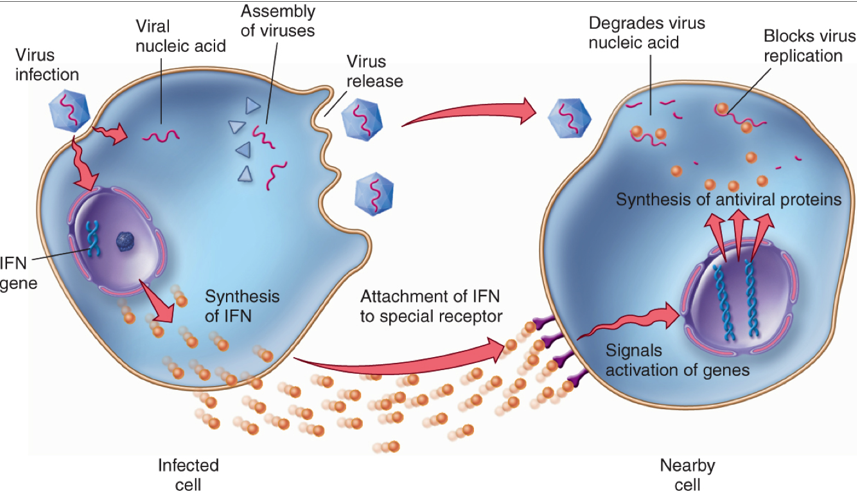
whats interferon?
Type of cytokine released by virus-infected cells.
Interferon is a protein that signals to neighbouring cells.
It helps to prevent viruses spreading to uninfected cells.
Functions of interferon:
Stimulates neighbouring cells to produce antiviral proteins that inhibit viral replication and protein synthesis.
Prevents viruses from entering healthy cells.
Activates cells involved in the specific immune response to kill infected cells.
Activates other mechanisms of the non-specific immune response.
Promotes inflammation, bringing more immune cells to the infection site.
what are phagocytes?
Phagocytes are white blood cells that engulf and digest bacteria and other foreign material (pathogens).
Types of phagocytes include:
Neutrophils – short-lived, first to respond to infection.
Monocytes – develop into macrophages in tissues.
Function: carry out phagocytosis as part of the non-specific immune response.
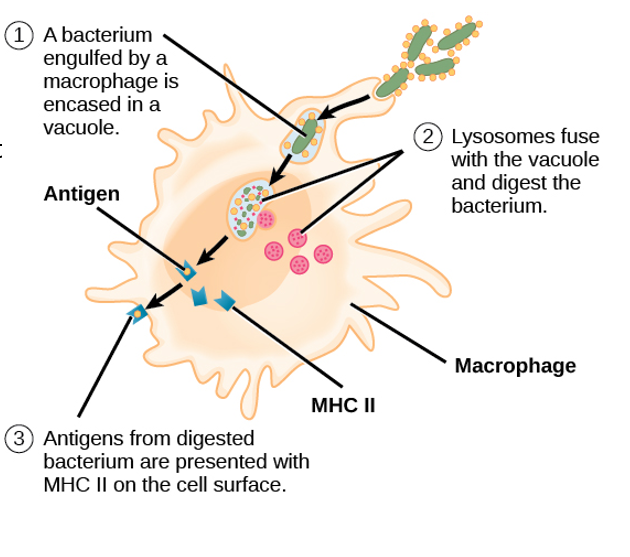
what happens in phagocytosis?
1.Chemicals released by bacteria, attracting the phagocytes
2.Neutrophils and macrophages extend their cell membrane around the bacteria, enclosing them in a phagosome
3.The phagosome fuses with the phagocyte’s lysosome
4.Allowing hydrolytic enzymes such as protease to destroy the pathogen
5.Antigens are presented on the cell surface after the pathogen is broken down
6.antigen presenting cell
how does the lymphatic system prevent spread of infection?
1.Fluid leaks out of capillaries…
2 .It forms tissue fluid that surrounds the cells of all tissues.
3.Pathogens can escape into the blood despite the inflammatory and phagocytic responses
4.Tissue fluid is collected into a network of vessels called the lymphatic system
what are antigens?
• Molecules on a cell-surface membrane which identify the cell → specific to each type of cell and each individual organism of a species
• Often proteins or glycoproteins
• Cells with foreign antigens stimulate an immune response → can be on pathogens, abnormal cells (e.g. cancer cells or cells infected with a virus), cells from other organisms of the same species
• Viruses are not cells, but still have foreign antigens on their surface.
whats specific immune response?
its antigen specific it produces responses aimed at specific pathogens
what are lymphocytes?
•Lymphocytes are a type of white blood cell.
•Their response is called the specific immune response.
•They circulate in the blood and lymph.
•Tissue can swell depending on the location of the infection – as lymphocytes get into ‘action’.
T and B cells
whats B cells?
•Made in bone marrow.
•Each B cell has a unique antigen receptor on its surface.
•A B cell will activate upon binding to an antigen with the complementary shape.
•B cells will then secrete antibodies in response to the antigen.
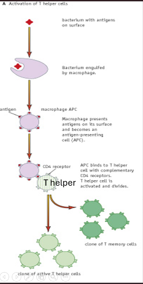
how do phagocytes activate T cells?
When a pathogen is engulfed by a macrophage (phagocytosis), the antigen from the pathogen is displayed on the macrophage’s surface, turning it into an antigen-presenting cell (APC).
The antigen on the APC binds to the CD4 receptor on a T helper cell — they are complementary.
This activates the T helper cell, which then divides by mitosis to form:
Active T helper cells → release cytokines that activate B cells, phagocytes, and T killer cells.
T memory cells → remain in the body for future immunity.
how do T helper cells activate B cells?
B cells are a type of white blood cell formed and matured in the bone marrow.
The surface of each B cell is covered with antibodies that are complementary to one specific antigen.
When a pathogen with a matching antigen binds to a B cell’s surface antibody, an antigen–antibody complex (APC) forms on the B cell membrane.
The B cell then acts as an antigen-presenting cell (APC).
A T helper cell with a complementary receptor binds to the antigen on the B cell.
The T helper cell releases cytokines, which activate the B cell.
The activated B cell divides by mitosis and differentiates into:
B effector cells → differentiate into plasma cells that secrete antibodies specific to the pathogen.
B memory cells → remain in the body and provide immunity for future infections by the same pathogen.
This entire process is known as clonal selection.
What do cytokines released by the T helper cell do?
The help stimulate division and differentiation of the B cell.
They form B effector cells and B memory cells.
This is known as clonal selection.
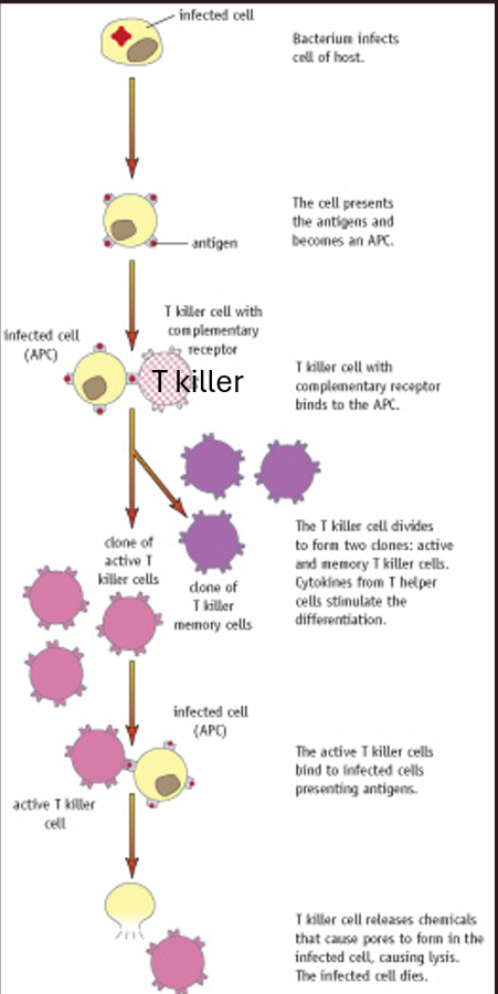
how is Tkiller activated
Active T helper cells release cytokines, which:
→ Activate phagocytes and macrophages.
→ Activate T killer cells.
→ Stimulate clonal selection of B cells.Infected host cells (e.g. by a bacterium or virus) display foreign antigens on their cell-surface membrane, becoming antigen-presenting cells (APCs).
T killer cells have receptors complementary to the antigen on the infected cell’s surface.
→ The T killer cell binds to the infected cell (APC).
→ It releases perforin, a protein that creates pores in the infected cell membrane.
→ This causes the infected cell to burst (lysis), destroying it along with the pathogen inside.→ T killer cells therefore kill virus-infected or tumour cells, not free pathogens.
T memory cells remain in the body after infection and provide immunity by allowing a faster secondary response upon reinfection.
whats TB?
TB (tuberculosis) is a communicable disease caused by the bacterium Mycobacterium tuberculosis.
The most common and contagious form is pulmonary (respiratory) TB.
Transmission:
Spread by inhalation of droplets from an infected person’s cough or sneeze.
Droplets containing the bacteria enter the lungs.
Symptoms (on reactivation):
Early: Cough, fever, fatigue, inflammation, weakness.
Progressive: Lung damage, respiratory failure.
Severe/Untreated: Spread to brain or kidneys, leading to organ failure and death.
whats primary infection with TB?
•Mycobacterium tuberculosis triggers a inflammatory response.
•Macrophages arrive and engulf the bacteria.
•Leads to the formation of granuloma tissue – acts to “wall off” the bacterium.
•In tuberculosis the tissue masses are called tubercles.
•The tissue is anaerobic, and since the bacteria requires oxygen to survive they die.
•3-8 weeks later infection is controlled and infected region of lung heals.
•Most people are infected as children and over 90% heal with no issues.
whats latent TB?
•Normally, macrophages engulf and destroy the bacteria.
•However in some cases, Mycobacterium tuberculosis can survive inside macrophages.
•They resist mechanisms which occur after being engulfed (due to their thick waxy cell walls making it difficult to break them down).
•They can therefore remain alive but inactive inside the tubercles for years.
•The infected person presents no symptoms of infection.
•T killer cells destroy any infected macrophages outside of the tubercles, preventing the spread beyond the tubercle.
•But the live and inactive bacteria remain can within the them.
•The antigens remain hidden to the rest of the white blood cells in the body.
•If the immune system is weakened, the bacteria may no longer be contained inside the tubercles.
whats active TB?
•Active tuberculosis occurs if the patient’s immune system cannot contain the bacteria upon its arrival.
•Could be due to large numbers of bacteria or previous controlled infection reactivates (80% of cases).
•Reactivation can occur due to:
•Old age
•Young (0-5)
•Malnutrition
•AIDS (virus targets w.b.c’s)
•Bacteria can multiply and destroy lung tissue causing cavities.
•Lung damage will eventually cause death.
the role of the fever in TB?
During TB infection, neutrophils and macrophages release chemicals that act on the hypothalamus, raising body temperature.
The resulting fever (around 40.5 °C) can enhance immune function and phagocytosis, helping the body fight infection.
High temperatures can also slow down or stop the growth of Mycobacterium tuberculosis, since it’s temperature-sensitive and stops functioning above 42 °C.
However, very high fevers (42–43 °C) are dangerous, as human enzymes denature, which can be life-threatening.
whats HIV?
humanimunodeficiency virus
infects and destroys a type of white blood cell called a T helper cell which act as a host cell for the virus. T helper cells activate other immune system cells so they’re hugely important in immune response
spread through sexual intercourse
whats the structure of HIV?
•HIV is a complex virus, an example of an enveloped virus.
•It consists of RNA surrounded by a 20 sided (icosahedral) protein capsid.
•The lipid envelope is formed from the host cell membrane as the new virus particles emerge from the cell cytoplasm.
•Sticking through the envelope are viral glycoprotein (gp) molecules.
how does HIV invade T helper cells?
•HIV invades T-helper cells within the immune system.
•Glycoprotein molecules called gp120, which are located on the virus surface, bind to the CD4 receptors on the surface of the T helper cell.
•The gp120 molecule also binds to the CCR5 co-receptor on the surface of the T-helper cell.
•The envelope surrounding the virus can then fuse with the cell membrane of the T-helper cell.
•This enables the virus to release the capsid containing the genetic material into the target cell’s cytoplasm.
•Macrophages also have CD4 receptors so can also be infected by HIV.