First Aid - GI - Pathology - Part 2
 Call Kai
 Learn
Learn Practice Test
 Spaced Repetition
 Match
Match Flashcards
 Knowt Play
1/152

There's no tags or description
Looks like no tags are added yet.
Name | Mastery | Learn | Test | Matching | Spaced | Call with Kai | Chat |
|---|
No analytics yet
Send a link to your students to track their progress
153 Terms
Acute mesenteric ischemia presentation
Abdominal pain out of proportion to physical exam findings due to bowel necrosis.
May see “Currant jelly” stools

Acute mesenteric ischemia etiology
Thrombosis
Embolism
Acute mesenteric ischemia risk factors
AFib
Peripheral Artery Disease
Recent MI
CHF
What is Angiodysplasia
It refers to tortuous dilation of blood vessels, leading to hematochezia
Where along the GI tract is angiodysplasia more common
Right-sided colon
Which age groups is angiodysplasia more common in
Older patients
How can you diagnose angiodysplasia
With Angiography
Angiodysplasia risk factors
End-stage renal disease
von Willebrand disease
Aortic Stenosis
What is chronic mesenteric ischemia
It’s aka “Intestinal Angina”, and it works similarly to stable angina
It represents atherosclerosis of the Celiac Artery, or SMA (most common), or IMA → Intestinal hypoperfusion
Chronic mesenteric ischemia presentation
Postprandial epigastric pain → Food aversion & Weight loss
How does colonic ischemia present
Presents as Crampy abdominal pain followed by HematocheziaC
Colonic ischemia frequently affects which areas of the colon
Watershed areas: Splenic Flexure & Rectosigmoid junction
Colonic ischemia seen in which age groups
Typically in older adults
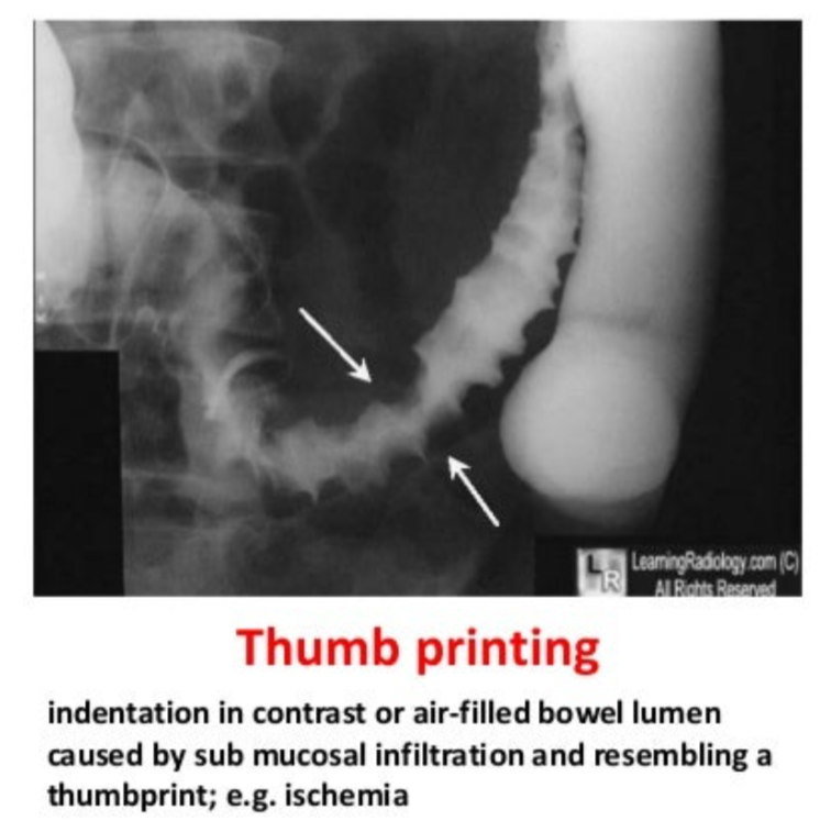
Colonic ischemia on imaging
Thumbprint sign
Ileus vs Pseudo-obstruction
Ileus refers to a functional/ non-mechanical obstruction of the small bowel. If large bowel it's called a pseudo-obstruction instead
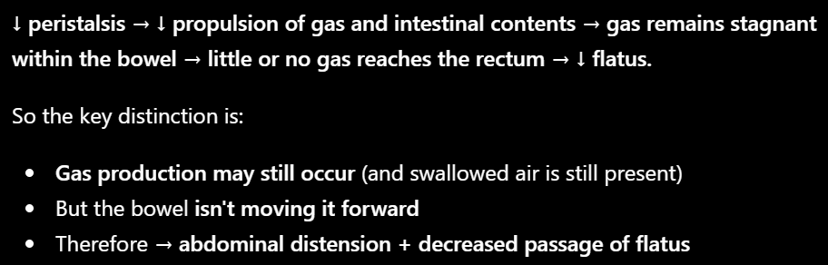
Ileus symptoms and presentation
Symptoms: Constipation, and Decreased flatus
Presentation: Distended/tympanic abdomen with decreased bowel sounds
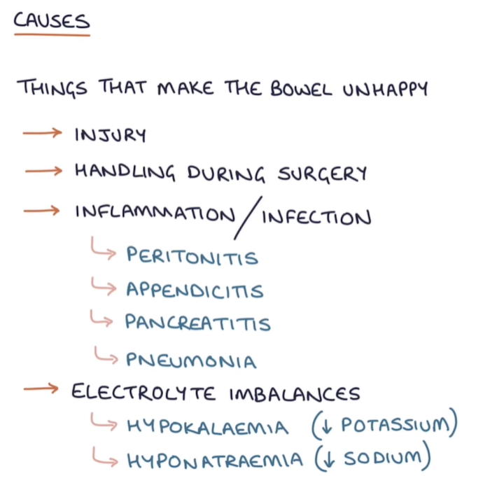
Ileus risk factors
Abdominal surgeries
Opiates
Hypokalemia
Sepsis
Ileus Imaging
No transition zone on imaging
Ileus treatment
Bowel rest
Electrolyte correction
Cholinergic drugs
Necrotizing enterocolitis is seen in which groups and why
Premature, formula-fed infants with immature immune system
(In preterm babies, Gut barrier and Gut immune system not fully matured -> easier for bacteria to invade)
What can necrotizing enterocolitis lead to
Pneumatosis intestinalis (Pneumatosis Intestinalis = Gas in the Bowel wall. Bacterial gas can enter the wall and accumulate in the submucosa/subserosa. So on imaging you can see little bubbles/linear streaks of gas within the bowel wall.
Pneumoperitoneum
Portal venous gas
Proctitis common causes
Infection (N gonorrhea, Chlamydia, Campylobacter, Shigella, Salmonella, HSV, CMV),
IBD
Radiation.
Proctitis symptoms
Tenesmus
Rectal bleeding
Rectal pain
Proctoscopy findings in proctitis
Proctoscopy reveals inflamed rectal mucosa (ulcers/vesicles in the case of HSV). Rectal swabs are used to detect other infectious etiologies.
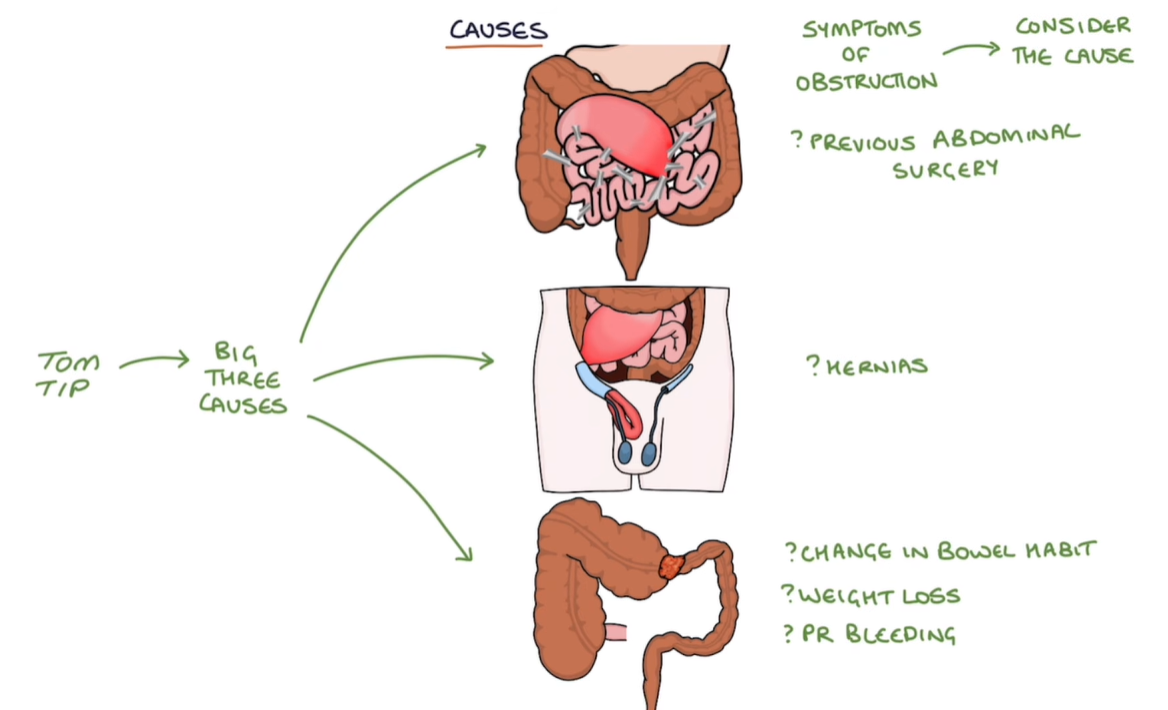
Small bowel obstruction risk factors/etiologies
Intraperitoneal adhesions (fibrous band of scar tissue)
Tumors
Hernias
Meconium plug in newborns → meconium ileus).
Small bowel obstruction symptoms/presentation
Abrupt onset of abdominal pain, nausea, vomiting, abdominal distension
A 25-year-old man presents with acute abdominal pain, nausea, and bilious emesis. His only medical history is of a reported “lump” in his groin area that has been present since birth. The abdominal pain is crampy, severe, initially intermittent, and now persistent. He is passing less gas and has not had a bowel movement in a few days. On exam he is tachycardic and mildly hypotensive. Abdominal auscultation reveals “tinkling”high-pitched bowel sounds. His abdomen is distended, tympanic to percussion, and tender to palpation. There is a nonreducible and extremely tender inguinal mass with overlying normal appearing skin. Evaluation/Tests: Upright chest/abdominal X-ray shows dilated loops of small bowel with multiple horizontal lines that are radiolucent above and radiopaque below.
Small-bowel obstruction secondary to incarcerated hernia
Short bowel obstruction X-Ray findings
Air-fluid levels
SBO management
Gastrointestinal decompression
Volume resuscitation
Bowel rest
SIBO Risk factors
Altered pH (e.g. achlorhydria, PPI use)
Anatomical (e.g. small bowel obstruction, adhesions, fistula, gastric bypass surgery, blind loop)
Dysmotility (e.g. gastroparesis)
Immune mediated (IgA deficiency, HIV)
SIBO Symptoms/presentation
Bloating
Flatulence
Abdominal pain
Chronic watery diarrhea
Malabsorption (vitamin B12) in severe cases.
SIBO diagnosis
CHO breath test
Small bowel culture
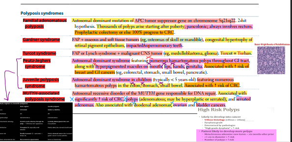
Hamartomatous polyps Description + Transformation risk
Solitary lesions do not have significant risk of transformation. Growths of normal colonic tissue with distorted architecture. Associated with Peutz-Jeghers syndrome and juvenile polyposis.
Hamartomatous polyps are usually pedunculated. They can cause painless rectal bleeding if they auto-amputate, this can rarely cause Iron-deficiency Anemia

Polyp stuff
Lynch syndrome
Hereditary nonpolyposis colorectal cancer (HNPCC).
It’s caused by an autosomal Dominant mutation in mismatch repair genes (MLH1/MSH2), Which causes Microsatellite Instability: 80% CRC progression
In Lynch syndrome, which part of the colon is always involved
Proximal Colon
Lynch syndrome is associated with which non-GI cancers
Endometrial
Ovarian
Skin
Lynch has many CEOS (C for colon)
Risk factors for CRC
Adenomatous and serrated polyps
familial cancer syndromes
IBD
Tobacco use
Diet of processed meat with low fiber
Right sided vs left-sided/Sigmoid CRC
Ascending—exophytic mass, iron deficiency anemia, weight loss.
Descending—infiltrating mass, partial obstruction (pencil-thin stools), colicky pain, hematochezia.

What should you do if a patient presents with S.bovis Endocarditis
Colonoscopy to check for colorectal cancer
What’s a condition that raises CRC suspicion in men over 50 and Postmenopausal women
Iron-deficiency anemia
CRC common X-Ray finding
Apple core lesion
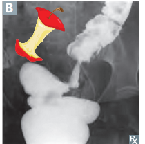
CRC screening stuff

Why NSAIDs in CRC
Because CRCs have elevated COX-2 activity for some reason
Molecular pathogenesis in CRC
Chromosome instability pathway: APC → Risky → KRAS → Adenoma → p53 → Carcinoma
Microsatellite instability pathway: MLH1 or MSH mutations
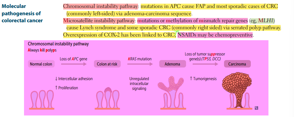
Cirrhosis etiologies
alcohol
nonalcoholic steatohepatitis
chronic viral hepatitis
autoimmune hepatitis
biliary disease
genetic/metabolic disorders.
Portal hypertension etiologies
Cirrhosis (most common)
schistosomiasis
portal vein thrombosis.
Ascites test
Serum-to-ascites albumin gradient (SAAG) analysis of ascitic fluid sample determines between etiologies of ascites.
SAAG ≥ 1.1 = portal hypertension.
Cirrhosis manifestations

Hepatic encephalopathy cause and treatment in cirrhosis
Cause: bacterial NH3
Treat: Lactulose, antibiotics (Rifamixin), Low protein diet
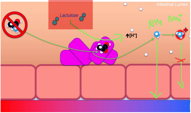
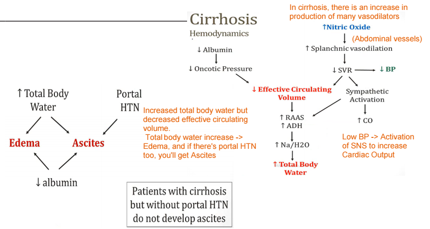
Cirrhosis hemodynamics stuff
What’s Budd-chiari
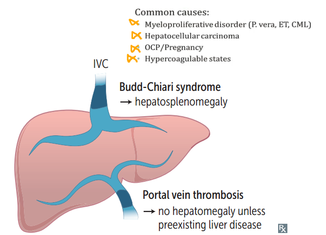
Thrombosis of the hepatic veins (hepatic outflow tract)
Budd-chiari symptoms
Centrilobular congestion and necrosis → congestive liver disease:
hepatomegaly
ascites
varices
abdominal pain
liver failure
***Absence of JVD unlike in Right heart failure where backup into liver is a consequence of the RHF itself***
Budd-chiari risk factors
hypercoagulable states
polycythemia vera
postpartum state,
HCC
(In HCC my have compression of centrilobular veins)
Portal vein thrombosis risk factors
Cirrhosis
Malignancy
Pancreatitis
Sepsis
Complications in Portal vein thrombosis
May lead to bowel ischemia if thrombosis extends to superior mesenteric vein
Which liver zone will be affected first in Buddchiari
Zone 3

What is SBP
SBP aka Primary bacterial peritonitis is a common and potentially fatal bacterial infection in patients with cirrhosis and ascites
Spontaneous bacterial peritonitis pathogenesis
Cirrhosis → portal HTN + intestinal barrier dysfunction → ascites + bacterial translocation from gut → ↓ hepatic complement/opsonization & impaired immunity → bacterial seeding/proliferation in ascitic fluid → peritoneal inflammation.
SBP Symptoms
Often asymptomatic, but can cause:
Fevers
chills
abdominal pain
Ileus
Worsening encephalopathy
Which bacteria typically cause SBP
Commonly caused by gram ⊝ organisms (eg, E coli, Klebsiella) or less commonly gram ⊕ Streptococcus
How is SBP diagnosed
Paracentesis with ascitic fluid absolute neutrophil count (ANC) > 250 cells/mm3
Empiric first-line treatment for SBP
Ceftriaxone
Reye etiology & pathophysiology
Aspirin in viral infection in children (Especially VZV & Influenza)
Aspirin-associated mitochondrial injury → ↓ oxidative phosphorylation + ↓ fatty-acid β-oxidation → microvesicular hepatic steatosis and impaired urea-cycle function → hyperammonemia + hypoglycemia → astrocyte dysfunction/swelling → cerebral edema → encephalopathy

Findings in reye syndrome
High ICP
Mitochondrial abnormalities
Microvesicular fatty liver changes
Hyperammonemia
Hypoglycemia
Vomiting
Hepatomegaly
Cardiac failure
Renal failure
Coma
High morbidity & mortality
AFLD
Excess NADH production → decreased fatty acid oxidation and increased lipogenesis
Microvesicular vs Macrovesicular fatty liver changes
Microvesicular: many small lipid droplets → usually mitochondrial dysfunction / ↓ β-oxidation. Think Reye, valproate, acute fatty liver of pregnancy.
Macrovesicular: large lipid droplet displaces nucleus peripherally → excess triglyceride accumulation/storage. Think alcohol, obesity/MASLD, insulin
Memory: Micro = Mitochondria. Macro = Massive fat storage.
When does Alcoholic hepatitis tend to present
Occurs after heavy binge drinking on top of long history of alcohol consumption
Alcoholic hepatitis findings
Swollen and necrotic hepatocytes with neutrophilic infiltration.
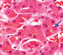
Mallory bodies B (intracytoplasmic eosinophilic inclusions of damaged keratin filaments)
Alcoholic cirrhosis
Final and usually irreversible form of AFLD
Regenerative nodules surrounded by fibrous bands in response to chronic liver injury → portal hypertension and end-stage liver disease
MASLD

Autoimmune hepatitis populations
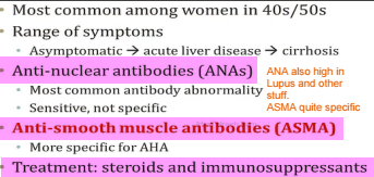
More common in females
Autoimmune hepatitis presentation
It’s a chronic inflammatory liver disease
It presents with:
fatigue
nausea
pruritus.
Autoimmune hepatitis labs & serologies
Serology: ⊕ for anti-smooth muscle or anti-liver/kidney microsomal-1 antibodies
Labs: Very high ALT and AST.
Autoimmune hepatitis histology
Portal and periportal lymphoplasmacytic infiltrate.
What’s a hepatic hemangioma
Most common benign liver tumor (Venous malformation, full of blood vessels)
Hepatic hemangioma typical age of onset
30-50
Hepatic hemangioma constraindication
Do NOT biopsy, risk of hemorrhage
What’s a hepatic focal nodular hyperplasia
Second most common benign liver tumor
hepatic focal nodular hyperplasia typical age onset
35-50
hepatic focal nodular hyperplasia etiology
Hyperplastic reaction of hepatocytes to an aberrant dystrophic artery
Common sign in hepatic focal nodular hyperplasia
Central stellate scar
hepatic focal nodular hyperplasia symproms
Usually asymptomatic, detected incidentally
Hepatic adenoma
Rare, benign tumor, often related to oral contraceptive or anabolic steroid use
Hepatic adenoma progression
May regress spontaneously or rupture, causing abdominal pain and shock
HCC epidemiology
Most common primary liver tumor in adults
HCC associations
HBV with or without cirrhosis + all other causes of cirrhosis:
HCV
AFLD
MASLD/MASH
Autoimmune liver disease
Hemochromatosis
Wilson disease
α1-antitrypsin deficiency
Aflatoxin exposure (Aspergillus)
HCC findings
Anorexia
Jaundice
Tender hepatomegaly
Decompensation of previously stable cirrhosis → Ascites (Ya3ni if there’s some functional tissue it destroys it)
Portal vein thrombosis
How does HCC metastasize
Hematogenously
HCC diagnosis
Ultrasound (screening) or contrast CT/MRI (confirmation); biopsy if diagnosis is uncertain
HCC monitoring
Monitor recurrence with serum AFP
Hepatic angiosarcoma
Rare malignant tumor of endothelial origin He
Hepatic angiosarcoma risk factors
Exposure to Arsenic or Vinyl chloride
Metastatic liver disease
Most common malignant liver tumors overall
Originate from:
GI cancers
Breast cancers
Lung cancers
A1AT liver involvement
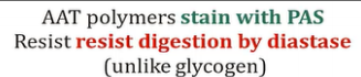
Misfolded protein aggregates in hepatocyte ER leading to Cirrhosis with PAS ⊕ globules in liver

A1AT typical presentation
Young patient with liver damage & dyspnea (panacinar emphysema) without a history of smoking
Diastase test on A1AT liver
Biliary Atresia
Fibro-obliterative destruction of bile ducts → cholestasis
Biliary atresia epidemiology
Most common reason for pediatric liver transplantation
Biliary atresia accompanying findings
Associated with absent/abnormal gallbladder