Lec - Bacteria and Bacterial Diseases of WLDF
1/19
There's no tags or description
Looks like no tags are added yet.
Name | Mastery | Learn | Test | Matching | Spaced | Call with Kai |
|---|
No analytics yet
Send a link to your students to track their progress
20 Terms
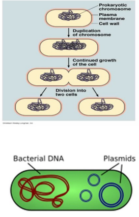
Bacteria
exist almost everywhere
contain a rigid cell well, cell membrane, cytoplasm, single double-stranded circular loop of DNA, flagella, pili, and ribosomes (only organelles)
lack nuclei
reproduce by binary fission
Bacterial Shapes and Arrangement
Cocci (singles, pairs, clusters, chains)
Rods (bacilli, pairs, chains, coccobacilli)
Spirals (incl. spirochetes)
Pleomorphic

Gram Positive Bacteria
have a thick peptidoglycan layer that absorbs violet stain and retains it during “destaining”
violet masks the pink counterstain

Gram Negative Bacteria
have a lipopolysaccharidae outer membrane, don’t retain the violet stain, and are counterstained pink by saffranin

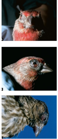
Mycoplasma spp.
lacks a cell wall, smallest bacteria, intra/extracellular
several species cause disease in birds and other vertebrates
unapparent infections to acutely severe disease and chronic carriers are common
Songbirds: upper-respiratory signs include - nasal discharge, conjunctivitis, decreased weight gain, death
Avian Cholera (Pasteurella multocida - in birds)
directly transmitted (+ no IH)
transmission is density-dependent and the largest outbreaks occur during winter on the staging areas
acute death (6-12 hrs; 1-2 days common) caused by toxin
lots of strains; all diff w/ some affecting either birds, mammals, or both
Mgmt - hazing to decrease densities, carcass removal, refuge management to separate snow geese from other susceptible species
Transmission of Pasteurella multocida among birds
many ways to transmit, ingestion > aerosols > direct contact
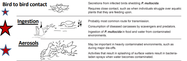
Bird to bird contact (transmission of Pasteurella
multocida)
secretions from infected birds shedding P. multocida
requires close contact
Ingestion (transmission of Pasteurella
multocida)
most common route of transmission
consumption of diseased carcasses by scavengers and predators
ingestion of P. multocida in food and water from contaminated environments
Aerosols (transmission of Pasteurella
multocida)
may be important in heavily contaminated environments, such as during major die-offs
activities that result in splashing of surface waters result in bacteria-laden sprats when water becomes contaminated
Bacterial Pneumonia (+/- Septicemia) in ungulates
most cases have little or no impact on wildlife population dynamics
epidemics of pneumonic or septicemic pasteurellosis can cause significant mortality in susceptible species
domestic sheep are chronic carriers of P. multocida, M. haemolytica and Mycoplasma ovipneumoniae that only develop disease occasionally
contact between bighorn and domestic sheep increases risks of bighorn die-offs
bighorn sheep may harbor these bacteria and may suffer severe disease with significant population declines
Bacterial Pneumonia in Ruminants
Gram negative bacteria that infect most species of wildlife (many spp. w/ dif diseases)
infections of respiratory tracts and other organs
respiratory signs = bronchopneumonia, interstitial pneumonia, pleuritis, purulent discharge from the nose and mouth, coughing, fever, toxic shock, and death
toxic shock
lipopolysaccharide toxin that compromises macrophages and other leucocytes
Brucellosis (Brucella abortus)
transmission = direct contact, bodily fluids, ingestion of milk or meat, licking aborted tissues
bison = abortion, w/ retained placenta, fever, infertility, etc.
elk = abortion, no retained placenta
moose = weakness, debilitated, fever, etc
predators = non-diseased carriers (coyotes, bears, birds)
zoonosis + risk to livestock
Brucellosis in bison
current mgmt: haze the bison back into the park, capture and test those not hazed, slaughter those that test positive and send meat to tribes, shoot those that can’t be hazed back or herded into facilities for testing
federal agencies currently spend at least 2.4 million dollars/ yr to implement the plan
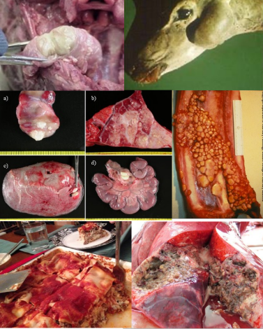
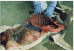
Tuberculosis (Mycobacterium bovis)
bacteria causes TB in most mammals and some birds
Other strains:
M. tuberculosis - is the number one cause of TB in humans
M. avium - avian tuberculosis and Johne’s disease of ungulates
M. leprae - leprosy (non-TB)
Signs of Bovine TB
most infected animals appear healthy, signs may not be noticed, chronic, progressive, disease
signs commonly include fever, chronic, progressive, emaciation, dyspnea, swollen lymph nodes
tuberculous lesions appear yellow caseous/necrotic areas in greyish fibrous nodules
no practical treatment
Animal to Animal Transmission of TB
disease transmitted from infected animals by aerosols (coughing), ingestion, and environmental contamination with feces and urine or via bite wounds
density-dependent transmission
bacilli survive in the environment for days to many months especially in cool weather
Tuberculosis in White-tailed Deer
Michigan, Minnesota, and Manitoba
Management: wildlife disease surveys and deer management to minimize risks of human infections, cull infected animals, carcass and meat inspection, proper food handling
TB in Badgers in the UK
badgers transmit M. bovis bacilli by respiratory droplets, urine, and bites to other badgers, and probably via urine to cattle via environmental contamination
infected large portions of cattle herds, major culling projects that were VERY expensive
new vaccine licensed