Blood Part 2
 Call Kai
 Learn
Learn Practice Test
 Spaced Repetition
 Match
Match Flashcards
 Knowt Play
1/56

There's no tags or description
Looks like no tags are added yet.
Name | Mastery | Learn | Test | Matching | Spaced | Call with Kai | Chat |
|---|
No analytics yet
Send a link to your students to track their progress
57 Terms
Defends against eveyrthing without targetting specific pathogens. Included examples are intact skin, enzymes in saliva, tears, mucus, acidic gastric secretion (stomach), WBCs (mainly granulocytes and monocytes/macrophages).
Non-specific defenses (innate immunity)
The body’s targetted, adaptive immune responses that recognize/eliminate unique pathogens and foreign molecules (includes B- and T-cells).
Specific defenses (acquired immunity)
non-specific
no memory
fast (effect in seconds/minutes/hours)
handled by phagocytes (neutrophils and macrophages)
uses the complement system to defend (a system of proteins that circulate in the blood in an inactive state until triggered)
innate/natural immunity
specific
has memory
slow (days/weeks)
handled by lymphocytes (B- and T-cells)
uses antibodies and cytotoxic molecules (ex. killer T-cells) to fight off pathogens
acquired/adaptive immunity
List the pros (3) and cons (2) of the immune system
pros:
defends against foreign invaders (bacteria, viruses)
removes our own old/damaged/abnormal cells
identifies and destroys abnormal or mutant cells
cons:
exaggerated responses to “harmless” substances (allergies)
autoimmune diseases (attacking “self immune system”)
___is a nonspecific, innate response to tissue injury. It can be triggered by cuts on skin surfaces, bullet wounds, injuries due to sun burn, infected sutures, during surgery, or infection of tonsils by cold virus.
inflammation
infammation usually has 3 purposes:
healing
destruction of “non-self” cells/pathogens
repair by fibrosis/scar tissue
list the 5 cardinal signs of inflammation and what causes them
redness (rubor)=a result of increased blood flow caused by histamines, etc.
swelling (tumor)=a result of increased blood flow caused by histamines, etc.
heat (calor)=a result of increased blood flow caused by histamines, etc.
pain (dolor)=a result of pressure on nerve endings caused by bradykinin and prostaglandin (PGE2)
loss of function
What are the effects of histamine on the vasculature (blood vessel walls)?
release of histamine
arteriole dilation (increased blood flow+heat to the area) due to histamine’s release.
expansion of capillary bed to accommodate for increased blood flow.
as a result the capillary bed becomes leaky; proteins (bradykinin, prostaglandins, complement proteins) and fluids in the vessels escape into the extracellular matrix to fight infection
as proteins leave the blood vessels, they alter the osmotic pressure in the interstitial space, drawing water out of the vessels and edema (swelling) results.


What are the cellular events associated with inflammation?
margination of WBCs: leukocytes move from the center of the blood vessel towards the edges (endothelium) due to increased blood flow.
tethering and rolling of WBCs: carbohydrate molecules on the WBC surface catch onto endothelial surface proteins (selectins). This tethers the WBCs to the vessel wall, causing them to roll.
activation of WBCs and endothelial cells: inflammatory chemicals (cytokines and chemokines) activate the endothelial cells+the WBCs, causing them to express new surface proteins and prepare for strong binding.
arrest/firm attachment of WBCs to endothelial cells: activation signal triggers anchoring proteins on the WBC to lock, stopping the WBC against the force of blood flow.
emigration/diapedesis: wBC squeezes betweeen adjacent endothelial cells by temporarily breaking/rearranging the tight junctions OR through transcellular migration.
chemotaxis of WBCs: once outside the blood vessel, the WBC migrates through the tissue towards the site of injury/infection.
Recognition and phagocytosis of foreign/”nonself” agents by WBC

Migration of neutrophils (macrophages) to the site of infection
chemotaxis and chemo-attractants
ability of WBCs to move against a concentration gradient (low→high) in respones to chemical factors (chemotactic factors).
chemotaxis
chemotactitc factors can include (4)
complement products (C5a)
chemokines (IL-8)
bacterial products
damaged membrane products (arachidonic acid metabolites)

What are PRRs?
Pattern Recognition Receptors. PRRs can be found on macrophages, epithelial cells, and dendritic cells. They help the phagocytes recognize common patterns.
What is the process of phagoctosis?
recognition of foreign body (using PRRs)
attachment to the foreign body (opsonization).
internalization (engulfing)
destruction of the “non-self” substance
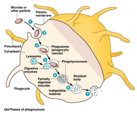
the process of adding host factors to the outside of bacteria to enhance attachment and speed up the process of phagocytosis
opsonization

what are host factors, aka opsonins
factors made by the “self” or own body

what are the two types of opsonins
antibodies
complement proteins
____are the WBC that manufacture and release antibodies to fight infection.
plasma cells
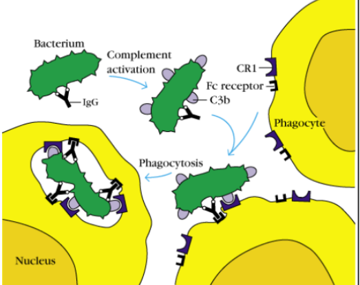
how do antibodies work (3 steps)
antibody attaches to the bacterium (IgG)
antibody brings the bacterium to the phagocyte
the phagocyte recognizes the antibody covered bacterium and eats it.
___is the primary organ that produces and secretes most of the body's complement proteins into the blood.
the liver
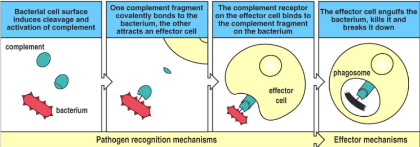
how do complement proteins work (4 steps)
bacterium presence causes the complement protein to split into 1 big piece and 1 small piece
the big piece bonds to the bacterium, the small piece attaches to an effector cell
the complement-bacterium complex binds to the complement receptor on the effector cell.
the effector cell eats the bacterium and kills it.
There are 3 different ways of killing by neutrophils
oxygen dependent killing (inside neutrophils)
oxygen-independent enzymatic killers
suicidal killing (outside neutrophils)
what method refers to corrosive free radicals being produced to destroy a foreign body (superoxide anion O2-, hydrogen peroxide H2O2, myeloperoxidase produces HOCl. these act like bleach to kill the bacteria.
oxygen dependent killing.
give 3 examples of bactericidal proteins and enzymes that are oxygen-INDEPENT killers
lysozymes
lactoferrin
defensins
___act inside the cell by breaking bacterial cell walls
lysoyzmes
___act in the extracellular space by binding to iron in the plasma, preventing bacteria from using it to grow
lactoferrin
___acts outside the cell by poking holes into bacteria to kill it
defensins
____acts by bursting open, projecting its sticky DNA onto the bacteria and gluing it into place. eventually the bacteria that is glued in place breaks down.
Neutrophil extracellular traps (NETs)
Neutrophilic killing is destructive and indiscriminate (causes collateral damage to surrounding tissues). products produced during phagocytosis are also released extracellularly (lysosomal enzymes and oxygen-derived activated metabolites) as neutrophils die off, they accumulate at the infection site which appears as pus.
Functions of activated complement proteins can be represented by the acronym OIL:
Opsonization (coating pathogens)
recruits Inflammatory mediators (aka immune cells)
Lysis (killing by a membrane attack complex formation (MAC)) punches a hole into the bacterium

Outline the vascular and cellular events of the innate immune system during infection/inflammation
Vascular
bacteria triggers macrophage to release cytokines and chemokines
vasodilation and increased permeability of vessels cause redness, heat, swelling
inflammatory cells move into tissues, releasing inflammatory mediators that cause pain
cellular events
neutrophils in the bone marrow are released when needed to fight infection (can sense inflammation)
neutrophils enter the infected tissue to kill bacteria. they die in the tissue and are eaten by macrophages

there are two different lymphoid tissues that are made during lymphocyte development and selection, what are they called
primary lymphoid tissue: the structures in which b and t cells originated (bone marrow+thymus)
secondary lymphoid tissue: lymph nodes
in primary lymphoid tissue, lymphocytes differentiate and are educated. explain this further.
lymphocyte eduaction=stages where lymphocytes are exposed to the body’s own proteins. two sets of b and t cells are produced:
the first set binds to the body’s own proteins and are flagged as dangerous and destroyed
the second set recognize the body’s cell and proteins without binding to them
explain secondary lymphoid tissue
in the lymph nodes, lymphocytes encounter foreign antigens and matured b and t cells who recognize the antigen become activated
All lymphocytes (b and t cells) must be able to do the following 3 R’s
recognize antigens (foreign agents)
respond to the antigens (destroy them)
remember the first encounter so they can better respond next time they see the same antigen
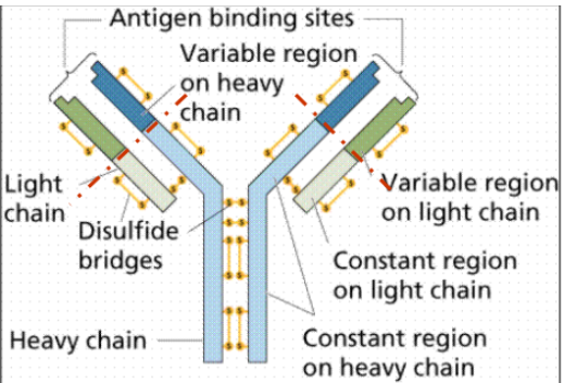
Describe the general structure of an antibody
the two arms are called antigne binding sites. they bind to proteins and carbohydrates.
the stem of the antibody is the constant region. it is where the macrophage binds in opsonization

What happens to a B cell after it encounters its specific antigen? (HUMORAL immunity)
B cell recognizes antigen→makes copies of itself→copies become either plasma cells or memory cells
What is the fate of a B cell which turns into a plasma cell (fights infection) in HUMORAL immunity
antigen enters the body
antigen binds to the matching b cell receptor on a b cell (membrane bound recognition)
b cell become activated
b cell undergoes cloning
b cells differentiate/mature into plasma cells
plasma cells produce and secrete antibodies (soluble recognition)
antibodies circulate through the body and bind to the specific antigen
what is the fate of a b cell which turns into a memory cell (lives in lymph nodes) in HUMORAL immunity
antigen enters the body
antigen binds to the matching b cell receptor on a b cell
b cell becomes activated
b cell undergoes cloning
b cells differentiate/mature into memory cells
memory cells remain in the body for a long time
if the same antigen appears again, memory cells rapidly multiply and produce many plasma cells
plasma cells produce antibodies
plasma cells fight current infection, memory cells remain in the lymph notes for years or even decades
After a b cells produce an antibodies, the antibodies function in 3 ways in HUMORAL immunity
opsonization (mark for eating)
complement activation (OIL)
neutralization (blocking toxins)

decsribe how antibodies neutralize bacterial toxins in HUMORAL immunity
some bacteria produce toxins which bind to receptors on healthy cells and changes/damages the cell.
antibodies defend by binding to the toxin FIRST, which blocks it from binding to receptors on healthy cells.
then a phagocyte can engulf the antibody-toxin complex and destroy it.

There is another branch of acquired immunity called cellular immunity =___
acquired immunity involving t cells
t cells are especially important for (3)
viruses
cancer
transplants
the types of t cells involved in cellular immunity are (3). these are responsible for recognizing, responding, and remembering pathogens
helper t cells (recognize)
cytotoxic t cells (respond)
memory t cells (remember)
unlike b cells, t cells do not recognize free-floating antigens directly. they require the pathway to be:
antigen→antigen-presenting cell (APC) displays a piece of the antigen→t cell recognizes the displayed antigen→t cell becomes activated and responds
Describe the steps of antigen presentation to t cells
foreign antigen enters the body
an antigen presenting cell (APC) like a macrophage takes up the antigen
APC processes the antigen into smaller pieces
APC displays an antigen piece using a MHC protein
helper t cell recognizes the antigen-MHC complex

Describe the steps for a APC and helper t cell to trigger an immune response
APC displays an antigen on MHC which the t cell receptor recognizes
handshake: co-stimulatory molecules on APC bind to receptors on the t cell
APC releases cytokines which activate the t cell and direct its response
helper t cell is fully activated
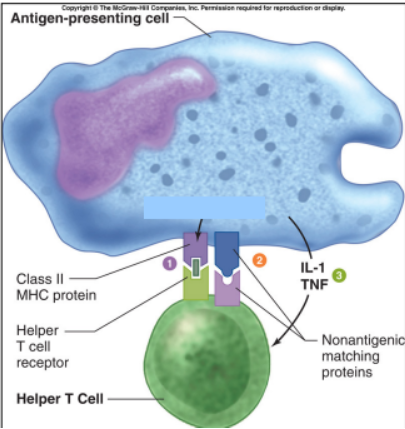
Describe the two types of MHC proteins and where they are present
MHC I = on the membrane of cells with a nucleus
MHC II = on the membrane of specialized antigen-presenting cells like macrophages or dendritic cells
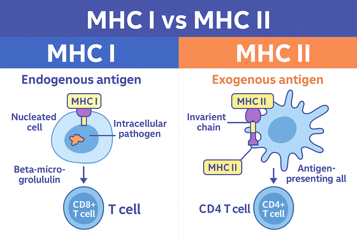
Differentiate between innate immunity, adaptive immunity, humoral immunity, and cellular immunity
Innate immunity: rapid, nonspecific protection from birth
adaptive immunity: targeted, long-lasting memory against specific pathogens. two types:
humoral (antibody-mediated, b cells; is extracellular so occurs in body fluids/blood)
cellular (cell-mediated, t help/cytotoxic cells; is intracellular so occurs inside infected host cells)

What are the two types of immune response
primary vs secondary

describe the pathway of primary immune response
foreign antigen A enters
matching b/t cells recognize it
cloning
plasma+memory cells develop
antibodies increase SLOWLY

describe the pathway of secondary immune response
familiar antigen A centers
memory cells recognize it
cloning
LOTS of plasma cells+antibodies are produced QUICKLY

what is the difference between active and passive immunity?
active=your immune system does the work
passive=you recieve antibodies that someone/something else already made and gave to you
describe the characteristics of active immunity
acquired from exposure to the actual disease or an injection of an altered antigen (vaccine)
immune system produces b/t cells→plasma+memory cells
antibodies are self-generated
takes weeks for the primary response but days for the secondary response
can last months to years thanks to memory cells
describe the characteristics of passive immunity
acquired from antibodies transferred through the placenta or breast milk
antibodies are pre-formed in the mother’s body and work immediately (no cell cloning necessary)
passive immunity only lasts a few weeks because no long-term immune memory can be transferred (memory cells only from acitve immunity)