Gastrointestinal System - Stomach, Intestines, and Accessory Organs
1/12
There's no tags or description
Looks like no tags are added yet.
Name | Mastery | Learn | Test | Matching | Spaced |
|---|
No study sessions yet.
13 Terms
Stomach (1)
- Dilated portion of the digestive tract
- Varies significantly between species depending on their diet
- Carnivores: small, simple stomach
- Herbivores: may have a small, simple stomach or a large, complex stomach, depending on what part of their GI tract breaks down complex carbohydrates (vegetation)
- Distal to the esophagus, proximal to the small intestine
Simple (Monogastric) Stomach
- Fully within the ribcage
- Receives food from the esophagus
- Area where the esophagus opens into: cardia (1)
- Typically toward the left side of the body
- Large blind sac portion that rises above the body: fundus (2)
- Main area from the cardia toward the "bend": body (3)
- Tubular part: pyloric portion (4)
- Typically toward the right side of the body
Layers of the simple Stomach Wall
Very similar to the esophageal layers
- Serosa: outermost layer
- Smooth muscle
- Three overlapping layers of muscle in different directions
- Submucosa
- Aids in creating the characteristic mucosal folds
- Muscularis mucosa (very thin)
- Mucosa: innermost layer
Mucosal Layer of Simple Stomach
- Contains 3 types of glands
- Cardiac - produce mucus to protect the mucosa
- Proper gastric - produce hydrochloric acid for digestion
- Pyloric - produce mucus to protect the mucosa
- The location of the glands do NOT always correspond with their namesake
Inside the Simple Stomach
- 2 muscular sphincters to control flow in/out of the stomach
- Cardiac sphincter - controls flow associated with the esophagus; "entry"
- Pyloric sphincter - controls flow associated with the small intestine; "exit"
- Rugae = folds
- Provide extra surface area for secretion and digestion
- Stretch out when the stomach is fully distended
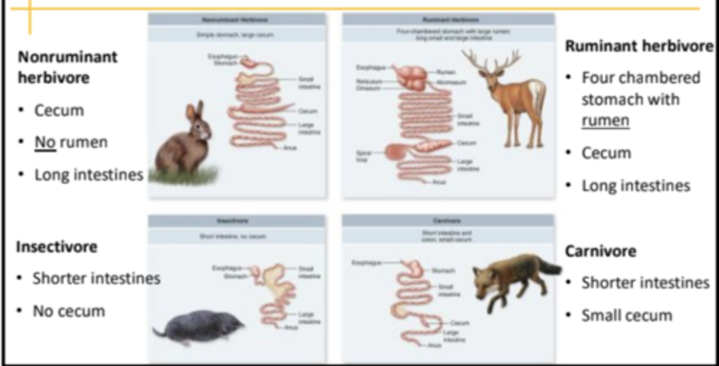
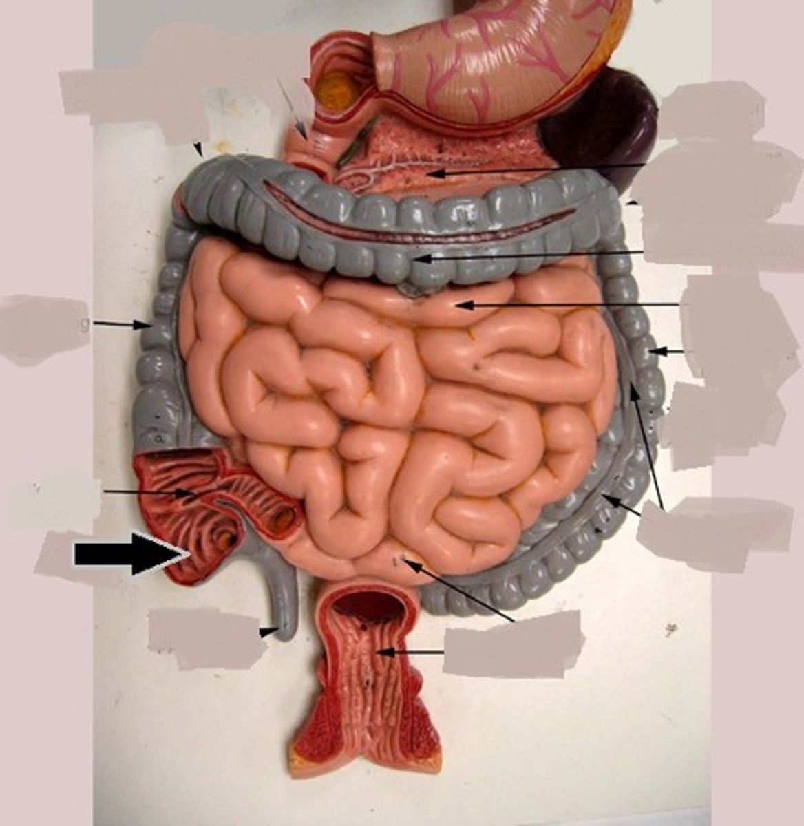
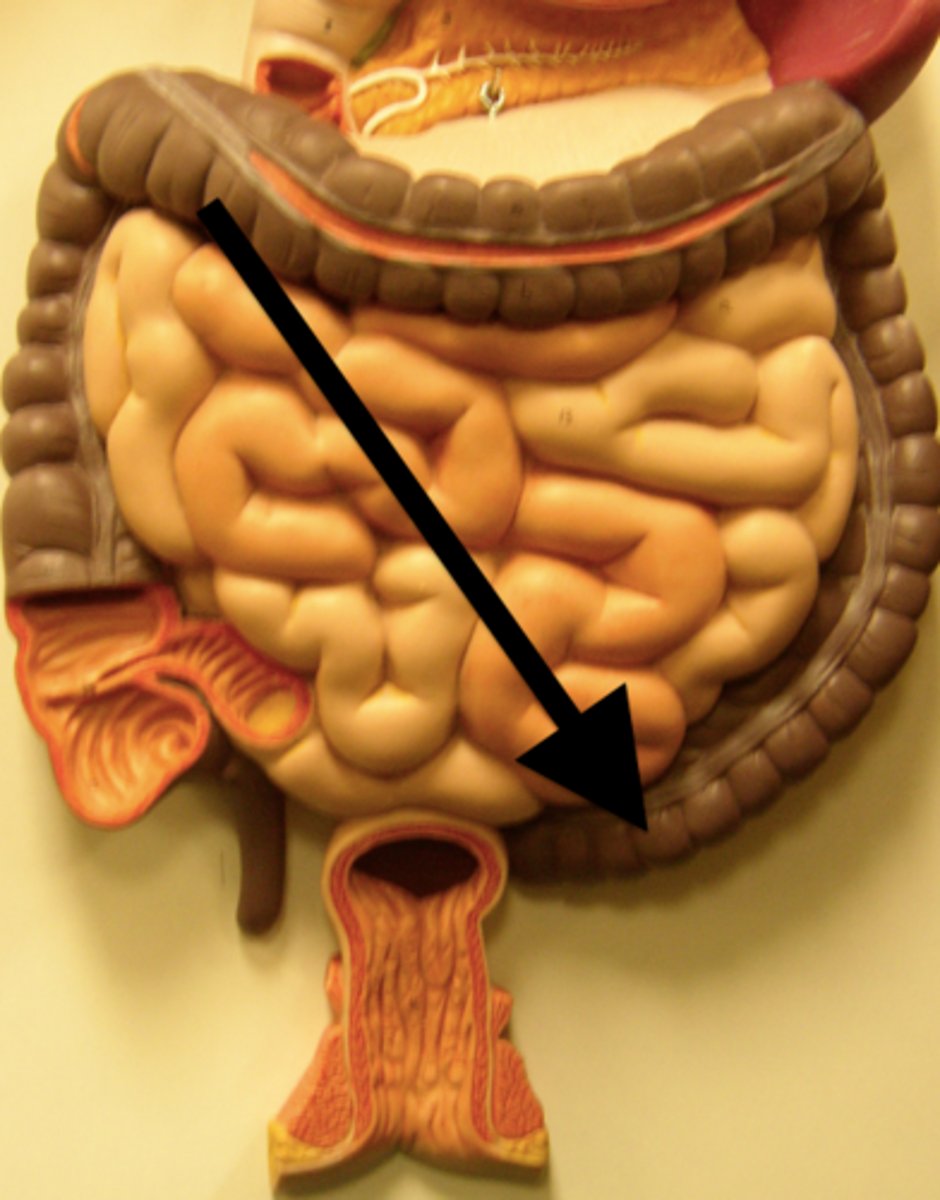
The Intestine
- Begins at the pylorus and ends at the anus
- Shorter in carnivorous animals, and longer in herbivores
- Small intestine: proximal portion, small in diameter but very long
- Duodenum: most proximal
- Jejunum• Ileum: most distal
- Large intestine: distal portion, large in diameter but often shorter
- Cecum
- Colon
- Rectum
Small Intestine
- Primary function is to be the location of enzymatic digestion and nutrient absorption
- Has the usual 4 layers of tissue
- Serosa (4)
- Muscular - 2 layers, running indifferent directions (3)
- Submucosa (2)
- Mucosa (1)
- Both the liver and the pancreas have ducts that enter the small intestine to aid with digestion
Small Intestinal Mucosa
- Contains many intestinal villi (finger- like projections) that greatly increase the surface area available for nutrient absorption
-Also contains crypts (glands that open up into the surface of lumen) that secrete mucous and enzymes to aid in digestion
Sections of the Small Intestine
- Duodenum (2-4)
- Relatively short
- A continuation of the pylorus of the stomach
- Jejunum (5)
- Very long and relatively free in the abdomen -very long mesenteric attachment
- Ileum (6)
- Mostly a direct path from the jejunum to the large intestine
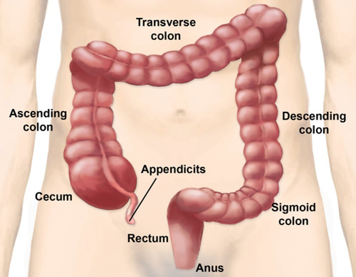
Large Intestine
- Divided into the cecum, colon, and rectum (in that order)
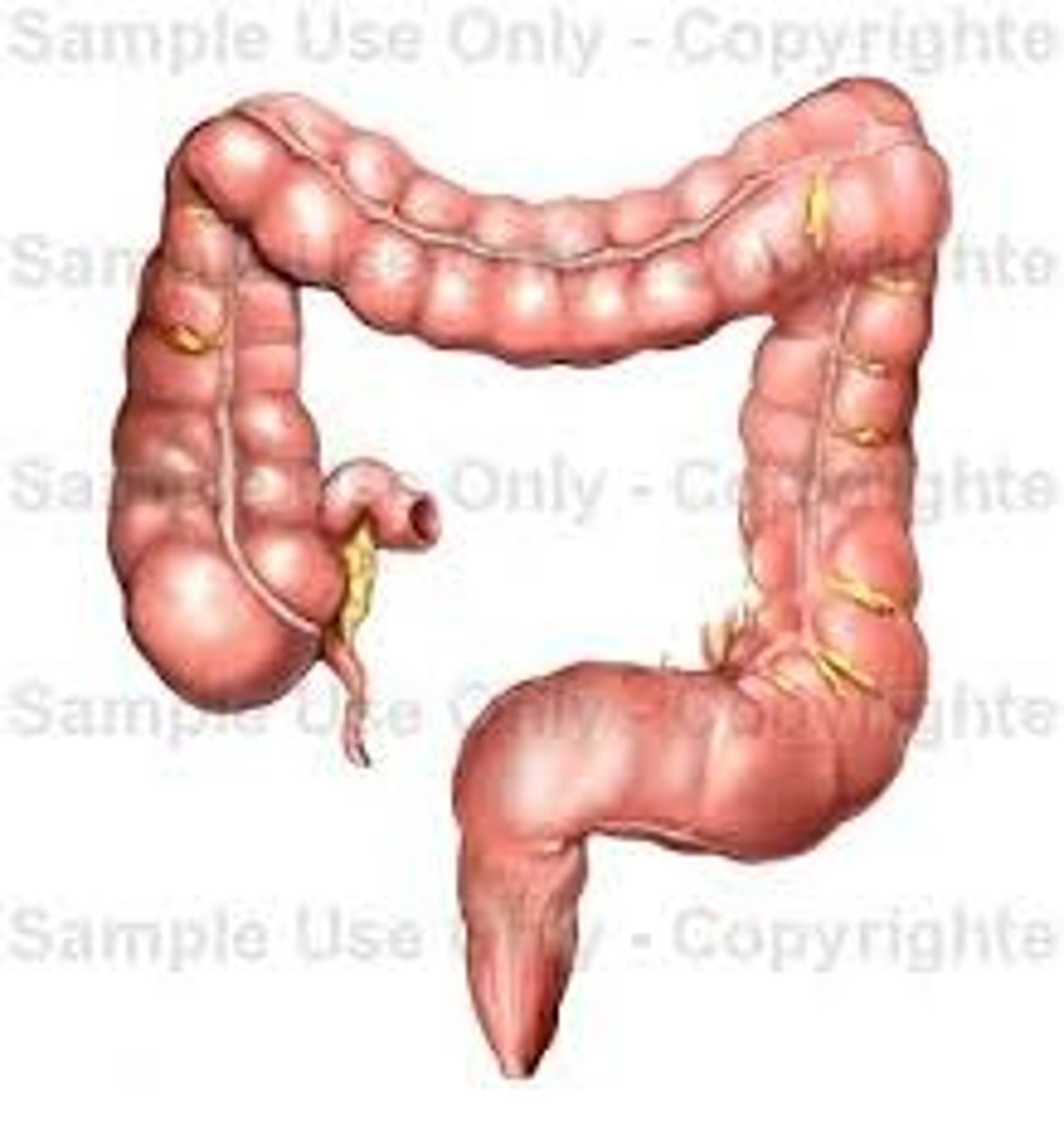
Cecum
- Blind-ended sac protruding from the continuous tube of the large intestine
- First portion of the large intestine
- In carnivores & omnivores it is relatively small and unimpressive
- In the ruminant, it is larger and is continuous with the colon
- Division of the cecum & colon is marked by the entrance of the ileum
Colon
- Generally has a larger diameter than the small intestine, and the mucosa is smooth(no villi)
- Suspended within the abdomen and receives its blood supply via the mesocolon(mesentery specifically for the colon)
- Both an ileal orifice (4) and a ceco-colic orifice (5) open into the colon from their respective structures
- Where exactly these openings are relative to one another depends on the species
Colon
- Divided into ascending (5),transverse (6), and descending (7) parts, based on location in the abdomen