Lecture 4 - Joint Classifications & Skeletal Movement
 Call Kai
 Learn
Learn Practice Test
 Spaced Repetition
 Match
Match Flashcards
 Knowt Play
1/54

Earn XP
Description and Tags
Vocabulary practice flashcards covering joint classifications, structures, types of movements, and neuroarticular receptors from Dr. Kim Adie's lecture.
Name | Mastery | Learn | Test | Matching | Spaced | Call with Kai | Chat |
|---|
No analytics yet
Send a link to your students to track their progress
55 Terms
Joint
A site of the junction or union of two or more bones, also called an articulation
Kinesiology
The scientific study of movement.
Proprioception
The body's sense of self-movement, force, and body position, mediated by mechanosensory neurons located within muscles, tendons, and joints.
Degenerative Joint Disease (DJD)
The breakdown of cartilage and joints, also commonly referred to as Osteoarthritis (OA).
Crepitus
A repeatable clicking or grinding sensation produced within a joint during movement.
Cavitation
The popping sensation or sound in a joint caused by the formation of air bubbles within joint fluid.
What are the 3 types of joint structures?
*Fibrous joint - fixed, immovable and no joint cavity ie. cranial suture, tib/fib - jointed by strong ligament (syndesmosis) and periodontal ligaments & tooth (gomphosis)
*Cartilagenous - contain cartilage, no cavity, slightly moveable.
-2 types: synchondrosis (contain hyaline cartilage) & a symphysis (contain fibrocartilage)
eg. epiphyseal growth plate, costochondral junctions, joints between mandible, pubic symphysis & vertebrae & IVD (fibrocartilagenous joint)
*Synovial - joint cavity with synovial fluid

Fibrous Joint & give example
A fixed or immovable joint connected via fibrous connective tissue that lacks a joint cavity.
ie. cranial suture, tib/fib - jointed by strong ligament (syndesmosis) and periodontal ligaments & tooth (gomphosis)
Cartilaginous Joint & give example
- contain cartilage, no cavity, slightly moveable.
-2 types: synchondrosis (contain hyaline cartilage) & a symphysis (contain fibrocartilage)
eg. epiphyseal growth plate, costochondral junctions, joints between mandible, pubic symphysis & vertebrae & IVD (fibrocartilagenous joint)
Synovial Joint
A joint characterized by a fluid-filled joint cavity lined with synovial membrane, reinforced by a capsule and ligaments, providing free movement and shock absorption.
How are joints classified by function?
Joints are classified by function into three categories: synarthroses (immovable), amphiarthroses (slightly movable), and diarthroses (freely movable). This classification reflects the degree of movement allowed at each type of joint.

Synarthrosis
An immovable or nearly immovable union of rigid components of the skeletal system, including fibrous joints, synchondroses, and synostoses.
Amphiarthrosis
A functional classification of joint that allows slight movement, such as the symphysis pubis.
Diarthrosis
A specialized articulation allowing free movement, where bony elements are enclosed by an articular capsule lining a cavity with synovial membrane.
What are the types of synarthroses joints?
The types of synarthroses joints include syndesmoses (immoveable or barely moveable), synchondroses (cartilageouns, no joint cavity, slightly moveable), and synostoses (bones united by ossification of a fibrous joint or synchrondosis between them).

Syndesmosis & give example
An immovable or barely movable fibrous joint lacking a joint cavity.
eg. cranial sutures, bones joined by strong ligaments e.g. metacarpus of horse, joint between tooth & socket (gomphosis)
Synchondrosis & give example
A cartilaginous joint with no joint cavity that is slightly movable and often temporary, such as epiphyseal growth plates.
Other examples: joint between jaws, pelvis (symphysis), and between vertebral bodies (IVD)
Synostosis & give example
A bony union formed by the ossification of a fibrous joint or synchondrosis between bones.
Eg. between ilium, ischium, and pubis to form acetabulum
Uniaxial Joint. What is a hinge & pivot joint & give examples.
A joint classification permitting movement in only one plane, encompassing hinge and pivot joints.
Hinge joint allows for flexion and extension, exemplified by the elbow and knee. A concave surface glides around a convex surface.
Pivot joint permits rotational movement, as seen in the atlantoaxial joint between the first and second cervical vertebrae.
Biaxial Joint. What is a condyloid & saddle joint?
A joint classification permitting movement in two planes, such as saddle & condyloid joints.
A condyloid joint (ellipsoid) is an ovoid surface with and elliptical cavity that allows for flexion, extension, adduction, and abduction, exemplified by the radiocarpal joint in the wrist & MCP joints. Does not allow for axial rotation.
A saddle joint has reciprocally concave & convex surface like a saddle. Allows flexion, extension, adduction, abduction, and some rotation, exemplified by DIP joints in dogs/cats
Multiaxial Joint
A ball and socket joint classification permitting movement in several planes.
Eg. hip joint
Translation & example
A sliding motion where flat surfaces move over each other, producing no change in orientation of the bodies.
Eg. vertebral facet joint & epophyseal joints
Rotation & example
A movement in which a moving bone turns around an axis perpendicular to the articulation.
Eg. At should if you extend the arm out and move the palm up and down
Swing & example
A movement in which a moving bone turns around an axis parallel to the articulation.
Eg. femur during locomotion
Flexion
A movement that reduces the angle between two segments of a limb; also termed palmar flexion.
Extension
A movement that increases the angle between two segments of a limb.
Drawing arm forward is extension of the forelimb in the quadruped.
Overextension
Extension beyond normal anatomical position, also known as dorsal flexion, such as the posture of an equine fetlock standing at rest.
Adduction
A pendular movement directed toward the median plane.
Abduction
A pendular movement directed away from the median plane.
Circumduction
A circular movement achieved through a combination of flexion and extension.
What are the characteristics of a synovial joint?
Synovial joints are characterized by a fluid-filled joint cavity, articular cartilage covering the bone surfaces, a fibrous joint capsule, and synovial fluid that lubricates and nourishes the joint. Nutrient cross via diffusion. Some of these joints have a disc (e.g. TMJ) or a meniscus (e.g. stifle)
What are the functions of synovial fluid?
Lubrication, nourishment & shock absorption
What are the sense organs located in the capsules of synovial joints?
Sense organs such as Ruffini & Pacinian corpuscles are located in the capsules of synovial joints and are sensitive to pressure & may be involved in kinaesthesia & golgi tendons in the tendons.
Articular Cartilage
Wear-resistant tissue covering bone ends in a joint to reduce friction; characterized as avascular, anervic, and alymphatic.


Synovial Membrane
A vascular and sensitive structure lining the joint capsule that secretes synovial fluid to nourish and lubricate the joint.

Ligaments
Tough, elastic bands of connective tissue surrounding a joint to provide support and limit motion.

Tendons
Tough connective tissue structures on each side of a joint that attach muscles to bones to control joint movement.

Bursae
Fluid-filled sacs located between bones, ligaments, or adjacent structures that help cushion friction in a joint.
Synovial fluid
A viscous, lubricating fluid found within synovial joints lubricates, nourishes cartilage, and aids in shock absorption. Weeping is the process where synovial fluid seeps into cartilage during movement, enhancing its cushioning effect.

Proproceptive fibers
Endings in the capsules & ligaments that are very sensitive to position and movement.
What are the synovial joint types?
Synovial joint types include hinge, ball-and-socket, pivot, saddle, condyloid, and plane joints, each allowing different ranges of motion.
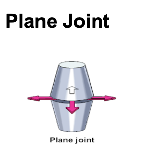
Plane Joint & example
A synovial joint with flat or nearly flat surfaces that slide across one another, producing translation or gliding movement.
Eg. articular facets & costovertebral joints
Hinge Joint & example
A uniaxial joint formed by a cylindrical bone fitting into a grooved bone, permitting pendular motion in one plane.
Eg. elbow joint & equine fetlock joint

Pivot Joint & example
A uniaxial joint where a peg fits into a ring or cavity, allowing rotational movement around an axis.
Eg. atlas and axis joints & proximal radioulnar joint

Condylar Joint & example
A joint where two condyles on one bone articulate with corresponding grooves on another, permitting primary movement in one direction with slight rotation and sliding.
Eg. femorotibial joint. Stifle is a complex condylar joint

Ellipsoidal Joint & example
A joint featuring a convex surface fitting into a concave surface, allowing flexion, extension, abduction, adduction, and slight rotation.
Eg. cranial radoiocarpal joint and metacarpophalangeal joints

Saddle Joint & example
A joint formed by maximum concavity on one surface and maximum convexity on another meeting at a 90∘ angle, offering greater range of motion than ellipsoidal joints. Flexion/extension, abduction & adduction.
Eg. DIP joints in dogs

Spheroidal Joint & example
A ball and socket joint where a sphere fits into a cup, providing the greatest movement versatility among joint types.
eg. hip joint

Muscle Spindle Fibers
Intrafusal muscle structures wrapped with sensory receptors that monitor lengthening and trigger the stretch reflex to prevent muscle tearing. Prevents over stretching by contraction of the reflex - known as the myotonic or stretch reflex.
Golgi Tendon Organs (GTOs)
Sensory receptors interwoven with collagen fibers in tendons that monitor muscle tension and trigger reflex relaxation when excessive force endangers the muscle.
Type 1 Mechanoreceptor
A slow-adapting postural receptor located in the superficial capsule that provides tonic stabilization and responds to end-range oscillations.
Stimulated by joint play.
Type 2 Mechanoreceptor
A fast-adapting dynamic receptor in the deep capsule that fires as movement begins to sense dynamic motion and mid-range oscillations.
Type 3 Mechanoreceptor
An inhibitive receptor located in ligaments that provides reflex muscle inhibition similar to Golgi tendon organs. Proprioceptive.
Type 4 Receptor
A nociceptive receptor present in most joint tissues that responds to excessive pressure or tissue damage to produce protective muscle guarding.

Joint Receptor Morphologies and Innervation
An anatomical schematic (Figure 6.10) illustrating knee joint receptor distribution and the specific structural morphologies of Type 1, Type 2, Type 3, and Type 4 receptors.