NEUR200 Exam 4
 Call Kai
 Learn
Learn Practice Test
 Spaced Repetition
 Match
Match Flashcards
 Knowt Play
1/84

There's no tags or description
Looks like no tags are added yet.
Name | Mastery | Learn | Test | Matching | Spaced | Call with Kai | Chat |
|---|
No analytics yet
Send a link to your students to track their progress
85 Terms
circadian rhythm
- 24 hour biological cycle
- influences regulation of sleep
- hypothalamus = circadian clock
suprachiasmatic nucleus (SCN)
- groups of neurons in hypothalamus that regulate sleep-wake cycle
- influenced by light
- controls melatonin release (pituitary gland)
- watched video on hamster getting a different hamster's SCN --> completely changes sleep-wake cycle
reticular formation
arousal
locus coeruleus (LC)
main synthesizer of norepinephrine (arousal)
histamine
- hypothalamus
- must be low to sleep well
- antihistamines make you sleepy
orexin
- hypothalamus
- necessary to stay awake
- people with narcolepsy lack
NREM stage numbers
1, 2, 3, 4
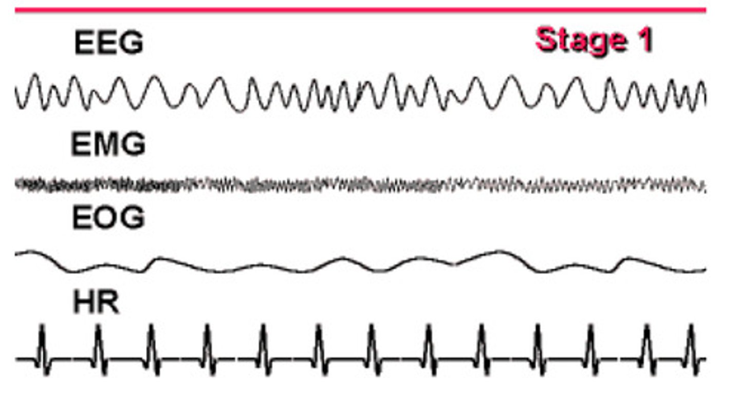
stage 1 sleep
- lasts 5-10 minutes
- theta waves
stage 2 sleep
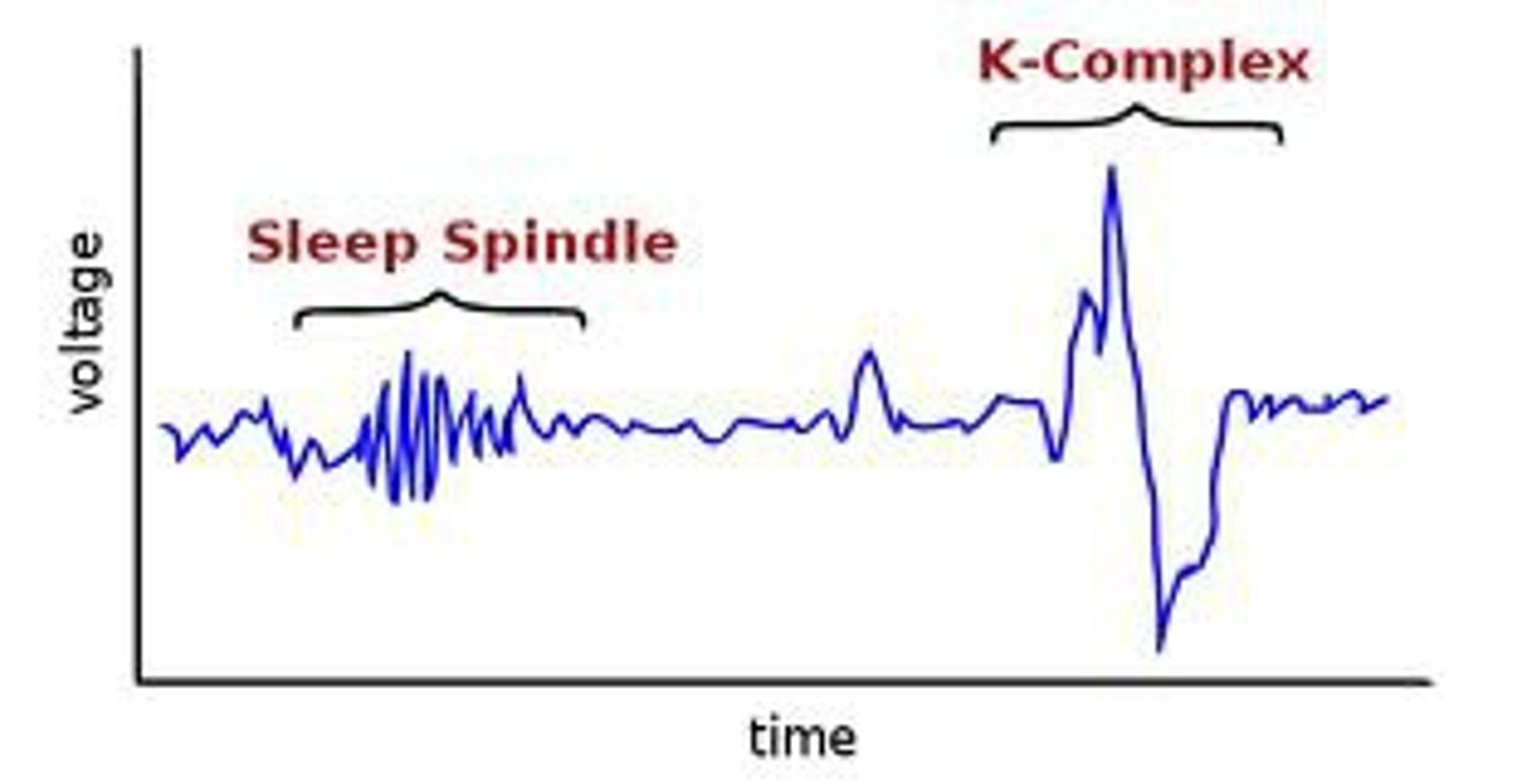
- brief bursts of higher frequency brain waves
- k-complex: sharp waves, helps to keep you asleep through noise
- sleep spindles: generated in thalamus, more = better sleep
stage 3 and 4 sleep
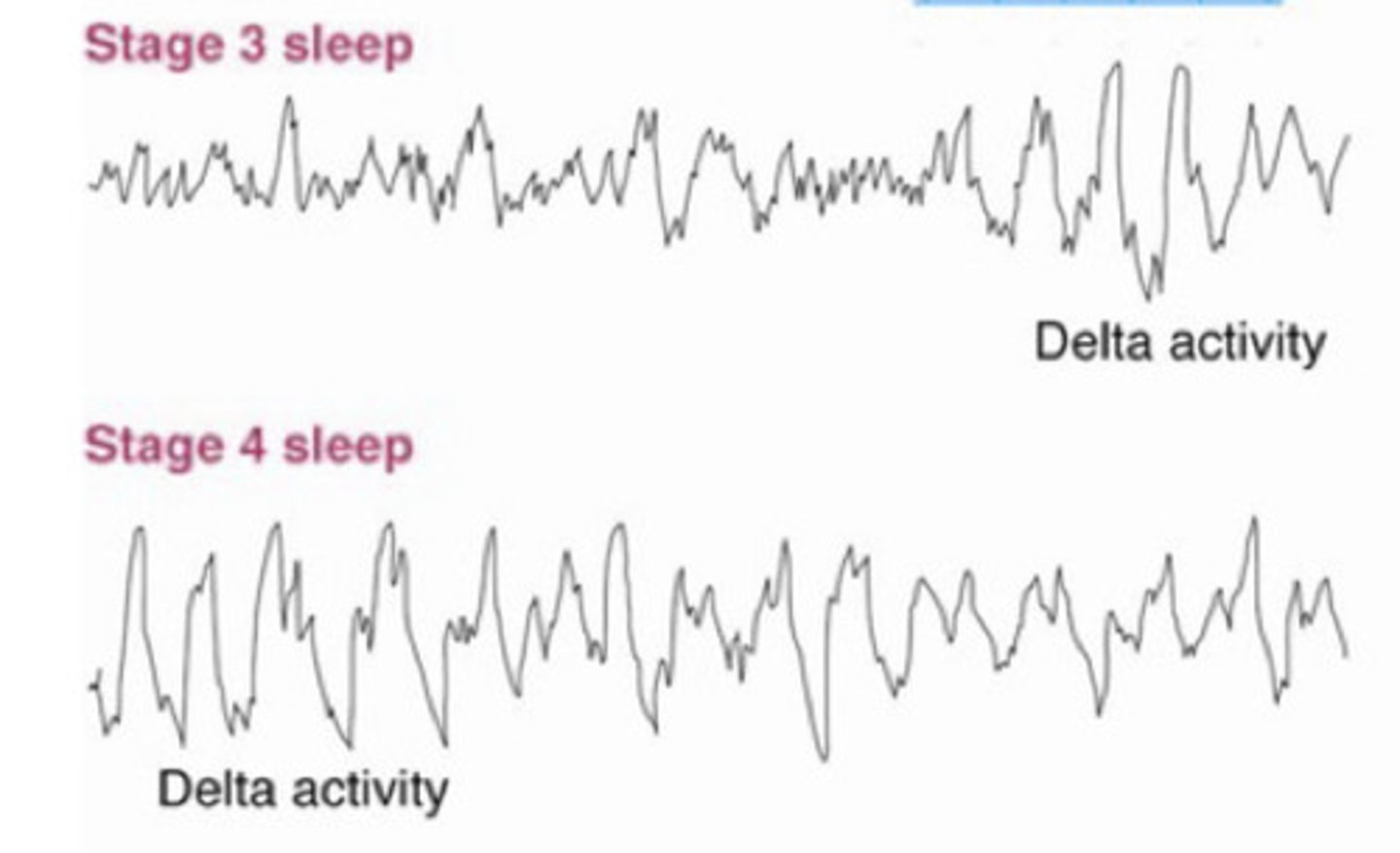
- slow-wave sleep, high amplitude
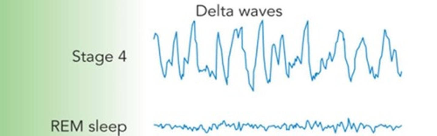
- synchronized low frequency delta waves
- memory consolidation
- some dreams
REM sleep (stage 5)
- rapid eye movement
- high frequency beta waves
- dreams
- amount of REM increases as night goes on
insomnia
- chronic problems in getting adequate sleep
- causes: anxiety (adrenaline), tension, depression
narcolepsy
- irresistible onsets of sleep during normal waking periods
- causes: lack of orexin, Huntington's
sleep apnea
- frequent, reflexive gasping for air that awakens a person
- causes: obesity, genetics, hormones, old-age
night terrors
- abrupt awakenings from NREM (stage 2) sleep
- accompanied by intense autonomic arousal and feeling of panic
- common in children
nightmares
anxiety-arousing dreams that lead to awakening
REM behavior disorder
- people who move vigorously during REM, acting out dreams
- causes: GABA deficiency
sleepwalking
motor cortex is awake
lucid dreaming
monitoring areas (frontal lobe) are awake
sleep paralysis
waking up while you still can't move
fatal familial insomnia
- inherited rare disease
- initially looks like insomnia
- causes: large amounts of PrPres --> degeneration of thalamus
- consequences: absence of sleep spindles and k-complexes, deep sleep is reduced, weight loss, hallucinations, seizures, coma, death
differences in sleep among species
- fishes that don't sleep live in caves with no difference between day and night
- humans have changed sleep habits (used to wake up for period of time in night)
- hibernation is analogous to how we sleep to conserve energy
EEG during sleep
- resembles those that occur during learning
- sleep spindles increase after new learning
dorsal pathway
- parietal --> prefrontal = ACTION
ventral pathway
temporal --> prefrontal = PERCEPTION
encoding
- from sensory system --> STM
- using memory to learn something
- visual objects: right prefrontal and right parahippocampal cortex
- concepts/words: left prefrontal and left parahippocampal cortex
consolidation
- STM --> LTM
- be able to talk about things in the future
- medial temporal lobe
- key area = hippocampus and "what" pathway (ventral)
retrieval
- bringing memories to consciousness using working memory
- requires attention (frontal areas)
- retrieval from LTM make memories plastic again and can be updated
explicit memory
- conscious
- episodic and semantic
episodic memory
memory of personal experiences
semantic memory
memory of facts and general knowledge
implicit memory
- unconscious
- priming, procedural, perceptual learning, and classical conditioning
priming
past experiences influences thoughts of responses
procedural memory
memory for how to do skills and tasks thru repetition (hard to consciously change once learned)
perceptual learning
improved perception thru experience
classical conditioning
learning connections between stimuli
neural substrates for memory
- temporal-frontal lobes
- ventral stream
- temporal lobe (hippocampus and rhinal cortex)
neurotransmitters for memory
- acetylcholine
- serotonin
- noradrenaline
spatial memory
- place cells: hippocampus, directions and navigation (rainbow video), neuron fires when in same spot
- grid cells: entorhinal cortex, learning unknown space or room (black dots in grid), mapping the environment
contextual memory
- memory for context surrounding an event
- hippocampus: links everything together
- parahippocampal cortex: processes scenes and places
fear conditioning
- depends on amygdala (fear/anxiety)
- damage to amygdala abolishes emotional memory
- gut feeling like something is wrong but you don't know what (haunted house)
procedural learning
- depends on basal ganglia (habits/skills)
- people with Parkinson's have this impaired
basal ganglia
- includes striatum, globus pallidus, and substantia nigra
- gradual learning of habits (procedural learning)
- reinforcement- based teaching
- early in learning we rely on prefrontal cortex, but with practice the control shifts to basal ganglia (driving a car)
prefrontal cortex
- used when learning new things for the first time before habits are developed
- can be changed more easily than procedural
long-term potentiation
- glutamate receptors: AMPA and NMDA
- AMPA regularly allows sodium ions thru (causes depolarization)
- NMDA can allow sodium and calcium but is blocked by magnesium
- glutamate released --> sodium enters thru AMPA --> large depolarization --> magnesium unblocks NMDA --> sodium and calcium enter thru NMDA --> strengthens synapse
anterograde amnesia
inability to form new memories
retrograde amnesia
loss of memories formed before onset of amnesia
time-dependent amnesia
- type of retrograde amnesia
- lose most recent memories first, childhood memories last
HM patient
- had severe epilepsy --> surgeons removed hippocampus and part of amygdala
- suffered anterograde and retrograde amnesia
- severe impairment of episodic memory (personal events) and LTM
- difficult imagining the future and making plans
- intact STM/working memory, but when distracted info is lost
- intact procedural memory (video games)
attention
narrowing or focusing awareness on specific part of environment
alertness
state of arousal and readiness to respond to stimuli (part of attention)
consciousness
primary level of awareness (yourself and surroundings)
top-down processing
start with expectations/memory --> interpret what you're seeing
bottom-up processing
start with what you see/hear --> brain figures out what it is
alerting network
- function: maintains alertness / readiness to respond
- region: reticular activating system (RAS)
- neurotransmitter: noradrenaline (from locus coeruleus)
- prepares prefrontal and posterior parietal to detect stimuli quickly
orienting network
- function: helps with prioritizing stimuli
- region: parietal and frontal cortex
- neurotransmitter: acetylcholine
- more stimulus-driven (bottom-up)
dorsal attention system
- function: helps you stay focused on a task (goal-directed attention)
- region: frontal eye fields and intraparietal sulcus
- more top-down visuospatial
- right-lateralized (damage = neglect syndrome)
neural synchrony
- neurons fire at same time --> timing makes signals more impactful and easier to transmit
- attentional system increases synchrony which boosts important signals and filters out noise
resting potential
- sodium outside, potassium inside (-70mV)
- inside of neuron is more negative
- sodium-potassium pump moves 3 Na out, 2 K in
- creates electrical gradient that stores energy for AP
action potential
- depolarization: Na enters neuron
- threshold = -55 mV
- at peak: Na channels close, K channels open
- K exits cell --> repolarization restores negativity
refractory period
- neuron cannot immediately fire again
- Na channels closed, K channels open
- AP moves forward, triggers adjacent channels
- no backfiring due to hyperpolarization
all-or-none law
if threshold is reached, full AP fires
myelin sheath
- fatty insulation around axons
- oligodendrocytes (CNS) and Schwann cells (PNS)
EEG
- measures electrical activity in brain
- high temporal (time) resolution
- poor spatial (place) resolution
neuroimaging
- measures brain activity indirectly
- high spatial (place) resolution
- poor temporal (time) resolution
primary motor cortex (M1)
- located in frontal lobe
- responsible for executing voluntary movements
premotor cortex
- located in front of M1
- responsible for planning and organizing movements
somatic nervous system
- part of PNS
- controls voluntary/conscious movement
- acetylcholine
- ex. walking, writing, waving
autonomic nervous system
- part of PNS
- works automatically/unconsciously
- acetylcholine and norepinephrine
- ex. heart rate, digestion, pupil dilation
visual field disorders
- hemianopsia: loss of half of visual field (opposite sides)
- blindsight/cortical blindness: no conscious visual perception but can respond to stimuli unconsciously
- cortical color blindness: cannot perceive color (V4)
object agnosias
- apperceptive: cannot form perception
- associative: can perceive but not identify
other agnosias
- prosopagnosia: face blindness (right FFA)
- alexia: word blindness (left FFA)
simultanagnosia
- cannot perceive more than one object at a time
- damage to dorsal stream or Balint's syndrome
amusia
- impairment in pitch discrimination
- Heschl's gyrus (musicians have larger)
auditory hallucinations
- perception of sound without external stimuli
- caused by spontaneous activity in A1
Wernicke's aphasia
- word deafness
- affects language comprehension
Geschwind syndrome
- temporal-lobe personality
- egocentric, aggressive, pedantic speech
central executive network
- present moment attention
- when central executive more active --> default mode is less active
default mode network
- thinking about past, future, or mind-wandering
- when default mode more active --> central executive is less active
salience network
- most active when behavioral change is needed
- snaps you out of mind-wandering
nonfluent aphasias
Brocas
Transcortical motor (TMA) - good repetition
fluent aphasias
Wernicke
Transcortical sensory (TSA) - good repitition
Conduction
disconnection between Broca and Wernicke areas
can understand and produce language but can’t relay info between
pure aphasias
agraphia: writing
alexia: reading
anarthria: speech production problem
meditation
reduces activity in default mode and increases salience and central executive
dual executive networks
frontoparietal: active when you first get instructions, adjust strategies
congulo-opercular: active throughout entire rask, sustained attention