BIOL 218 - Ch. 15: Autonomic Nervous System
 Call Kai
 Learn
Learn Practice Test
 Spaced Repetition
 Match
Match Flashcards
 Knowt Play
1/28

There's no tags or description
Looks like no tags are added yet.
Name | Mastery | Learn | Test | Matching | Spaced | Call with Kai | Chat |
|---|
No analytics yet
Send a link to your students to track their progress
29 Terms
List the similarities and differences between the SNS and the ANS
SOMATIC NERVOUS SYSTEM (SNS)
- includes processes perceived or controlled consciously
- somatic sensory portion
> detects stimuli from special senses, skin, and proprioceptors
> sends information to CNS
- somatic motor portion
> transmits nerve signals from CNS to control skeletal muscles
AUTONOMIC NERVOUS SYSTEM (ANS)
- includes processes regulated below conscious level
- visceral sensory portion
> detects stimuli from blood vessels and internal organs
- autonomic motor portion (visceral motor)
> transmits nerve signals to cardiac muscle, smooth muscle, and glands
- functions to maintain homeostasis
> constant internal environment
- regulates:
> heart rate and blood pressure
> respiratory rate, sweating, and digestion
- keeps these variables within optimal ranges
Compare and contrast neurons in the SNS and ANS
SNS
- single lower motor neuron
> extends from CNS to skeletal muscle fibers
> cell body within brainstem or spinal cord
> exits CNS in cranial nerve or spinal nerve
> myelinated axons with large diameter
>> fastest conduction
> always release acetylcholine (ACh) from synaptic knob
ANS
- chain of two motor neurons
> first neuron, the preganglionic neuron
> cell body within brainstem or spinal cord
> exits CNS in cranial nerve or spinal nerve
> projects to autonomic ganglion in peripheral nervous system
> myelinated axons with small diameter
>> slower conduction
> releases ACh from synaptic knob
> second neuron, the postganglionic neuron
> cell body within autonomic ganglion
> exits ganglion to effector (cardiac muscle, smooth muscle, gland)
> unmyelinated axons with even smaller diameter
>> slowest conduction
> releases ACh or norepinephrine (NE) from synaptic knob
Describe how the two-neuron chain in the ANS facilitates communication and control
- Allows for increasing communication and control
> has neuronal convergence
>> multiple preganglionic neurons synapsing with single cell
> has neuronal divergence
>> axons from one preganglionic cell synapsing with numerous cells
Describe the general functions of the parasympathetic and sympathetic divisions of the autonomic nervous system
PARASYMPATHETIC
- functions to maintain homeostasis at rest
- energy conservation and replenishing stage
- "rest-and-digest" division
SYMPATHETIC
- prepares the body for emergencies
- "fight-or-flight" division
- increased alertness and metabolic activity
- "three E's": emergency, exercise, or excitement
Compare and contrast the anatomic differences in the motor neurons and associated ganglia of the parasympathetic
and sympathetic divisions
PARASYMPATHETIC
- preganglionic neuron in brainstem or S2-S4 spinal cord
- termed craniosacral division
- ganglionic neuron innervating muscles or glands
- preganglionic axons longer
- postganglionic axons shorter
- few preganglionic axons
- ganglia close to or within effector
SYMPATHETIC
- preganglionic neuron in lateral horns of T1-L2
- termed thoracolumbar division
- ganglionic neuron innervating muscles or glands
- preganglionic axons shorter
- postganglionic axons longer
- many preganglionic axons
- ganglia relatively close to spinal cord (in sympathetic trunk ganglia or prevertebral ganglia)
Explain why parasympathetic activation is local and discrete, and sympathetic activation can result in mass activation
PARASYMPATHETIC
- local response
- due to long preganglionic neurons with limited branches
SYMPATHETIC
- usually many structures activated simultaneously
> termed mass activation
> sometimes only single effector activated
- due to short preganglionic neurons with many branches
- especially important in response to stress
> e.g., multiple changes during exercising
> increased heart rate, blood pressure, breathing rate, pupil dilation, etc.
Name the four cranial nerves associated with the parasympathetic division, and describe their actions
First three convey parasympathetic information to the head
OCULOMOTOR
- control focusing of the lens
- allow less light into the eye
FACIAL
- increases secretion of gland
GLOSSOPHARYNGEAL
- increases secretion of gland
Vagus - parasympathetic information for thoracic and most abdominal organs
VAGUS
- causes increased mucus production and decreased airway diameter
- causes decrease in heart rate and force
- causes increased smooth muscle motility and secretory activity
Explain the actions of the pelvic splanchnic nerves
- Distal portion of large intestine, rectum
- Bladder, distal ureter
- Most reproductive organs, and others
- Causes:
> smooth muscle motility
> secretory activity in digestive tract
> contraction in bladder wall
> erection of clitoris and penis
Give the location of the sympathetic preganglionic neuron cell bodies
Housed in lateral horn of T1-L2
Describe the left and right sympathetic trunks and ganglia
- Left and right trunks anterior to spinal nerves
- Lateral to the vertebral column
- Resemble string of pearls
> "string" composed of axons
> "pearls" composed of sympathetic trunk ganglia
- House sympathetic ganglionic bodies
- One ganglion associated with each spinal nerve
- Cervical portion of trunk partitioned into three ganglia:
> superior, middle, inferior
Compare and contrast white and grey rami with respect to their location and composition
WHITE RAMI COMMUNICANTES
- carry preganglionic sympathetic axons from T1-L2 nerves to trunk
- associated with T1-L2 spinal nerves
- myelination providing whitish appearance
GREY RAMI COMMUNICANTES
- carry postganglionic sympathetic axons from trunk to spinal nerve
- connect to all spinal nerves
- unmyelinated , so they have a greyish appearance
- similar to highway "exit ramps"
Explain the differences between the sympathetic trunk ganglia and the prevertebral ganglia
- Sympathetic trunk ganglia are part of the sympathetic trunks, which are located lateral to the vertebral column.
- Prevertebral ganglia are located anterior to the vertebral column and cluster around the origins of major abdominal organs
Describe the four pathways of sympathetic neurons:
SPINAL NERVE PATHWAY
- Preganglionic neuron synapsing in sympathetic trunk ganglion
- Travels through grey ramus at the same "level"
e.g., synapsing in L4 ganglion
> Postganglionic axon exit through L4 ramus
- Enters the spinal nerve and extends to target organ
- Skin of neck, torso, and limbs
- Sweat glands, arrector pili muscles, and blood vessels in skin

POSTGANGLIONIC SYMPATHETIC NERVE PATHWAY
- Preganglionic neuron synapsing in sympathetic trunk ganglion
- Postganglionic axon does not leave trunk via grey ramus
- Extends from ganglion and goes to target organ
- Esophagus, heart, lungs, and thoracic blood vessels
- Structures in the head
> e.g., blood vessels, sweat glands, dilator pupillae, superior tarsal

SPLANCHNIC NERVE PATHWAY
- Uses splanchnic nerves
- Preganglionic axons passing through sympathetic trunk without synapsing
- Extend to prevertebral ganglia
> synapse with ganglionic neuron
- Travels to effector organ
- Abdominal and pelvic organs

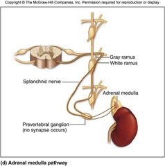
ADRENAL MEDULLA PATHWAY
- Internal region of adrenal gland, adrenal medulla
- Directly innervated by - Preganglionic sympathetic axons
preganglionic neuron
> extends through sympathetic trunk and prevertebral ganglia
- Upon stimulation, epinephrine and norepinephrine produced in medulla
> circulate within blood
> help prolong fight-or-flight response
> prolong effects of sympathetic stimulation
Compare and contrast which general effector organs are innervated by each pathway
SPINAL NERVE
- skin of neck, torso, limbs
- sweat glands, arrector pili muscles, blood vessels in skin
POSTGANGLIONIC SYMPATHETIC NERVE
- esophagus, heart, lungs, thoracic blood vessels
- structures in head
> e.g., blood vessels, sweat glands, dilator pupillae, superior tarsal
SPLANCHNIC NERVE
- abdominal and pelvic organs
ADRENAL MEDULLA
- upon stimulation, epinephrine and norepinephrine produced in medulla
> circulate within blood
> help prolong fight-or-flight response
> prolong effects of sympathetic stimulation
Identify the targets of the cholinergic and adrenergic neurotransmitters of the ANS
ACETYLCHOLINE
- synthesised and released by cholinergic neurons
> all sympathetic and parasympathetic preganglionic neurons
> all parasympathetic ganglionic neurons
> neurons innervating sweat glands and blood vessels of skeletal muscle
- bound by cholinergic receptors (two types)
NOREPINEPHRINE
- bound by adrenergic receptors
- synthesised and released by adrenergic neurons
> most other sympathetic ganglionic neurons
> form network of swellings at target organ
>> termed varicosities
>> contain stored NE
>> NE released along length of axon
Describe the two types of cholinergic receptors and the action of each when a neurotransmitter binds to them
NICOTINIC RECEPTORS
- sensitive to the drug nicotine
- found on all ganglionic neurons and adrenal medulla
- when bound:
> open ion channels
> greater movement of Na+ into cell than K+ out of cell
> excitatory postsynaptic potential produced
> always produces a stimulatory response
MUSCARINIC RECEPTORS
- responsive to muscarine, a mushroom toxin
- found in:
> all target membranes in parasympathetic division
> selected sympathetic cells
> e.g., sweat glands in skin, blood vessels in skeletal muscle
- different subtypes with different effects
> either stimulated or inhibited by binding ACh
> e.g., binding of ACh in GI tract
>> results in stimulation and increased motility
> binding on cardiac muscle
>> decreases heartbeat rate
List the neurotransmitters categorised as catecholamines
Alpha (α)
Beta (β) receptors
Name the four adrenergic receptors and give the locations of each
α1 receptors
- located in most smooth muscle cells
- stimulate smooth muscle contraction
- found in most blood vessels (vasoconstriction)
- arrector pili muscle (contraction)
- uterus (contraction)
- ureters, internal urethral sphincter (closing)
α2 receptors
- located in pancreas
- inhibit insulin secretion
- involved with contraction of GI tract sphincters
- facilitate blood clotting
β1 receptors
- primarily stimulatory
- found in heart (increase heart rate and force)
- found in kidney (stimulate renin secretion)
β2 receptors
- primarily inhibitory effects
- in smooth muscle of vessels to heart, liver, and skeletal muscle
- cause smooth muscle relaxation and vessel dilation
- lung (bronchodilation)
- uterine and GI tract smooth muscle (relaxation)
- detrusor muscle of bladder (relaxation)
Discuss the nature of autonomic tone and its effects
- E.g., diameter of most blood vessels in a partially constricted state
> due to sympathetic tone
- Decrease in stimulation below tone
> causes vessel dilation
- Increase above sympathetic tone
> causes greater vessel constriction
Explain what is meant by dual innervation
= organ receives input from both the sympathetic and parasympathetic divisions
Describe the antagonistic and cooperative effects of dual innervation
ANTAGONISTIC
- parasympathetic and sympathetic effects usually antagonistic
e.g., control of heart rate
> parasympathetic stimulation slowing heart rate
> sympathetic stimulation increasing heart rate
> same cells with both muscarinic and adrenergic receptors
e.g., control of muscular activity in GI tract
> parasympathetic stimulation accelerating rate of contraction and motility
> sympathetic stimulation decreasing motility
> same cells with both types of receptors
e.g., control of pupil diameter in the eye
> parasympathetic stimulation of circular muscle layer of iris
>> causes pupil constriction
> sympathetic stimulation of radial muscle layer of iris
>> causes pupil dilation
> different effectors innervated
COOPERATIVE
- when both parasympathetic and sympathetic produce single result
e.g., male sexual function
> penis erect due to parasympathetic innervation
> ejaculation due to sympathetic innervation
Describe the systems innervated only by the sympathetic division and how they function
E.g., blood vessels innervated by sympathetic axons only
- cause increased smooth muscle contraction and blood pressure
- vasodilation achieved by decreasing stimulation below autonomic tone
E.g., sweat glands in the trunk and arrector pili muscles in the skin
- cause sweating and "goosebumps"
E.g., neurosecretory cells of adrenal medulla
- release epinephrine and norepinephrine, prolonging fight-or-flight effects
Describe some major types of autonomic reflexes
CARDIOVASCULAR REFLEX
- stretch receptors stimulated in blood vessel walls with pressure elevation
- signals propagated to cardiac center in medulla oblongata
- inhibit sympathetic and activate parasympathetic output to heart
- slows heart rate and decreases volume ejected
- decreases blood pressure
GASTROINTESTINAL REFLEX
- control proximal GI tract
> stimulates secretion of gastric glands by parasympathetic stimulation
> stimulated by sight or smell of food
- control rectum
> stretch of rectum by fecal matter walls
> parasympathetic reflex causing contraction to aid elimination
MICTURITION REFLEX
- mechanism leading to bladder emptying
- stretch receptors signaling when urine fills
- results in reflex contraction of smooth muscles in bladder
- results in relaxation of urinary sphincter
- in toilet-trained individuals
> urination follows voluntary relaxation of external urethral sphincter
OTHER REFLEXES
- alter respiratory rate and depth
- regulate digestive system activities
- change pupil diameter
Describe the CNS hierarchy that controls the autonomic nervous system
HYPOTHALAMUS
- integration and command centre for autonomic functions
- communicates with association areas of cortex
- affected by sensory processing in thalamus
- impacted by emotional states in limbic system
- also communicates with brainstem, cerebellum, and spinal cord
- central structure involved in drives that act through ANS
BRAINSTEM NUCLEI
- mediate major ANS reflexes
- control changes in blood pressure, blood vessel diameter, and digestion
- control changes in heart rate, pupil size, and eye lens shape for focusing
SPINAL CORD
- some autonomic responses controlled at level of spinal cord
e.g., defecation and urination (in children)
- may be inhibited by higher centres
Describe the structure and location of the five autonomic plexuses
CARDIAC
- consists of:
> sympathetic postganglionic axons from cervical and thoracic trunk
> parasympathetic preganglionic axons from vagus nerve
- increased sympathetic activity increasing heart rate and blood pressure
- increased parasympathetic decreasing heart rate
PULMONARY
- consists of:
> sympathetic postganglionic axons from cervical and thoracic trunk
> parasympathetic preganglionic axons from vagus nerve
- project to bronchi of lungs
- increased sympathetic activity causing bronchodilation
- increased parasympathetic activity causing bronchoconstriction
ESOPHAGEAL
- consists of:
> sympathetic postganglionic axons from cervical and thoracic trunk
> parasympathetic preganglionic axons from vagus nerve
- project to esophagus
- sympathetic activity inhibiting muscle motility
- parasympathetic activity controlling swallowing reflex
> innervates cardiac sphincter, a valve through which food and drink pass
ABDOMINAL AORTIC PLEXUS
- consists of:
> celiac, superior mesenteric, and inferior mesenteric plexuses
> sympathetic postganglionic axons from prevertebral ganglia
> parasympathetic preganglionic axons from vagus or pelvic splanchnic nerves
- innervates all abdominal and some pelvic organs
HYPOGASTRIC AORTIX PLEXUS
- consists of:
> sympathetic postganglionic axons from aortic plexus and sympathetic trunk
> parasympathetic preganglionic axons from pelvic splanchnic nerves
- innervates viscera within pelvic region
Discuss how autonomic reflexes help maintain homeostasis
(visceral reflexes)
- ANS helps maintain homeostasis through the involuntary activity of autonomic reflexes
- Involves the reduction of blood pressure