B3.2: Transport | IB Biology HL
1/62
Earn XP
Description and Tags
Name | Mastery | Learn | Test | Matching | Spaced | Call with Kai |
|---|
No analytics yet
Send a link to your students to track their progress
63 Terms
Describe how the structures of capillaries are adapted to capillary function. Include lumen diameter, branching, wall thickness, and fenestrations.
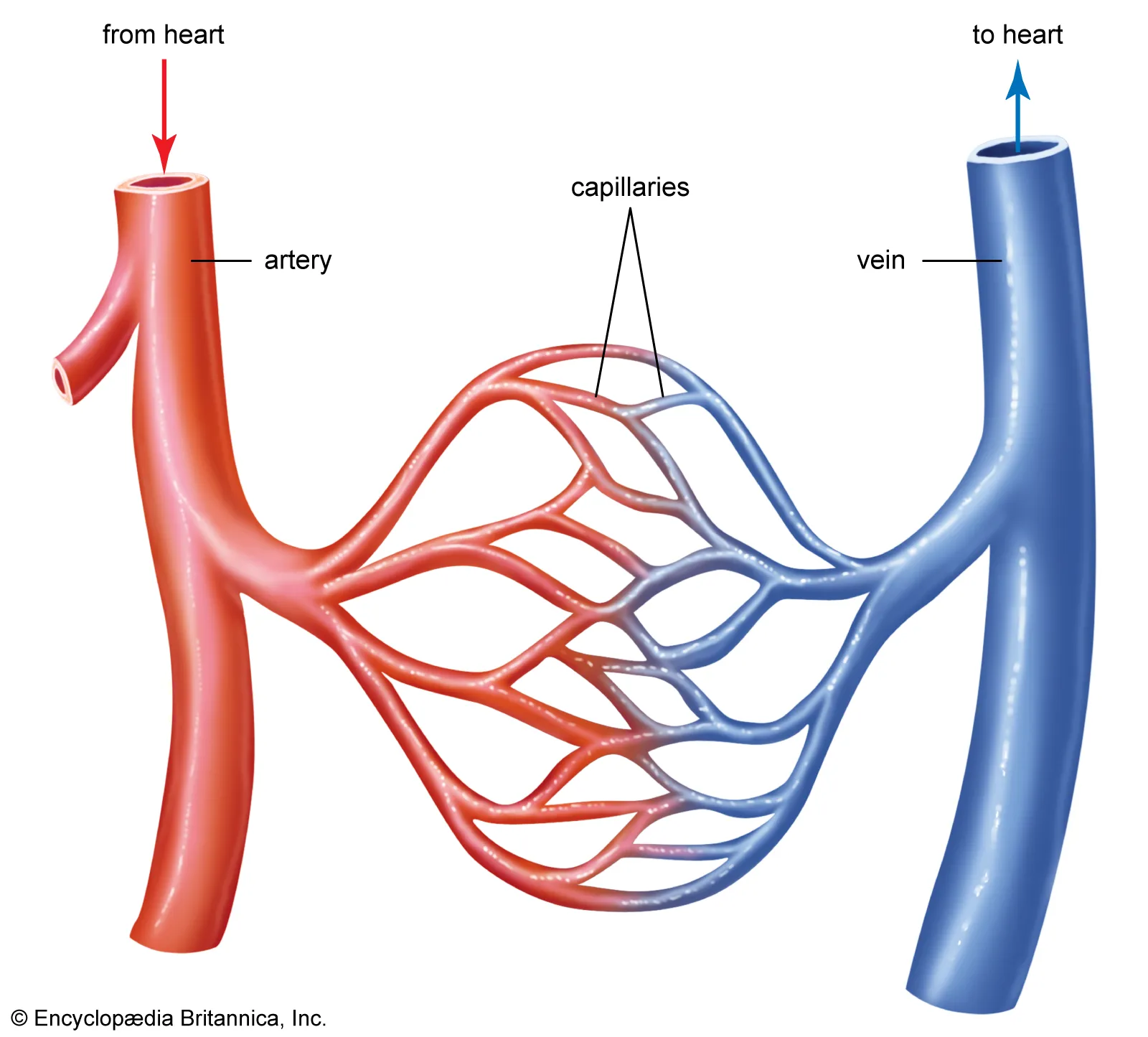
B3.2.1: Adaptations of capillaries for exchange of materials between blood and the internal or external environment.
Capillaries are small blood vessels that connect arteries to veins.
Capillaries have a narrow lumen (small lumen diameter) just wide enough for one red blood cell to pass through at a time. This allows gases to diffuse through and reach the RBC faster.
Capillaries are highly branched to have a larger surface area for more diffusion to occur at the same time.
Capillaries have very thin walls, typically one cell thick, that allows for rapid exchange of materials by diffusion.
Some capillaries have fenestrations (pores) that allow larger molecules to pass through them.
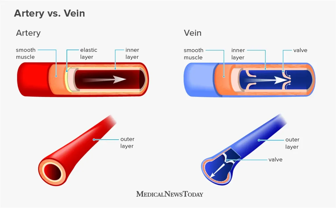
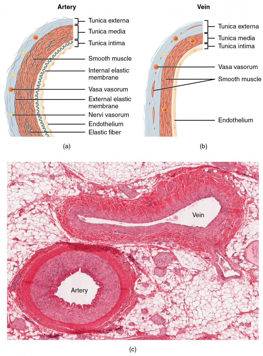
Compare the diameter, relative wall thickness, lumen size, number of wall layers, abundance of muscle and elastic fibers and presence of valves in arteries and veins.
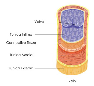
B3.2.2: Structure of arteries and veins.
Arteries have smaller diameters than veins. This is because arteries have a thicker wall, while veins have a much thinner wall. As a result, arteries have relatively narrow lumen, but veins have a much wider lumen. Both arteries and veins have 3 wall layers (tunica externa, tunica media, tunica intima). However, arteries have more smooth muscle and elastic fibers than valves do. Arteries do not have any valves, while veins do.
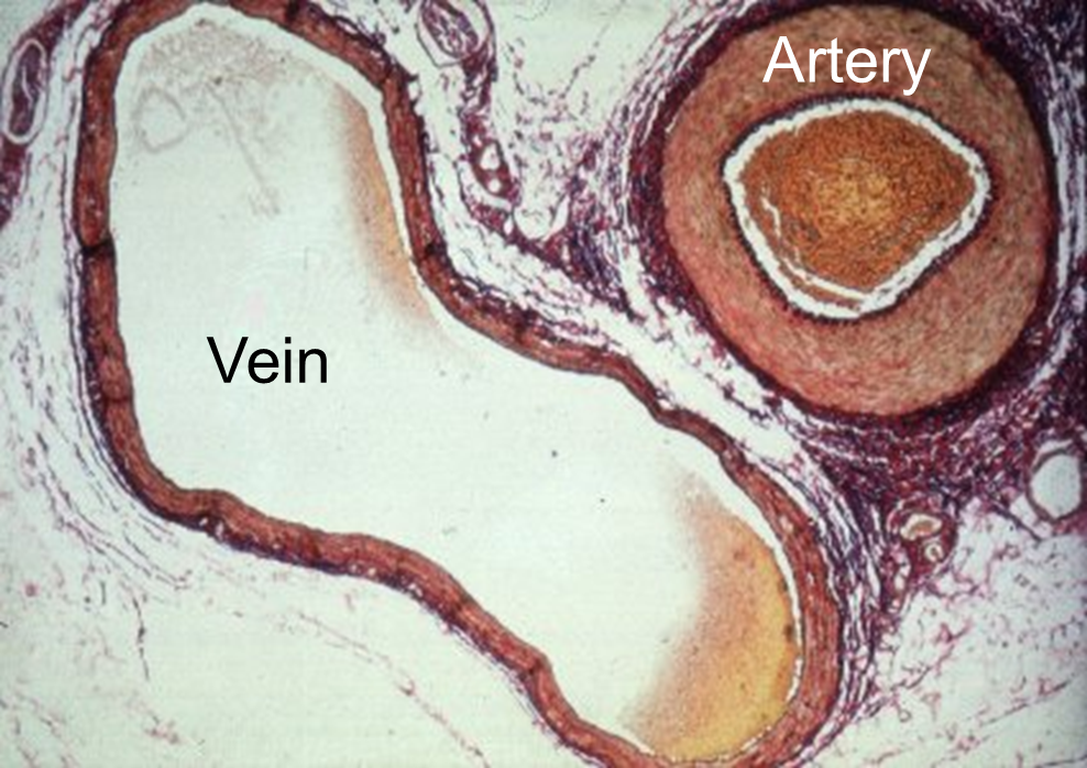
Given a micrograph, identify a blood vessel as an artery or vein.
B3.2.2: Structure of arteries and veins.
Arteries have thicker walls, a narrower lumen, and tend to be more circular.
Veins have much thinner walls, a relatively wider lumen, and may be more “squished” (not as perfectly circular).
State the function of arteries.
B3.2.3: Adaptations of arteries for the transport of blood away from the heart.
Arteries transport blood away from the heart, carrying oxygenated blood to tissues.
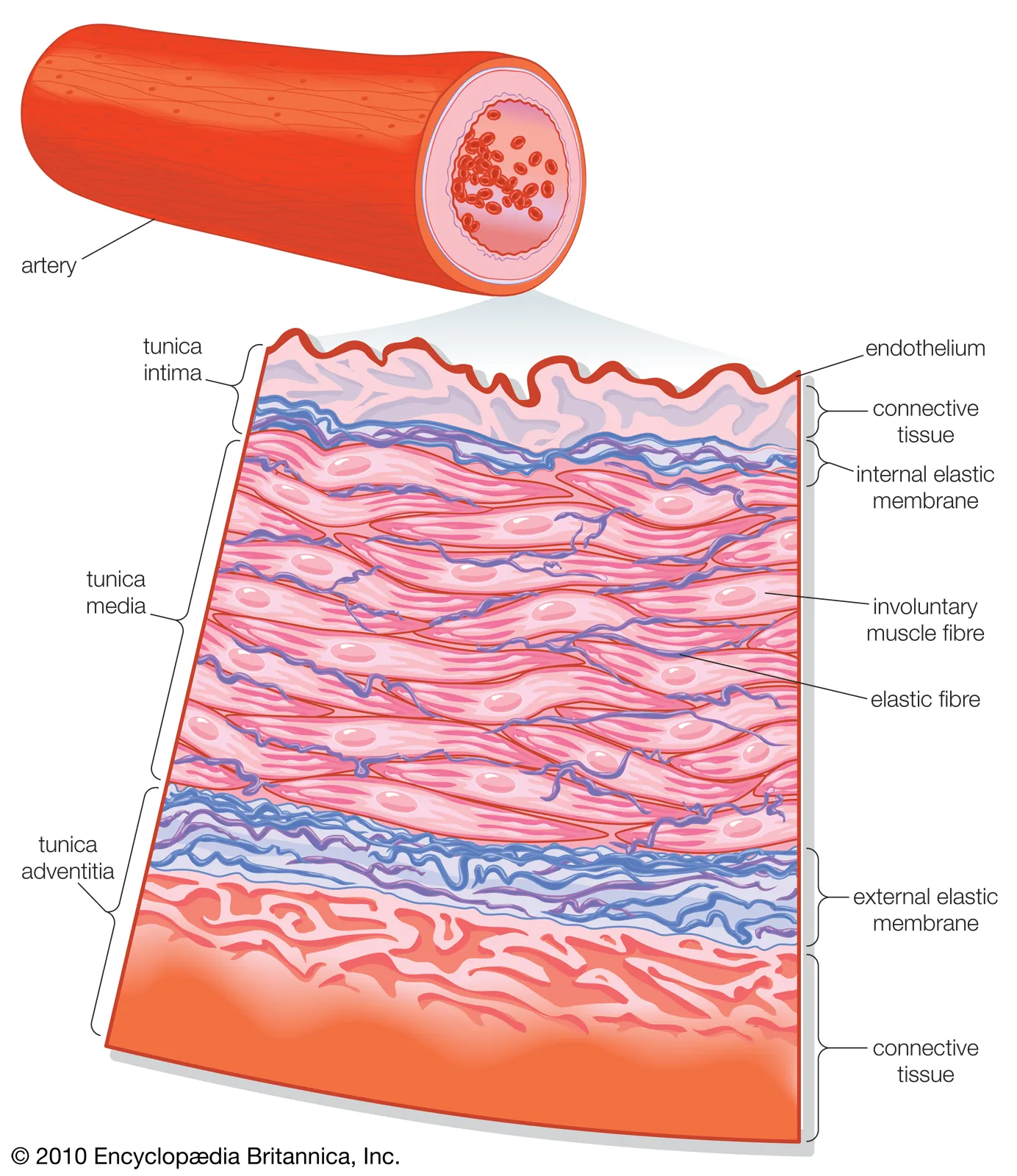
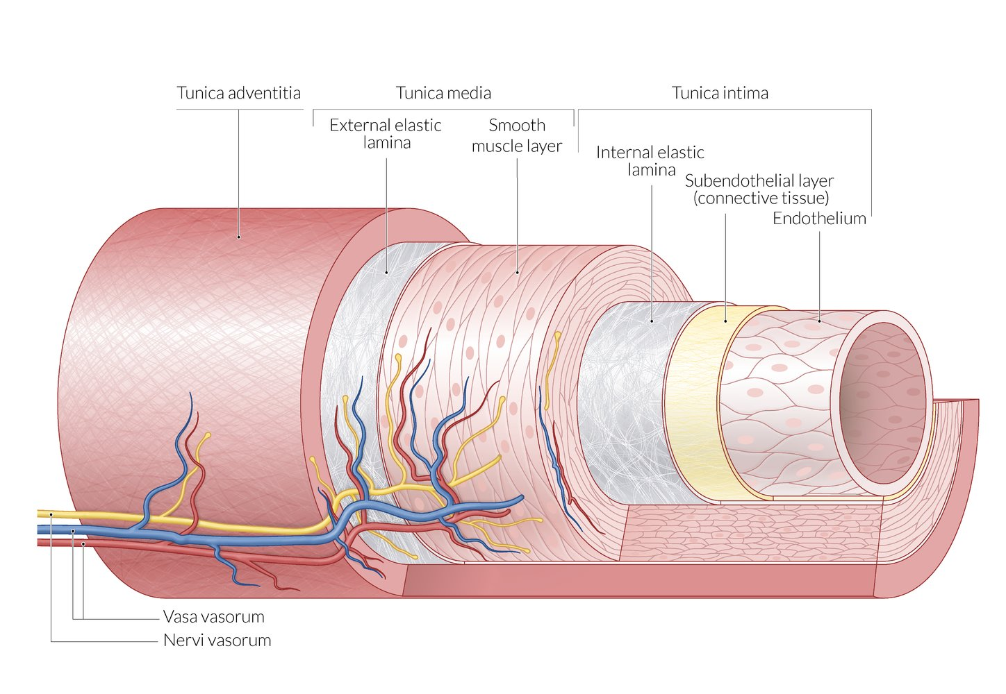
Describe the structures and functions of the three layers of the artery wall.
B3.2.3: Adaptations of arteries for the transport of blood away from the heart.
The tunica intima is the innermost layer of the artery. It is found throughout the cardiovascular system. It is made of a simple squamous epithelium surrounded by a connective tissue basement membrane with elastic fibers. It provides structural support to larger vessels.
The tunica media is the middle layer. It is typically the thickest, and is primarily smooth muscle. It provides support for the artery and changes artery diameter to regulate blood flow and blood pressure.
The tunica adventitia (externa) is the outermost layer. It is made of connective tissue with elastic and collagen fibers. It attaches the artery to the surrounding tissue.
Discuss how the wall thickness, lumen size, and muscle and elastic allow arteries to withstand and maintain high blood pressures.
B3.2.3: Adaptations of arteries for the transport of blood away from the heart.
The arteries’ thick walls allow them to withstand high blood pressure.
The narrow lumen helps maintain high blood pressure.
Smooth muscle in the arteries allow them to contract in order to maintain high blood pressure (between heart beats, etc.)
Elastic fibers in the artery walls allow the arteries to stretch and recoil as pressure increases and decreases due to heart beats. This recoil helps keep the blood moving in the artery.
Smooth endothelial cells lining the lumen reduces the friction as blood flows.
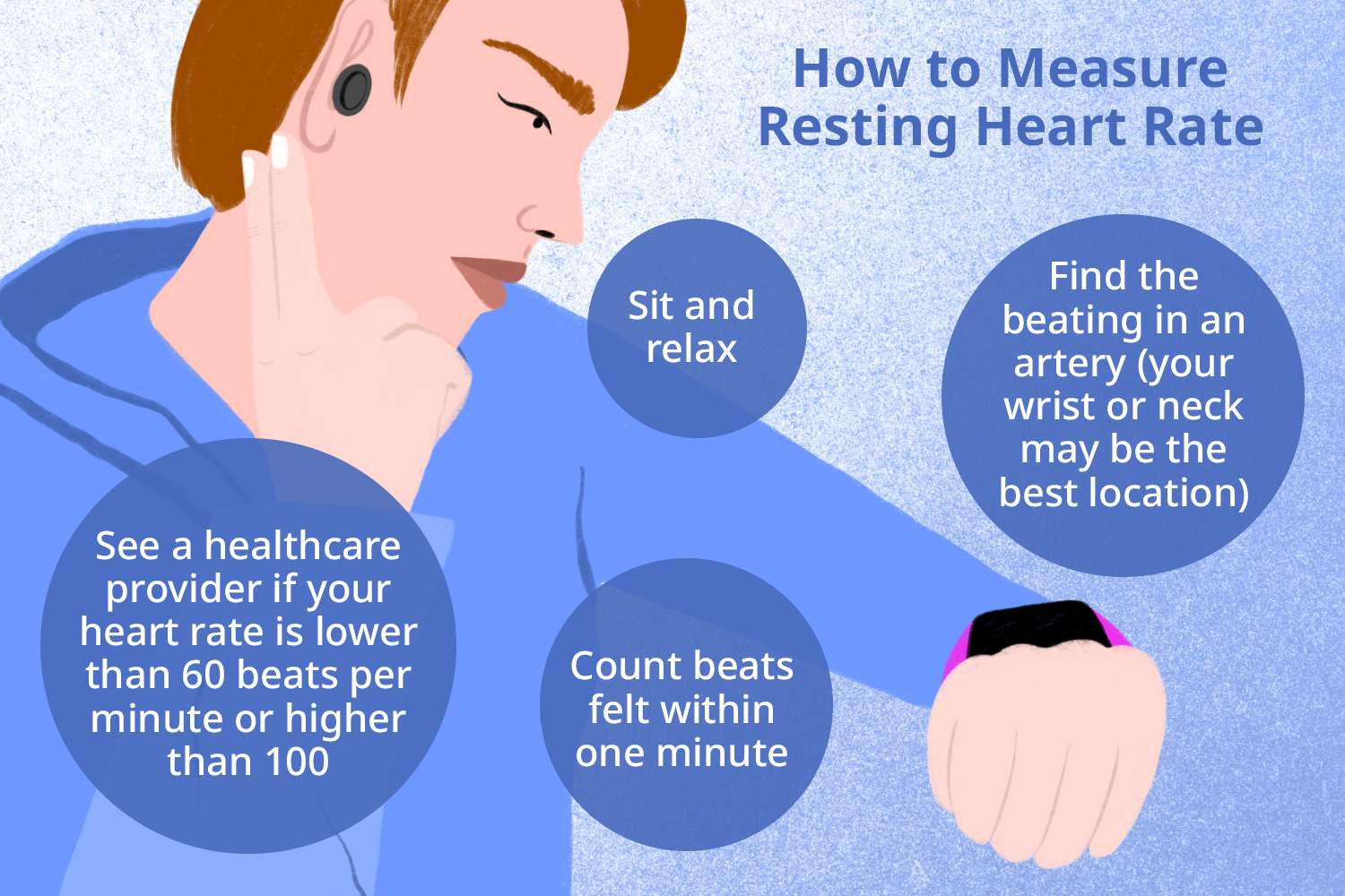
State the unit of measurement of the pulse rate.
B3.2.4: Measurement of pulse rates.
Pulse rate is measured in beats per minute (bpm).
Outline two methods for determining heart rate.
B3.2.4: Measurement of pulse rates.
Heart rate can be determined manually or with technology.
If determining manually, place two fingers by your neck OR on your wrist and count the number of beats per minute or per 30 seconds (in which case, multiply by 2).
Technology like smart watches and oximeters can determine the heart rate for you.
State the function of veins.
B3.2.5: Adaptations of veins for the return of blood to the heart.
Veins return deoxygenated blood back to the heart.
Discuss how pocket valves, thin walls and skeletal muscles maintain the flow of blood through a vein.
B3.2.5: Adaptations of veins for the return of blood to the heart.
Blood returning to the heart moves slowly and is not under high pressure, so the veins must maintain blood flow.
The valves of veins prevent the backflow of blood.
The veins’ thin walls allow the vein to be compressed by skeletal muscles. This compression helps move blood back to the heart.
When skeletal muscles contract, veins in the muscle are compressed. As a result, blood pressure increases, and blood flow is maintained.
The wide lumen of veins allow veins to carry a large volume of blood back.
State the function of the coronary arteries.
B3.2.6: Causes and consequences of occlusion in the coronary arteries.
The coronary arteries branch off of the aorta and wrap around the heart to supply the heart with oxygen and nutrients.
Outline the cause and consequence of a coronary occlusion.
B3.2.6: Causes and consequences of occlusion in the coronary arteries.
Coronary occlusion occurs when there is high blood pressure and the inner lining of an artery is damaged. As a result, macrophages (a type of white blood cell) are attracted to sites of damage within the arteries. Therefore, growth factors are released, which stimulates the development of fibrous tissue. When macrophages consume cholesterol, they form plaque. Over time, the plaque grows and may occlude the artery. The plaque may break away from the artery and cause a blood clot.
Coronary occlusion may lead to death of heart tissue and heart attacks.
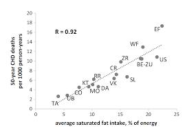
Evaluate correlations between diet and lifestyle variables and risk of coronary heart disease.
B3.2.6: Causes and consequences of occlusion in the coronary arteries.
A diet high in fats and cholesterol may increase the risk of coronary heart disease because it can lead to more cholesterol buildup. A lifestyle lacking physical inactivity can lead to obesity, which increases blood pressure and damages artery walls. Smoking also increases blood pressure, leading to a higher risk of coronary heart disease.
As saturated fat intake increases, there is an increase in the number of deaths caused by coronary heart disease.
List factors that are correlated with an increased risk of coronary occlusion and heart attack.
B3.2.6: Causes and consequences of occlusion in the coronary arteries.
Factors that are outside of our control:
Genetics: Several genes are associated with an increased risk of atherosclerosis
Age: The arteries of older people are more likely to be damaged
Gender: males are more likely to develop atherosclerosis
Factors we can control:
Obesity: Obesity increases blood pressure & can damage artery walls
Physical inactivity: Can lead to obesity
Smoking: Increases blood pressure
Diets high in fats and cholesterol
Define the Pearson correlation coefficient (r).
B3.2.6 NOS: Correlation Coefficients
The Pearson correlation coefficient is used to quantify correlations between variables and allow the strength of the relationship to be assessed.
If the correlation coefficient is closer to 0, it can be assumed that there is no relationship between the two variables.
More positive coefficients are more positively correlated. More negative coefficient means it is more negatively correlated.
Differentiate between correlation and causation.
B3.2.6 NOS: Correlation Coefficients
Correlation does not imply causation.
Correlation means two variables are associated. Causation, on the other hand, requires the change of one variable to be a direct cause of the change of another.
State that xylem tissue is used to transport water from roots to leaves in plants.
B3.2.7: Transport of water from roots to leaves during transpiration.
Xylem tissue is used to transport water from roots to leaves in plants.
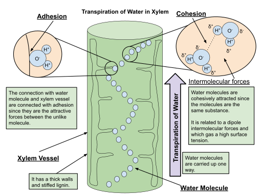
Outline the role of cellulose in the transport of water via capillary action.
B3.2.7: Transport of water from roots to leaves during transpiration.
The cellulose of the walls of the xylem are polar molecules, so they can form hydrogen bonds with polar hydrogen bonds (adhesion). This maintains the movement of water up the xylem as they stick to the walls, bringing more water molecules up (through cohesion).
Describe the cause and consequence of transpiration pull.
B3.2.7: Transport of water from roots to leaves during transpiration.
The transpiration pull occurs because there is negative pressure in the leaf, due to the loss of water (evaporation). When water enters the roots by osmosis, there is higher pressure.
The transpiration pull allows water to move through the xylem from the roots to the leaves.
State why transport of water relies on cohesion between water molecules.
B3.2.7: Transport of water from roots to leaves during transpiration.
The transport of water relies on cohesion between water molecules because water molecules need to stick together in order to be drawn up in a continuous column of water in the xylem.
State that transpiration is a passive process.
B3.2.7: Transport of water from roots to leaves during transpiration.
Transpiration is a passive process.
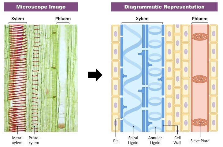
Describe how the structure of xylem vessels are adapted for the transport of water under low pressure.
B3.2.8: Adaptations xylem vessels for transport of water.
The xylem do not have end walls between cells, which allows for the column of water to move up the plant without impeded flow.
Similarly, xylem cells lack cell contents or plasma membrane, which also allows for unimpeded flow of water.
The xylem has pits that allow water to enter and exit the xylem between adjacent cells.
Lignin strengthen the cell walls of xylem, allowing xylem vessels to resist inward pressures created by transpiration.
Outline how xylem is able to maintain rigidity even under low pressure or mechanical disturbance.
B3.2.8: Adaptations xylem vessels for transport of water.
The xylem can maintain rigidity even under pressure because it has lignin. It strengthens cell walls and allow the vessels to resist pressure created by transpiration.
Draw a plan diagram to show the distribution of tissues in a stem, including vascular bundles, xylem, phloem, cambium, cortex, pith and epidermis.
B3.2.9: Distribution of tissues in a transverse section of the stem of a dicotyledonous plant.

Outline the function of tissues in a stem, including vascular bundles, xylem, phloem, cambium, cortex, pith and epidermis.
B3.2.9: Distribution of tissues in a transverse section of the stem of a dicotyledonous plant.
The vascular bundles transport materials up and down the plant. It contains the xylem and phloem.
The xylem transports water and minerals up the stem of the plant.
The phloem transports organic compounds (like sucrose and amino acids) up and down the stem of the plant.
The cambium is a circular layer of undifferentiated cells responsible for lateral growth of the stem.
The cortex provides structural support for the stem.
The pith is internal ground tissue that provides structure and helps transport and storage.
The epidermis provides protection for the stem by covering the outer layer, waterproofs it, and controls gas exchange.
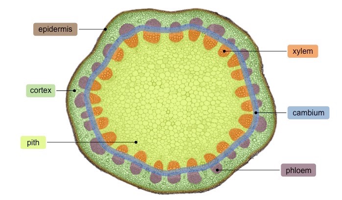
State two ways xylem and phloem can be differentiated in cross sections of stem.
B3.2.9: Distribution of tissues in a transverse section of the stem of a dicotyledonous plant.
The xylem is on the interior side of the bundle.
The phloem is more outward, closer to the epidermis and cortex.
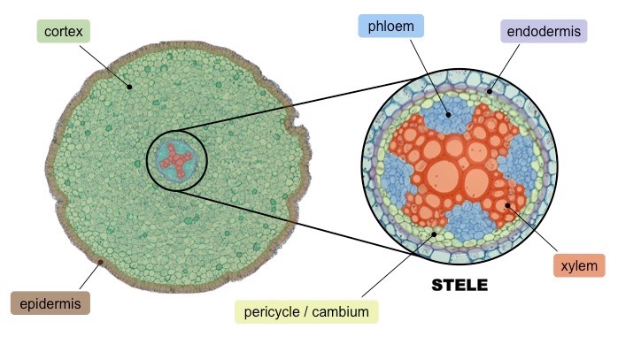
Draw a plan diagram to show the distribution of tissues in a root, including vascular bundles, xylem, phloem, cortex and epidermis.
B3.2.10: Distribution of tissues in a transverse section of the root of a dicotyledonous plant.
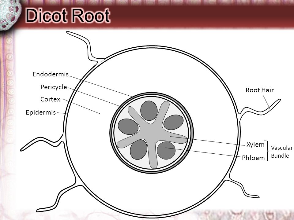
Outline the function of tissues in a root, including vascular bundles, xylem, phloem, cortex and epidermis.
B3.2.10: Distribution of tissues in a transverse section of the root of a dicotyledonous plant.
The vascular bundles are located centrally to withstand stretching forces and allow for material transport to be controlled.
The xylem transports water and minerals from the roots up to the stem of the plant.
The phloem transports organic compounds (like sucrose and amino acids) through the plant.
The cortex provides structural support for the stem and stores starch in the roots.
The epidermis provides protection and may have root hairs (protrusions) to increase available surface area for material exchange.
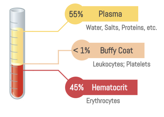
List components of blood plasma.
B3.2.11 (AHL): Release and reuptake of tissue fluid in capillaries.
Water (90%)
Proteins
Ions
Nutrients
Wastes
Define tissue fluid.
B3.2.11 (AHL): Release and reuptake of tissue fluid in capillaries.
Tissue fluid is the fluid between cells and blood that allows for cells to chemically exchange substances with blood. It is the fluid found in the space around cells.
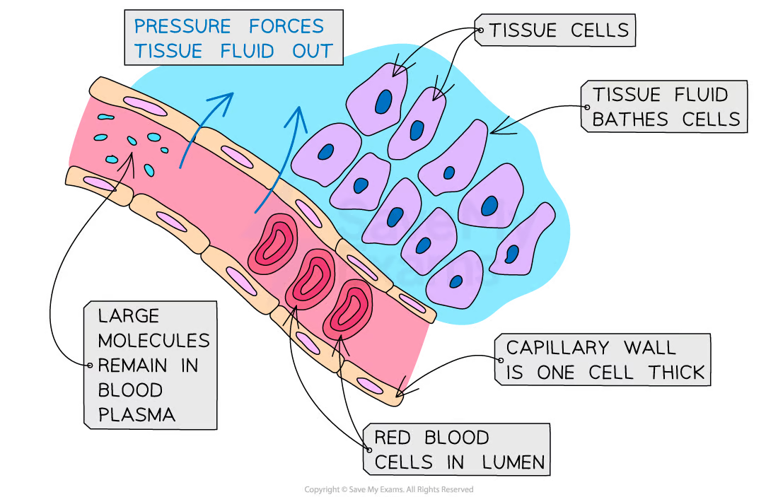
Describe the cause and effect of diffusion of blood plasma into and out of a capillary network from tissue fluid.
B3.2.11 (AHL): Release and reuptake of tissue fluid in capillaries.
The arteriole side of the capillary bed has very high pressure. Pressure filtration is the release of tissue fluid and occurs because the pressure at the arteriole end is high, which opens gaps between the cells that make up the wall of the capillary. Thus, tissue fluid can diffuse between cells.
The venules end of the capillary bed has relatively low pressure. This allows tissue fluid to drain back into the capillaries through diffusion.
Compare and contrast the substances found in blood plasma and tissue plasma.
B3.2.12 (AHL): Exchange of substances between tissue fluid and cells in tissues.
The chemical makeup of blood plasma and tissue fluid is very similar because of the unregulated passage of substances through porous capillary membranes and gaps under arteriole pressure.
There are some white blood cells in tissues fluid because some WBC can squeeze through capillaries into tissue fluid.
However, there are no red blood cells and large proteins in tissues fluid because they are too large to exit through capillary walls.
Outline the direction of transport of substances that are exchanged between tissue fluid and cells in the tissues.
B3.2.12 (AHL): Exchange of substances between tissue fluid and cells in tissues.
Oxygen and nutrients are transported from tissue fluid into cells.
Waste products like carbon dioxide and urea (waste products of amino acid metabolism) have natural concentration gradients and diffuse directly through plasma membrane or protein channels (facilitated diffusion).
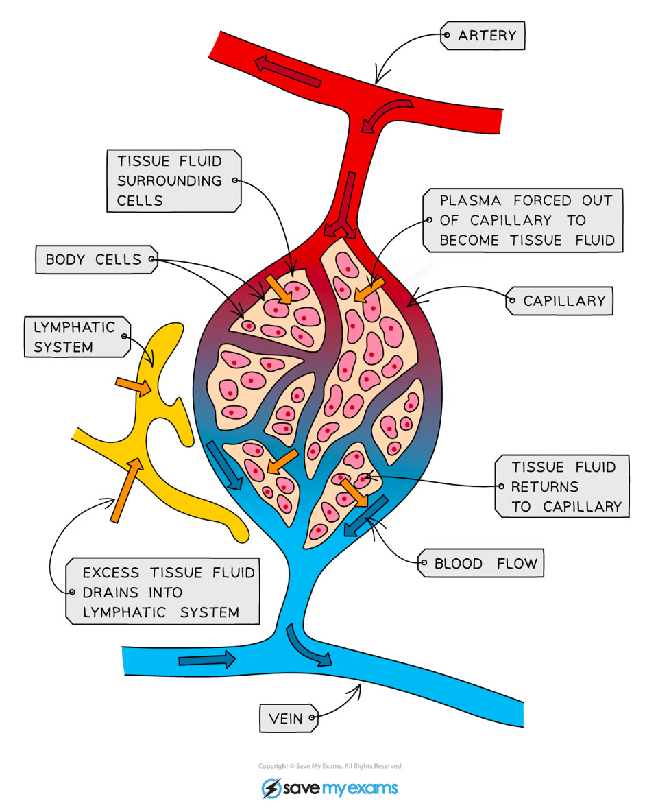
Outline why there is a need to drain excess tissue fluid into lymph ducts.
B3.2.13 (AHL): Drainage of excess tissue fluid into lymph ducts.
Excess tissue fluid must be drained into lymph ducts to prevent fluid build-up around body cells.
Outline the structure and function of lymph ducts.
B3.2.13 (AHL): Drainage of excess tissue fluid into lymph ducts.
Lymph ducts collect tissue fluid in its vessels to prevent fluid build-up around body cells.
Lymph ducts have small capillaries and vessels that join together into larger and larger lymph ducts. Its small capillaries have very thin walls and gaps between adjoining cells to facilitate easy movement of water and solutes. Lymph ducts have internal valves that prevent backflow. Like veins, lymph ducts rely on skeletal muscle contractions to squeeze vessels and keep lymph fluid moving.
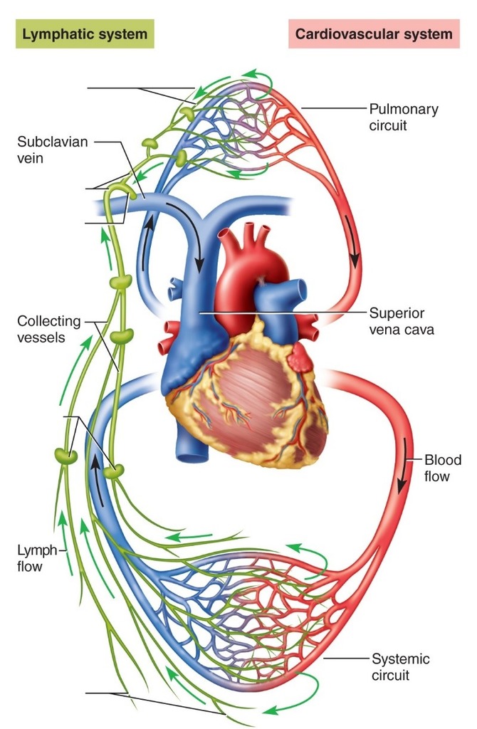
State how lymph is returned to the blood circulation.
B3.2.13 (AHL): Drainage of excess tissue fluid into lymph ducts.
Lymph vessels join together into larger and larger lymph ducts until it takes lymph fluid back to veins to become part of blood plasma again.
State the function of the heart and lungs/gills in the circulation of blood.
B3.2.14 (AHL): Differences between the single circulation of bony fish and the double circulation of mammals.
The heart pumps blood around the body.
The lungs/gills are where gas exchange occurs. It is where blood becomes oxygenated (picks up oxygen) and gets rid of carbon dioxide (as a waste products).
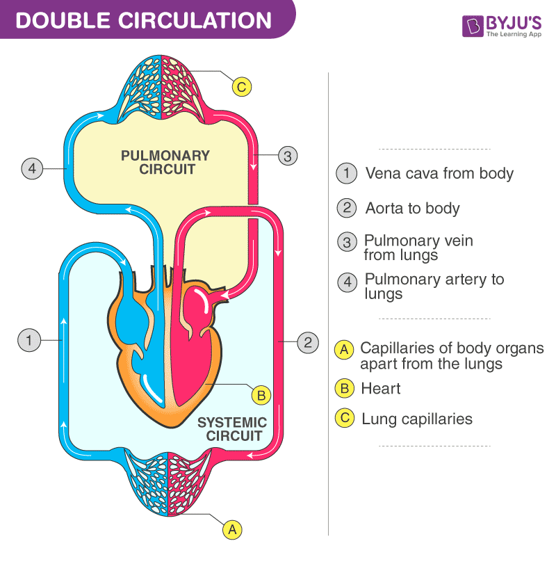
Draw a diagram to illustrate the double circulation system in mammals.
B3.2.14 (AHL): Differences between the single circulation of bony fish and the double circulation of mammals.
The mammalian double circulation pattern includes pulmonary circulation (to pump blood to capillaries for reoxygenation) and the systemic circulation (blood returns to be pumped out to capillaries in body tissues to supply oxygen.
Draw a diagram to illustrate the single circulation system in fish.
B3.2.14 (AHL): Differences between the single circulation of bony fish and the double circulation of mammals.
Fish have a single circulation system. They only have a 2-chambered heart. One chamber receives blood, and the other pumps blood to be sent to the gills. From the gills, deoxygenated blood is sent directly to capillary beds in body tissues.

Explain why the mammalian heart must function as a double pump.
B3.2.14 (AHL): Differences between the single circulation of bony fish and the double circulation of mammals.
The mammalian heart must function as a double pump to restore blood pressure. In a single circulation pattern, blood pressure is lost. However, the mammalian heart has four chambers. One side can be used for pulmonary circulation to pump blood to lung capillaries for reoxygenation. The other side is where the blood returns to be pumped out to capillaries in body tissues to supply oxygen (systemic circulation). This maintains blood pressure.
Label a diagram of the heart with the following structure names: superior vena cava, inferior vena cava, pulmonary semilunar valve, aorta, pulmonary artery, pulmonary veins, aortic semilunar valve, left atrioventricular valve, left ventricle, septum, right ventricle, left atrium, right atrium, septum and right atrioventricular valve.
B3.2.15 (AHL): Adaptations of the mammalian heart for delivering pressurized blood to the arteries.
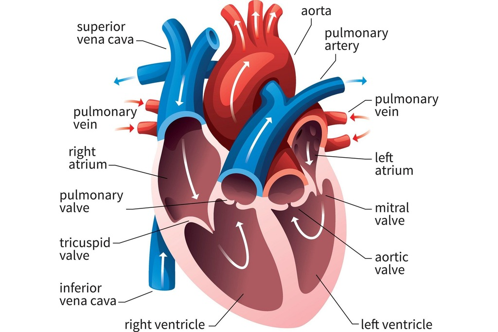

Outline how the following structures allow the heart to function in delivering pressurized blood to arteries: cardiac muscle, pacemaker, atria, ventricles, atrioventricular and semilunar valves, septum and coronary vessels.
B3.2.15 (AHL): Adaptations of the mammalian heart for delivering pressurized blood to the arteries.
Cardiac muscle allows the heart to contract to create high pressure. The cardiac muscle for the left ventricle is much thicker than the right ventricle because high pressure is required to move blood around the body.
The pacemaker (sinoatrial node) initiates and controls when the ventricles should contract to control heart beat.
The atria receive blood from the body and the lungs.
The ventricles contain a lot of cardiac muscle to be able to pump blood to the lungs and the body.
Atrioventricular valves prevent the backflow of blood from the ventricles to the atria.
Semilunar valves prevent the backflow of blood from the arteries to the ventricles.
The septum prevents oxygenated and deoxygenated blood from mixing.
The coronary vessels (arteries and veins) send and receive blood from the heart itself so the heart can function.

Define myogenic contraction.
B3.2.16 (AHL): Stages in the cardiac cycle.
Myogenic contraction is the contraction of a blood vessel that occurs when intravascular pressure is elevated. It contracts independently of neural stimulation.
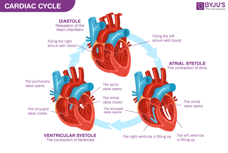
Define cardiac cycle.
B3.2.16 (AHL): Stages in the cardiac cycle.
The cardiac cycle is a series of events (pressure changes) in the heart commonly referred to as one heartbeat.
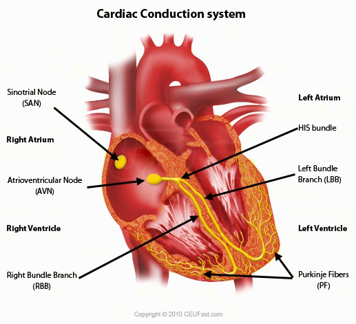
Outline the role of the pacemaker cells in the sinoatrial node.
B3.2.16 (AHL): Stages in the cardiac cycle.
The pacemaker cells in the sinoatrial node initiate action potentials (electrical signals) of the heart. This passes through cardiac muscles and causes contraction.
Describe the propagation of the electrical signal from the sinoatrial node through the atria and ventricles.
B3.2.16 (AHL): Stages in the cardiac cycle.
The sinoatrial node controls the heart beat. It signals the right and left atria at the same time through nerves that signal their contraction.
Then, the signal passes through a layer of fibrous tissue between the sinoatrial node and atrioventricular node to prevent the action potential from travelling directly to the ventricles. This pause prevents all 4 chambers from contracting at the same time.
After passing through, the atrioventricular node detects the signal from the sinoatrial node then initiates its own action potential. This spreads across the ventricles, causing ventricular systole (contraction).
The action potential then travels down the Purkinje fibers to the apex of the heart. The action potential travels up the walls of the ventricles, which initial ventricular systole from the apex. This pumps all of the blood out of the ventricles.
Explain the flow of blood during atrial and ventricular systole and diastole.
B3.2.16 (AHL): Stages in the cardiac cycle.
During diastole, the right atrium and left atrium fill with blood because there is low pressure.
An action potential from the sinoatrial node causes the atria to enter systole. As the atria contract, pressure increases, which forces all the blood into the ventricles.
An action potential from the atrioventricular node causes the ventricles to enter systole. Therefore, the ventricles contract, which increases the pressure in the ventricles. As a result, the atrioventricular valves close (because pressure is higher in the ventricles than in the atria) The high blood pressure in the ventricles increase until the semilunar valve open, and blood moves into the aorta OR until the pulmonary valve opens and blood moves through it.
After, the ventricles enter diastole and pressure in the ventricles decrease. The pressure in the aorta becomes greater than in the ventricles, so the semilunar valve closes.
When the pressure in the ventricle is less than the pressure in the atria, the atrioventricular valves open.

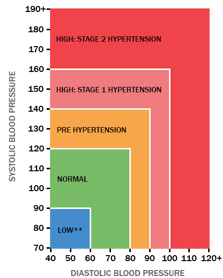
Define systolic and diastolic blood pressure.
B3.2.16 (AHL): Stages in the cardiac cycle.
Systolic blood pressure is the maximum blood pressure during ventricular systole (contraction).
Diastolic pressure is the minimum blood pressure recorded prior to the next contraction, between ventricular contractions.
State the cause of systolic and diastolic blood pressure.
B3.2.16 (AHL): Stages in the cardiac cycle.
Systolic blood pressure is caused by ventricular systole, or ventricular contractions.
Diastolic blood pressure is caused by ventricular diastole, or ventricular relaxation.
Interpret systolic and diastolic blood pressure measurements from data and graphs.
B3.2.16 (AHL): Stages in the cardiac cycle.
Elevated blood pressure is when readings consistently range from 120-129 systolic and less than 80 mm Hg diastolic.
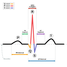
Define what an electrocardiogram is.
An electrocardiogram (ECG/EKG) is a test that records the electrical activity of the heart.
It is conducted by placing skin electrodes on different parts of the heart to test for irregularities in how the heart functions.
Define depolarization.
Depolarization is when a cell undergoes a shift in electric charge distribution. Positive ions rush into the cell, so there is less negative charge inside the cell. The cell’s membrane potential is the difference in charge.
Describe what each part of an EKG/ECG represents.
The P wave represents the depolarization of the atria.
The QRS complex represents the depolarization of the ventricles. It is much larger than the P wave because the atria are smaller than the ventricles, so there are fewer cells to depolarize in the atria.
The T wave represents the repolarization of the ventricles.
The U wave is not always seen, but it may be the repolarization of Purkinje fibers (in the ventricles of the heart).
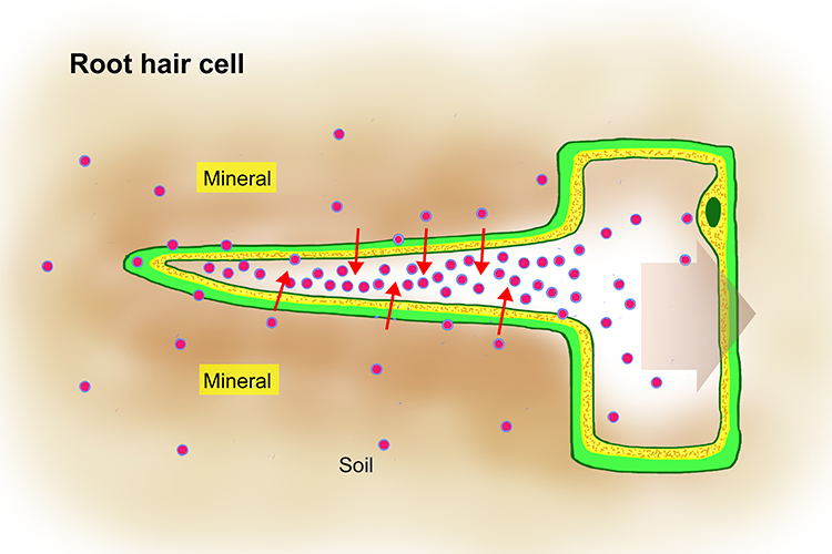
State that the transport of minerals is an active process.
B3.2.17 (AHL): Generation of root pressure in xylem vessels by active transport of mineral ions.
The transport of minerals from outside to inside of root hair cells is an active process.
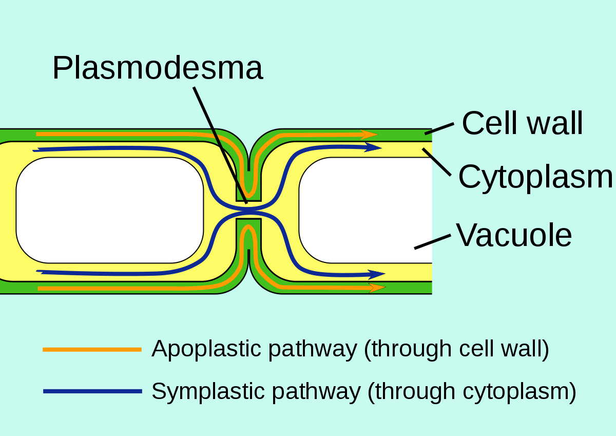
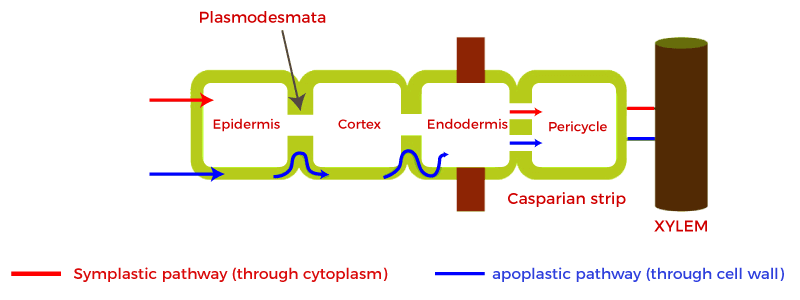
Compare symplastic and apoplastic pathways.
B3.2.17 (AHL): Generation of root pressure in xylem vessels by active transport of mineral ions.
Both pathways move water from the roots hairs to the xylem.
The symplastic pathway moves water through the cytoplasm of adjacent cells. The apoplastic pathway moves water through the cell walls of adjacent cells.
The symplastic pathway uses osmosis, while the apoplastic pathway uses capillary action.
The symplastic pathway is slower than the apoplastic pathway because there are things in the cytoplasm (organelles) that slow it down.
List conditions in which a plant may generate root pressure to transport water.
B3.2.17 (AHL): Generation of root pressure in xylem vessels by active transport of mineral ions.
Active transport of minerals
Outline the mechanism by which roots maintain a positive pressure potential when evaporation from leaves is insufficient to move water through a plant.
B3.2.17 (AHL): Generation of root pressure in xylem vessels by active transport of mineral ions.
Active transport brings minerals to the xylem. Roots maintain a positive pressure as water moves into the xylem by osmosis. This creates a positive pressure which moves water up the xylem. The root pressure allows water to move up the xylem of plants, even when transpiration rates are low.
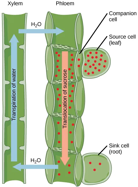
Define translocation, phloem sap, source and sink.
B3.2.18 (AHL): Adaptations of phloem sieve tubes and companion cells for translocation of sap.
Translocation is the movement of nutrients (like sucrose and amino acids) up or down the stem of a plant through phloem and tissue from source to sink.
Phloem sap is nutrient-rich fluid that flows in the phloem.
The source is any part of the plant that synthesizes the nutrients (photosynthesis).
The sink is the part of the plant that uses or stores the nutrients.
List example source and sink tissues.
B3.2.18 (AHL): Adaptations of phloem sieve tubes and companion cells for translocation of sap.
Source tissues
Leaves
Sink tissues
Roots
Fruit
Buds
Seeds
Flowers
State that phloem transport is bidirectional.
B3.2.18 (AHL): Adaptations of phloem sieve tubes and companion cells for translocation of sap.
Phloem transport is bidirectional (moves up AND down the phloem).
Outline the stages of phloem translocation including loading of carbohydrates at a source, transport of carbohydrates through the plant, and unloading of carbohydrates at a sink.
B3.2.18 (AHL): Adaptations of phloem sieve tubes and companion cells for translocation of sap.
First, sucrose (carbohydrates) are produced by the source.
Then, the sucrose is actively transported from the companion cells into the companion cells, and from the companion cells into the sieve tube elements.
There is high sucrose concentration in the phloem sieve tubes (phloem loading). As a result, water moves into the phloem sieve tubes from the xylem by osmosis.
Since water is incompressible, hydrostatic pressure builds up in the sieve tubes.
The increasing hydrostatic pressure moves water and nutrients from source to sink.
Sucrose is actively transported into the sink, where it is used either for respiration or stored as a starch.

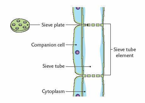
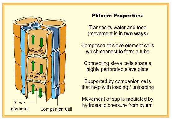
Outline the structure and function of sieve tube elements, with specific mention of the rigid cell wall, reduced cytoplasm and organelles, no nucleus and sieve plates.
B3.2.18 (AHL): Adaptations of phloem sieve tubes and companion cells for translocation of sap.
Sieve tube elements move dissolved sugars from source to sink.
Sieve tube elements have thick and rigid cell walls to withstand hydrostatic pressures (which facilitate flow).
Sieve tube elements have reduced cytoplasm and no nucleus in the cell, which allows the movement of cell sap.
Sieve tube element cells have plasma membranes with protein pumps to facilitate active transport.
Sieve plates are pores between cell walls that allow cell sap containing nutrients to flow from cell to cell
The plasmodesmata allows direct connections between the cytoplasms of the companion cells and sieve tube.
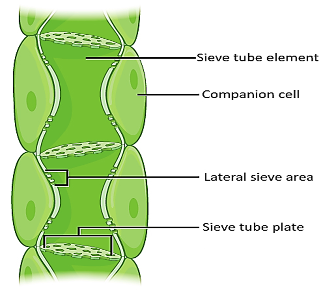
Outline the structure and function of companion cells, with specific mention of mitochondria and plasmodesmata.
B3.2.18 (AHL): Adaptations of phloem sieve tubes and companion cells for translocation of sap.
Companion cells provide metabolic support for sieve tube element cells.
Companion cells contain large numbers of mitochondria to provide sufficient ATP for active transport (loading) of nutrients into the phloem tissue.
The cytoplasm of the companion cells and sieve tube cells are connected via the plasmodesmata.
Companion cells contain transport proteins for loading nutrients into sieve tubes.