Cells- Anatomy
 Call Kai
 Learn
Learn Practice Test
 Spaced Repetition
 Match
Match Flashcards
 Knowt Play
1/74

Earn XP
Description and Tags
Slide Set 3
Name | Mastery | Learn | Test | Matching | Spaced | Call with Kai | Chat |
|---|
No analytics yet
Send a link to your students to track their progress
75 Terms
Cytology
The study of cells
Sex cells
germ cells or reproductive cells
(sperm + oocyte)
Somatic cells
Everything else
if it is not a sperm or egg cell
Plasma Membrane
acts as a barrier to separate what inside and what outside the cell
Phospholipid bilayer
containing proteins, cholesterol, and glycolipids
separates intracellular fluid (ICF) from extracellularr fluid (ECF)
Interstitial fluid (IF) is the ECF that directly surround most sells
Ion distribution differs across the membrane
ECF → high Na + and low K+
ICF → low Na + and high K+
The membrane its selectively permeable
regulates which substances enter and leave the cell
Membrane Lipids
Phospholipids (around 75%)
Glycolipids ( around 5%)
Cholesterol (around 20%)
Phospholipids (75%)
Form the basic lipid bilayer
Hydrophilic heads face the fluid
Hydrophobic tails face inward
Glycolipids (5%)
Glyco= think of carbs
Lipids with carbohydrate chains extending from extracellular surface
Important for cell recognition
Example: some blood group antigens on RBCs (A, B, and O)
Cholesterol (20%)
Located between phospholipids
Helps stabilize the membrane by regulating fluidity
Limits excessive phospholipid movement at higher temperatures
Prevents phospholipids from packing too tightly at lower temperatures
Cholesterol is hydrophobic, will mined itself in the hydrophobic enviorment
Functions of Membrane Proteins - Transport proteins
Move substances across the plasma membrane
Examples: ion channels, sodium- potassium pump
Receptors
bind chemical signals and trigger a cellular response
Example: acetylcholine receptors on skeletal muscle cells
Attachment proteins
anchor cells to the cytoskeleton or extracellular structures
Example: hemidesmosomes attach epithelieal cells to the basement membrane
Enzymes
Speed up chemical reactions at the membrane
example: digestive enzymes on intestinal epithelial cells
Intercellular joining
connect neighboring cells
Example: tight junctions, desmosomes, gap junctions, other adhesion molecules
Recognition proteins
help cells identify and distinguish one another
Example: MHC (Major histocompatibility complex) help immune cells recognize the body’s own cells, glycoproteins of red blood cells
Integral proteins
embedded within the phospholipid bilayer
Many extend completely across the membrane and are called transmembrane proteins
Hydrophobic regions interact with phospholipid tails
Hydrophilic regions are exposed to the ICF and / or ECF
Examples
ion channels
transporters
Receptors
some enzymes
Peripheral proteins
These do not stand the whole membrane
loosely attached to the inner or outer surface of the membrane
Do no extend into the hydrophobic interior of the bilayer
Often attach to integral proteins or phospholipid heads
Examples
Cytoskeletal proteins
Some enzymes
Six functions of membrane Proteins
Transport
Receptors for signal transduction
Attachment to cytoskeleton and basement membrane (basal lamina)
Enzymatic activity
Intercellular joining
Cell-cell recognition
Transport
Channel/pore
a protein that spans the membrane may provide a hydrophilic channel across the membrane that is selective for a particular solute ( left )
Some transport proteins hydrolyze ATP as an energy source to actively pump substances across the membrane (right)
In general:
allows rings to by pass the membrane that can’t do but on their own
Receptors for signal transduction
A membrane protein exposed to the outside of the cell may have a binding site that firs the shape of a specific chemical messenger, such as a hormone
When bound, the chemical messenger may cause a change in shape in the protein that initiates a chain of chemical reactions in the cell
In general:
when a receptor of activated, some type of signal (ligand) transduction will occur immediately
Ligand will only respond to respective receptor
Attachment to cytoskeleton and basement membrane (basal lamina)
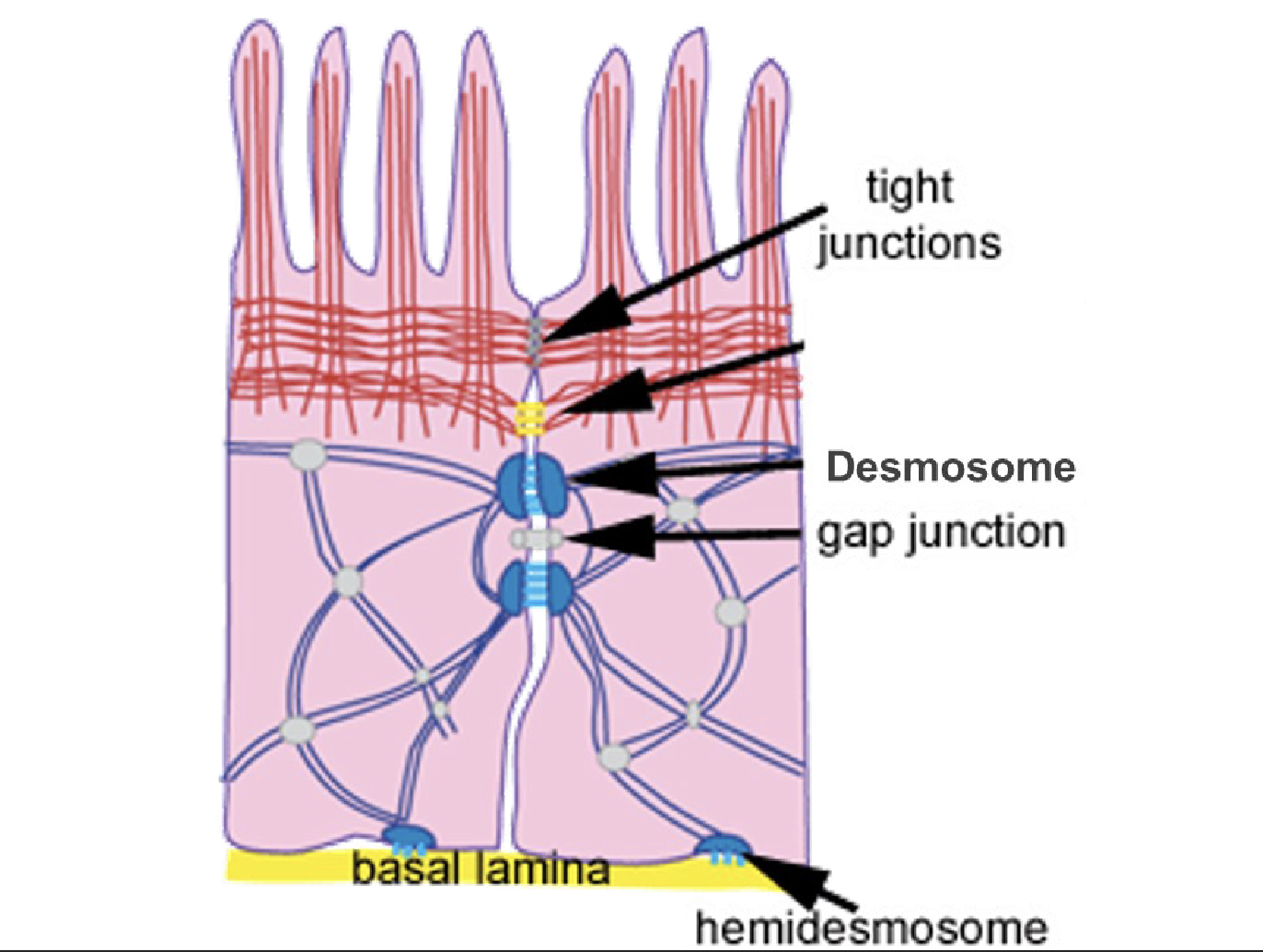
Hemidesmosome- desmosome that holds on to the basement membrane
Enzymatic activty
A membrane protein may be an enzyme with its active site exposed to substances in the adjacent solution
A team of several enzymes in a membrane may catalyze sequential steps of a metabolic pathway as indicated (left to right) here
Intercellular joining
attaches different types if cells interacting with each other
White blood cells (leukocytes) joining together to fight off infection
Intercellular joining part 2
Allow adjacent cells to attach
Common in epithelial cells
Limit fluids and most molecules from moving between cells
Tight junctions
Desmosomes
Tight Junctions
seal to prevent passage between cells (less strong)
Urinary bladder
like glue, easy to rip apart
Desmosomes
anchor for mechanical strength
Skin
keeps strong connection
Hemidesmosome- type of desmosome
Gap Junction
directly connects the cytoplasm of two neighboring animal cells, allowing ions, water, and small molecules to pass freely between them.
Cell-cell recognition
some glycoproteins (proteins bonded to short chains of sugars) serve as identification tags that are specifically recognized by other cells
Blood types are a identification type fir this
Example
- allow the immune system to recognize our cells as our own
Moving in and out of the cell (part 1)
Passive Transport (no energy)
No cellular energy (ATP) is required
Substances move down their concentration gradient from HIGH to LOW concentration
There are three types of passive transport
Simple diffusion
Facilitated diffusion
Osmosis
Simple
Substances move directly through the phospholipid bilayer
HIGH → LOW
primarily small, non polar or lipid soluble substances
Examples
O2
CO2
fat-soluble molecules
Faciliated
HIGH → LOW (down the gradient)
Substances cross the membrane using channel or carrier proteins
Channels transport smaller molecules, ions
Carriers transport substances such as glucose and amino acids
charged hydrophilic → they can’t just walk through the membrane
Osmosis
Diffusion of water across a selectively permeable membrane
Carrier- Mediated Facilitated Diffusion
uses transmembrane carrier proteins
Often transports larger polar molecules, such as glucose and amino acids
Substrate binds to carrier → carrier changes shape → substate is related on the other side
Molecules move DOWN their concentration gradient → ni cellular energy required
Passive
Transport rate is limited by the number of available carrier
Carrier become saturated when all binding sites are occupied
Key distinction:
Channels → substances pass through a pore
Carrier → substances bind to the protein, which changes shape to transport them
Passive Processes: Osmosis
Goes to the place with a higher solute concentration
net diffusion of water across a selectively permeable membrane
Membrane is permeable to water, but mostly impermeable to solutes
Water moves from an area of low solute concentration → higher solute concentration
Equivalently: from higher water concentration → lower water concentration
Occurs when there is a difference in solute concentration across the membrane
Passive process
Horseshoe test tube (osmosis)

Osmolality
Only number of solutes matter not size
Osmolality = total concentration of dissolved particles
Measured in mOsm/kg of water
Body fluids are about 285– 295 mOsm/kg
Preferred over osmolarity because it is based on mass, not volume
Tonicity
effect of a solution on cell volume
depends mainly on the concentration of non penetrating solutes
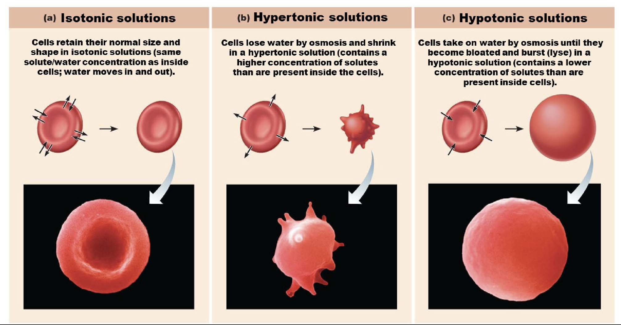
Tonicity of Solutions (isotonic)
Isotonic
No net water movement
Cell maintain normal volume
Example: 0.9 % NaCl
Hypertonic
Water moves out of the cell
Cell shrinks (crenation)
Example: 3% NaCl
Hypotonic
Water moves into the cell
Cell swells and may rupture'
RBC rupture = hemolysis
Example: 0.45% NaCl
Effects of Tonicity on Cell Volume
Osmosis causes cells to swell and shrink
Change in cells volume disrupts cell function, especially in Red Blood Cells and neurons
Aquaporins
Primary active transport
Moves substances against their concentration gradient
From low → high concentration
Requires energy
ATP directly provides the energy
Uses membrane pumps (carrier proteins)
Example
Na+/K+ pump
Pumps 3 Na + out of the cell and 2 K+ into the cell
Secondary Active transport
Moves a substances against its concentration gradient
Requires energy, but does not use ATP directly
Uses energy stored in the concentration gradient of another substance
One substance moves down its gradient
Prodives energy to move another substance against its gradient
The Na+ gradient is maintained by the Na+/K+ pump, which uses ATP
Uses cotransporter carrier proteins
Example: Na+ —Glucose Symporter
Na+ moves down its gradient into the cell
Glucose moves against its gradient into the cell at the same time
Endocytosis
Brings materials into the cell using vesicles
used to take in nutrients, fluids, or large particles
Phagocytosis
Cell eating
Takes in large particles such as bacteria or debris
Pinocytosis
Cell drinking
Takes in extracellular fluid and dissolved substances
Exocytosis
Moves material out of the cell using vesicles
Used to release substances such as hormones, enzymes, and neurotransmitters
Can also remove cellular waste products
Resting Membrane Potential
Voltage difference across the plasma membrane of a resting cell
Inside of the cell is negative relative to the outside
Na+ is high outside e, while K+ is high inside
At rest, the membrane is more permeable to K+, so K+ leaks out through leak channels
The Na+/K+ pump maintains these ion gradients by moving 3 Na+ out and 2 K+ in
Typical neuron rating membrane potential
-70 mV
Nucleus
Organelle that serves as the control center of the cell
Contains most of the cell’s genetic material (DNA)
directs many cellular activities bye controlling gene expression
Most cells contain one nucleus
Some cells are multinucleate
Example- skeletal muscle fibers
Some cells are anucleate and lack a nucleus
Example- mature red blood cells (RBCs)
Mitochondria
Double membrane organelles with inner membrane folds called cristae
Produce mosts of the cells ATP through aerobic cellular respiration
Requires oxygen
Uses energy from nutrients such as carbohydrates and fats
Contain their own DNA and ribosomes
Cytoskeleton
Network of protein fibers that provides structural support and helps maintain cell shape
Mircofilaments
Thin fibers composed mainly of actin
Cell contraction and movement
Support microvilli
Intermediate filaments
Provide mechanical strength and help stabilize cell structures
Help cells resist stretching and pulling
Microtubles
Hollow tubes that help organize and move structures within the cell
Acts as “tracks” for movement of organelles and vesicles
Important for cell division
Form the structural core of cilia and flagella
Microvilli
Small finger like extensions of plasma membrane
increase surface area for absorption
small intestine
Core of actin filaments for stiffening
Cilia
Cell surface projections supported by mircotubules and covered by plasma membrane
Primary cilia
Usually a single, nonmotile projection
Acts as a sensory structure that detects signals in a cells enviorment
Motile cilia
Less common
Beat in coordinated waves to move material across the cell surface
Respiratory tract
move mucus and trapped particles out of the air ways
Female reproductive tract
help move the oocyte through the uterine tube
Male reproductive tract
help move fluid and serum through parts of the reproductive tract
Brain ventricles
help circulate cerebrospinal fluid (CSF)
Flagella
also made off mircotubles
Whip-like extension of the plasma membrane
longer and beat run a wave-like fusion
Allow the cell to move
Only one human cell-type has this
SPERM
Ribosomes
Build proteins by lining amino acids together
Produce the primary structure of a protein
Only starts making the protein , the protein is not functioning yet
Rough ER
Studded with ribosomes
Synthesizes, folds, and modifies proteins
Especially important or proteins that will be secreted or inserted into cell membranes
Smooth ER
Synthesizes lipid, including steroid hormones
Helps detoxify drugs and other chemicals
Stores Calcium (Ca2+) in certain cells , especially muscle cells , bone
Golgi apparatus
Modifies , sorts, and packages proteins and lipis
Packages them into vesicles for delivery to their correct destinations
Acts like the cells “Post office”
Lysosomes
Contains digestive enzymes
breaks down
damaged organelles
Cellular debris
material borough in other the cell
helps recycle cellular components
Peroxisomes
Break down very long-chain fatty acids
oxidation reactions produce hydrogen peroxide (H2O2)
Contains catalase, which converts H2O2 into
Water and oxygen
helps protect the cell from oxidative damage
Damaging DNA
Apoptosis
programmed cell death that removes unneeded or damaged cells
Activates enzymes called caspases
These break down DNA and other cell components
Cells shrink and fragments → remnants are removed by phagocytosis (eating away at the cell)
Cell Division in Adults
Replaces short-lived cells and helps repair damaged tissues
Examples -
blood cells
epidermal cells
intestinal lining
hair follicles
Changes in Cell and Tissue Size
Hyperplasia
Hypertrophy
Atrophy
Hyperplasia
increase in the number of cells
Examples: endometrial growth during the menstrual cycle
Hypertrophy
increase in cell, tissue, muscle size
Example: skeletal muscle growth with exercise
Atrophy
decrease in the size of a tissue or organ
Usually involves decreased cell size and sometimes loss of cells
Example: muscle atrophy from disease or being bed-ridden
Neoplasm
abnormal, uncontrolled cell proliferation
May be benign or malignant
Cell Differentiation - intro
Most nucleated body cells contain essentially the same DNA, but they are not identical
Different cell types activate different genes
This causes cells to produce different proteins and develop specialized structures and functions
Cell Differentiation
Process by which cells develop specialized characteristics and functions
Stem cells
relatively undifferentiated cells that can divide and develop into specialized cell types