Phonatory System
1/138
There's no tags or description
Looks like no tags are added yet.
Name | Mastery | Learn | Test | Matching | Spaced |
|---|
No study sessions yet.
139 Terms
Larynx is made up of...
Bones, folds, muscles, joints, cartilages
Location of larynx
Deep in tissues of the neck
Biological function of larynx
To act as a valve to prevent air from escaping the lungs. Prevents foreign objects from entering the lungs, trachea, and glottis. While swallowing, epiglottis covers the opening.
Non-biological function of larynx
Larynx is in charge of producing sounds/phonation
Laryngeal skeleton
Made up of 1 bone and 9 cartilages. 3 unpaired/3 paired. Cartilage framework of larynx is suspended from hyoid bone
Characteristics of larynx
Larynx is flexible which allows for change in pitch/volume.
Phonatory system
Allows for vibration of vocal chords to produce sound
Hyoid bone
Top of larynx. Not a part of larynx/suspended from larynx. U-shaped/not attached to other bones - "floating". Greater cornu in back joins w/superior cornu of thyroid cartilage/lesser cornu extends from body/capped by cone-shaped cartilage. Thyrohyoid membrane is attached to inferior aspect of hyoid bone.
Thyrohyoid membrane
Larynx (thyroid) is suspended from hyoid bone by thyrohyoid membrane. Thyrohyoid membrane is attached to inferior aspect of hyoid bone. Allows entrance of superior laryngeal vessels/internal branch of superior laryngeal nerve.
Cartilage
A firm, elastic, flexible fibrous type of connective tissue.
What does each cartilage consist of?
1) Thyroid - Hyaline 2) Cricoid - Hyaline 3) Epiglottis - Elastic
4) Arytenoids - Hyaline 5) Corniculates - Elastic 6) Cuneiforms - Elastic
Hyaline cartilage ossifies over time
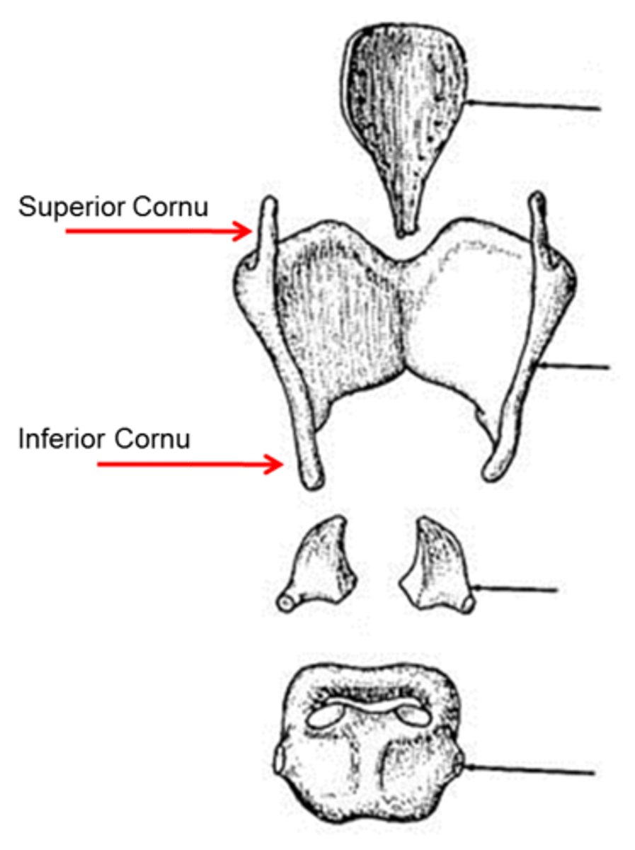
Thyroid
Rocks anteriorly/posteriorly. Articulates w/cricoid. Largest cartilage of larynx. Forms front/sides of laryngeal skeleton. Has housing for larynx/offers protection for many of its structures. Thyroid is directly attached to the thyrohyoid membrane and to the hyoid bone by 3 thyrohyoid ligaments. Forms the lateral and anterior walls of larynx by 2 plates (or laminae) fused at midline.
Thyroid notch
is where the laminae are fused at an angle. V-shaped depression.
Superior cornu of thyroid
project upward and attach to the hyoid bone by ligaments.
Inferior cornu of thyroid
project downward and join with the sides of the cricoid cartilage.
What do the facets provide?
formation of joints w/cricoid on lower inside surfaces which have facets that join
Cricoid
Narrow in the front/larger in the back. Inferior to thyroid and superior to trachea. Complete ring. Most inferior portion of larynx
Base of larynx. Attachment - trachea (inferiorly). Location - C6-C7 level in adults
Epiglottis
Broad, leaf-shaped structure. Superior end of larynx. Behind hyoid bone/tongue. During swallowing it folds downward over the entrance of larynx to prevent food from entering the trachea/direct food to esophagus. Sits posteriorly on cricoid
What does the epiglottis attach to?
Thyroid cartilage just below thyroid notch by thyroepiglottic ligament, body of hyoid bone by hypo epiglottic ligament, root of tongue
Arytenoids
Located on superior/posterior surface of cricoid. Vocal folds are attached to the vocal process. Many laryngeal muscles attach to the muscular process/move arytenoids
Cuneiform cartilages
Small rods that aid in stiffening the aryepiglottic folds. Sits anteriorly to corniculate
Corniculate cartilages
Part of arytenoids. Posterior/medial to cuneiforms. Work in conjunction w/cuneiforms to create supraglottic constriction
Cricoarytenoid joints
Allows the arytenoids to move medially and laterally and anteriorly and posteriorly. The arytenoid movement moves the vocal folds because the folds are attached to the Vocal Process. One of the main contributors to abducting and adducting the vocal folds.
Cricothyroid joints
Allows the thyroid to primarily tilt down.
One of the main contributors to changing pitch of the voice. When the cricoid and thyroid move toward each other in front, the distance between arytenoids and thyroid increases. Vocal ligaments will be stretched and tenses the VF. When this contracts, pulls thyroid forward/downwards
Articulations of cricothyroid joint
Articulations of laryngeal cartilages. Inferior horn of thyroid articulates w/facet on lateral aspect of cricoid.
Aryepiglottic folds
The most superior of the folds. Lateral and superior border of the epiglottis to the superior aspects of the arytenoid cartilage. Are contracted to pull the epiglottis backward and close the entrance of the larynx when swallowing.
False Vocal Folds
Inferior to the Aryepiglottic folds
Slightly superior and lateral to the Vocal Folds. Close during swallowing and effortful activities. Remain open during phonation.
True Vocal Folds
The Vocal Folds vibrate to create phonation.
At rest, they are open posteriorly and attach to the Vocal Process of the Arytenoids.
Anteriorly, they attach to the Thyroid just below the thyroid notch at the anterior commissure
The medial boarders of the VF are free to vibrate.
Layers of Vocal Folds
Epithelium
Superficial
Intermediate
Deep
Vocalis
Intermediate/ deep make up vocal ligament. Superficial, intermediate, deep make up lamina propria
Epithelium
thin membrane layer
Vocalis
Only muscle of Vocal Folds. VF is innervated/this nerve is innervating the VF via the vocalis
Change of VF composition
As VF goes from medial to lateral, composition changes. Most lateral: comprised of muscle, in that muscle it's not going to move as easily (not as compliant). More superficial: outermost portion of VF is going to be more combined, being able to move because not comprised of muscle, but elastic cartilage/colagen controls mucus in larynx.
Glottis
Space between VF/ at level of VF. Some of the space is between cartilage of arytenoids in posterior aspect of the space
Membranous glottis
The space that isn't bounded by cartilage. Makes up the greater portion of glottis 3/5. Most active during phonation. Vibrates more freely during phonation.
Cartilaginous glottis
Posterior, making up 2/5. For some, it may not completely close. May be a small opening in this glottis. Able to sustain good quality vowels.
When glottis is open during phonation...
it's going to expand even more when we're taking in air. During forced inhalation, it may open up beyond a 1/3 of an inch. During normal breathing, may be 1/3 of an inch. During forced inspiration, it may double to take in more air
Sub glottal cavity
Sub glottal pressure contributes to this. Underneath VF. Superior portion of sub glottal cavity would be at level of glottis. Sub glottal cavity would go past cricoid cartilage down to first tracheal ring. Most inferior.
Laryngeal ventricles
Above VF. Inferior to false VF. Within ventricles, there are glands that produce secretions which help keep VF moist. The moisture/hydration is going to keep VF sounding better (quality of one's voice is going to be dependent on that/ VF that don't have that are more prone to injury.
Example of moisture of ventricles
Taking meds because not feeling well> meds dry out sinuses/dry out larynx. That person may produce less secretions in their larynx. VF more prone to injury if talking a lot while taking meds.
Supra glottal cavity
most superior part of these cavities. This cavity is going to go down to level of glottis. Contributes to resonance. Cavity will extend up to tip of epiglottis/ around aryepiglottic folds.
True or false. Cricothyroid joints are located on the lateral aspect of the cricoid
True
True or false. VF are attached to vocal process of the arytenoids
True
Extrinsic muscles
One end of the muscle is attached to the hyoid bone or a laryngeal cartilage. The other end is attached to a structure outside the larynx (sternum, cranium, scapula).
Intrinsic muscles
Both ends (the origin and insertion) of the muscle are within the larynx. Inside larynx. Muscles help larynx move up/down. When you swallow, you can feel your larynx rise.
Cricoarytenoid name
Arytenoids move a lot. Cricoid is fixed and doesn't move that much.
Extrinsic muscles - suprahyoids
Above hyoid. Digastric, stylohyoid, mylohyoid, geniohyoid
Digastric
Has 2 bellies. Anterior/posterior belly. Elevates larynx. Intermediate tendon loops through anterior belly contracts/movies it a little more anterior.
Anterior belly of digastric
has an attachment to the inner surface of the mandible near the chin.
Posterior belly of digastric
Has an attachment to the mastoid process of temporal bone. Courses posteriorly. Posterior attachment of posterior belly is attached to mastoid process.
Attachment of stylohyoid
Posterior attachment/interior insertion of lateral aspects of corpus of hyoid.
Role of stylohyoid during swallowing
During swallowing, it pulls posterior/superior.
Mylohyoid
Deep to digastric. Fan-shaped muscle.. On top is digastric. Some muscle fibers run in midline
Stylohyoid
Originates from styloprocess of temporal bone. Mastoid, part of temporal bone. Styloid process, part of temporal bone. Thin projection comes off of temporal bone.
Attachment of mylohyoid
Attaches to inner surface of mandible. Courses down attaching to hyoid bone (corpus of hyoid)
Geniohyoid
The loop. Deep to mylohyoid. Medial hyoid is more deep to mylohyoid. Outside anterior belly, mylohyoid, genoihyoid (more deep).
Attachment of geniohyoid
Attaches to inner surface of chin similar to anterior belly. Attachment to medial aspect of corpus of hyoid.
How does supra hyoids relate to voice disorders
In those w/voice disorders where they have muscle tension, they carry a lot of muscle tension in supra hyoids (so much that it elevates larynx). Muscles tension can lead to dysphonia which is environmental
Extrinsic- infra hyoid muscles
Sternohyoid, omohyoid, sternothyroid, thyrohyoid
Thyrohyoids: infra hyoid
Smaller of the infra hyoids. Large muscle compared to intrinsics connecting to thyroid cartilage, anterior commissure, thyroid notch.
Attachment of thyrohyoid
On lamine, oblique line of thyroid. Oblique line extends from body of superior horn down to bottom of lamina of thyroid. the attachment of thyrohyoid running from oblique line of thyroid to greater cornu of hyoid there's a lateral aspect of hyoid bone.
What does the oblique line mark an attachment to?
Thyrohyoid, sternothyroid, and inferior constrictor muscle.
Sternothyroid
Where another muscle leaves off or stops another muscle picks up. Top part of sternum is where sternothyroid originates from
Attachment of sternothyroid
On oblique line this muscle appears which has an attachment to thyroid. Courses down to menubreum of sternum.
Sternohyoid
originating from menubreum of sternum/ inserting in corpus of hyoid. Lies superficial to sternothyroid.
Attachment of omohyoid
Attachment to scapula/side of corpus of hyoid.
Omohyoid
2 bellies: superior/inferior. Projects posteriorly to scapula. Bellies connected by tendon between bellies. Superior belly ascends from tendon/inserts on hyoid. Depresses/fixes hyoid
Where does the omohyoid originate from
Inferior belly originates on superior belly of scapula
How is the hyoid kept stable
Thyrohyoid keeps hyoid stable. Supra hyoid contracts. When hyoid is stable, thyroid lifts. Thyrohyoid contracts/there's a backwards attachment to oblique line which is lateral
Deep to superficial infra hyoid muscles
Geniohyoid, mylohyoid, anterior belly of omohyoid/ sternum (bilateral)
Names of Intrinsic muscles
Cricoarytenoid, Interarytenoid, cricothyroid, thyroarytenoid. Almost all are paired
Cricoarytenoid
Has lateral/posterior muscles. Separate muscles/share name because of attachment
Interarytenoid types
Transverse/oblique muscle.
Thyrohyoid ligament
where membranes come together more/is thicker. Superior horn connects via this membrane to greater horn of hyoid bong via ligament which is this thickening.
Vocal ligament
Lateral Cricoarytenoid
Superior/inferior orientation. Aids in VF adduction so closes VF. Projecting from arytenoid is cartilage/ vocal process of Arytenoid.
Origination of lateral cricoarytenoid
Originates on lateral aspect of cricoid. Exerts on arytenoid in muscular process of cricoid. Lateral aspect of cricoid to muscular process of arytenoid.
What does the lateral cricoarytenoid attach to?
Superior/anterior border of cricoid cartilage. Attachment of muscular process of arytenoids.
What does the apex articulate with?
Corniculate
Attachment of vocal process
vocal ligament
When lateral cricoarytenoid contracts...
It pulls and rocks arytenoids anteriorly/adducts them. It pulls muscular process and brings VF toward midline.
Recurrent laryngeal nerves
Lateral cricoarytenoid is innervated by RLN. It's a branch of vagus nerve which is a mixed nerve important for speech production/voice. Branch of cranial nerve #10 comes out/has many branches and the RLN is 1 of those branches
Anterior commisure
Thyroid notch is a part of anterior commisure. 2 lateral lamina come together. Where they come together anteriorly is anterior commissure.
Posterior cricoarytenoid
On the back. Pulls VC apart. Aids in abduction. When this contracts, it moves arytenoids posteriorly to open VF.
Attachment of posterior cricoarytenoid
Attachment to posterior part of cricoid cartilage. Attachment of muscular process of arytenoids.
Apex
Forms triangle shape.
Innervation of Posterior cricoarytenoid
RLN is a branch of vagus nerve cranial nerve #10. RLN innervates posterior cricoarytenoid.
What if there's damage to RLN cranial nerve #10
This is a wandering nerve/wanders around the body more than the other nerves. Has higher property to suffer injury. There can be difficulties with opening VF/closing VF and may lead to VF paralysis or VF paresis.
Opening of Vocal folds
Vocal folds open at rest
Interarytenoids
between arytenoid cartilages. 1 paired and 1 unpaired. Both contract and bring VF toward midline
Transverse Interarytenoid
Unpaired. Courses between interior aspect of arytenoids. Course from 1 arytenoid to the other/ muscle fibers run laterally of posterior surface of arytenoid. Innervated by RLN. Aid in VF adduction.
Movement of transverse Interarytenoid
Pulls on arytenoids to pull them more medially. If you close off the airway, the transverse/oblique interarytenoids are contracting to close VF. Both aid in adduction
Oblique Interarytenoid
Courses posteriorly from muscular process to arytenoid. 1 arytenoid goes to apex (top). Muscle fibers continue to extend up obliquely/continue to extend up to aryepiglottic folds/form aryepiglottic muscle.
Movement of oblique interarytenoid (VF)
If we're going to fully close airway, these fully approximate/ VF come together. If we're initiating voicing, we're not going to bring them together as much because we want some air to escape through. Depends on needs if we contract more or less.
General movement of oblique interarytenoid
Goes past arytenoids and extend up to aryepiglottic folds.
Cricothyroid
More inferior. Extending from superior border of cricoid exerting in inferior border on lateral aspect of thyroid.
Attachment of cricothyroid
Attachment to cricoid/thyroid. Lateral/superior surface of cricoid on lateral border of cricoid cartilage. Attachment to thyroid cartilage on inferior border/ lateral margin of thyroid extending posteriorly back to inferior horn.
Movement of cricothyroid
When contracts, pulls on thyroid cartilage. Tilt thyroid down to elongate VF thus increases pitch. Muscle pulls on thyroid/rock thyroid anteriorly/inferiorly. When it does this, it stretches thyroarytenoid. When this is stretched, mass of muscle decreases. Frequency of vibration increases contributing to increase in pitch
Innervation of cricothyroid
Not innervated by RLN. Innervated by superior laryngeal nerve which has an internal branch/external branch. External branch innervates cricothyroid.
Thyroarytenoid
Has 2 parts: thyrovocalis and thyromuscularis. Thyromuscularis is more lateral/deep to the thyrovocalis. 5th layer of VF because has vocals. Runs from thyroid cartilage (interior aspect) inserting into vocal process of arytenoid posteriorly. Vocals is most deep. Main body of VF.
Movement of thyroarytenoid
Thyroarytenoid can contract/increase or decrease pitch. It gets stretched out because of cricothyroid/increases pitch. Vocals adds tension to arytenoid/increase pitch