human phys final
 Call Kai
 Learn
Learn Practice Test
 Spaced Repetition
 Match
Match Flashcards
 Knowt Play
1/287

There's no tags or description
Looks like no tags are added yet.
Name | Mastery | Learn | Test | Matching | Spaced | Call with Kai | Chat |
|---|
No analytics yet
Send a link to your students to track their progress
288 Terms
what are the two types of receptors?
membrane-bound receptors: embedded to cell surface; binds to water-soluble ligands
trigger rapid, short-lived responses via signal transduction cascades
e.g., insulin, amino hormones-epinephrine, neurotransmitters-acetylcholine
intracellular receptor: located inside the cell (cytoplasm/nucleus); binds to lipid soluble ligands that can cross the membrane
slow, but long-lasting genomic effects to inhibit/facilitate transcription + gene expression
e.g., steroids-testosterone, estradiol, thyroid hormones, vitamin D
what is the difference between tight junctions and gap junctions?
tight junctions: creates a barrier to prevent leakage between cells
gap junctions: create channels to allow movement of molecules between cells (communication)
What is the functional division of the nervous system?
sensory (afferent) division: carries impulses from receptors to CNS; detects internal & external stimuli
motor (efferent) division: carries impulses from CNS to effectors
somatic nervous system (SNS): control voluntary movements (e.g., skeletal muscles)
autonomic nervous system (ANS): controls involuntary functions (e.g., smooth muscle, cardiac muscle)
sympathetic: “fight or flight”
parasympathetic: “rest and digest”
what are the types of glial cells in the CNS?
oligodendrocytes
microglia
ependymal cell
astrocyte
what are astrocytes?
maintains blood brain barrier
nourishes neurons w/ glycogen
reuptake of neurotransmitters
repair + regenerate neurons
communicate changes in blood
regulation of blood in extracellular brain
maintain ion concentrations (K+)
what is the blood brain barrier?
protective, selective barrier that separates the bloodstream from the brain tissue
structure: Blood → Endothelial cell with tight junctions → Basement membrane → Astrocyte end-feet → Brain tissue
what factors influence ion movement across the membrane?
concentration gradient (diffusion)
ion channels (leak channels, gated channels)
equilibrium potential (influenced by electrical gradient)
What is the resting membrane potential? What contributes to the resting membrane potential?
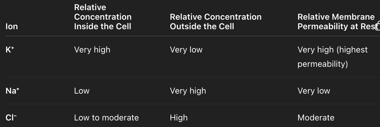
-70mV → excess positive ions outside of membrane
more Na+ and Cl- outside of cell
more K+ inside of cell
Na+/K+ pump: 3 Na+ out, 2K+ in
K+ leaky channel: move K+ out of cell down its gradient
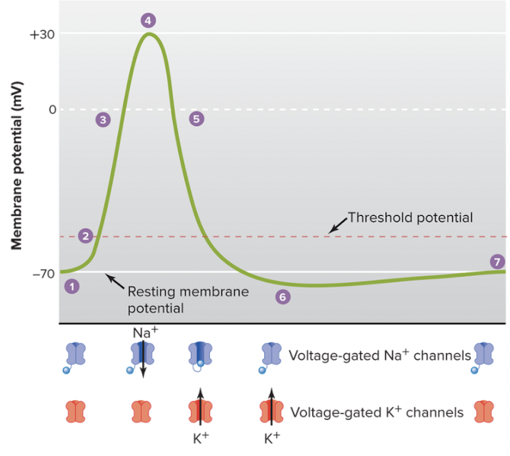
Describe the phases of action potential?
resting membrane potential (-70 mV)
stimulus causes membrane to reach threshold voltage (-50 mV)
depolarization: voltage-gated Na+ channels open causing influx of Na+ into the cell; membrane potential becomes more positive
at +30 mV: inactivation of Na+ channels & delayed opening of voltage-gated K+ channels
repolarization: voltage-gated K+ channel open causing K+ to leave the cell; helps membrane return towards -70mV resting potential
hyperpolarization (-80 mV): delayed closing of K+ channel causes membrane to be below resting membrane potential
voltage-gaated K+ channel closes & resting membrane potential is restored
what is the all-or-none principle?
applies to action potential
any stimulus that is strong enough to meet threshold potential will generate same strength action potential
strength of stimulus does not affect strength of action potential
what is the refractionary period?
refractionary period: regulates # of action potentials that can occur during repolarization & hyperpolarization
absolute refractionary period: NO action potential can occur during refractionary period bc Na+ channel is inactivated
relative refractionary period: action potential can occur during hyperpolarization if strong enough to reach threshold bc Na+ channels are closed, not inactivated
how is an action potential self-propagating?
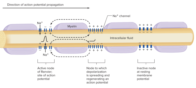
local depolarization from Na+ influx at one segment of the membrane triggers depolarization in the adjacent Na+ channels of the next segment
refractory period ensures action potential moving in one direction
myelination allows depolarizing current to travel longer distance and speeds up conduction
what is myelin?
insulator on axons that allows for fast propagation of action potential
insulation stops leakages of ions → causes longer effective distance of ions to activate next segment of Na+ channels (aka longer depolarization)
formed by oligodendrocytes (CNS) and schwann cells (PNS)
what is salutatory conduction?
axon potentials appears to jump from node to node due to Na+ channels located there
what are graded potentials? what are the key characteristics?
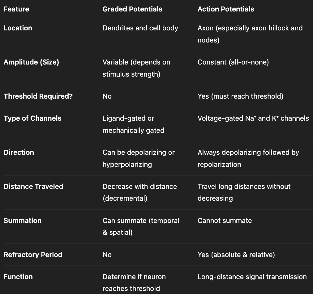
changes in membrane potential that are confined to relatively small region of plasma membrane (short-distance signaling)
direction: stimulus can cause depolarization or hyperpolarization
strength of stimulus affects intensity of graded potential
short distance propagation from leaky membrane + unmyelinated axon
summation of graded potentials can lead to action potentials
what are the types of graded potential summations?
temporal summation: consecutive depolarization events occurring at same location but diff times before neuron is able to return to resting membrane potential
spatial summation: depolarization events simultaneously occurring at diff locations but at same time
compare graded and action potentials
what is length constant? why might they differ between neurons?
distance that ions travel until their voltage reaches 37% of original value
increase in axon diameter = increase length constant
myelination = increase length constant
What are the steps from action potential to neurotransmitter release?
action potential reaches presynaptic terminal
depolarization opens voltage-gated Ca2+ channels
influx of Ca2+ in presynaptic terminal
Ca2+ directly activates synaptotagmin (protein involved in neurotransmitter release) → activates SNARE protein → leads to vesicle docking and fusion
exocytosis—vesicle releases neurotransmitter into synaptic cleft
neurotransmitter diffusion across synaptic cleft and binds to receptors on postsynaptic membrane
postsynaptic potential generated
termination of signal: reuptake/diffusion/inactivation
Limitations on the maximum action potential firing frequency of neurons are determined by ____.
length of refractory periods
how does botulism/botox work?
destroys SNARE protein → no vesicle docking + fusion → no acetylcholine release from motor neuron → no muscle action potential → no DHP activation → no Ca2+ release from SR → no contraction
results in muscle weakness/paralysis
How can neurotransmitter concentration be regulated in a synaptic cleft?
reuptake
enzymatic degradation
diffusion
what is the 3 step process of the somatosensory system?
reception: receptor detects stimulus
transduction: convert stimulus into signal (aka receptor potential)
perception: process + interpret signal in brain
what is a receptor potential?
graded potential that occurs in sensory receptors in response to stimulus
converts stimulus into electrical signal
can trigger action potential if reaches action potential
what is adaptation? what are the two types of adaptors?
adaptation: diminishing activity in response to repeated or sustained stimuli
phasic receptors: fast-adapting; burst of action potential when stimulus is applied and removed; receptor potential does not persist when stimulus is applied
e.g., slight touch (clothes on body), slight temperature (stepping in shower), smell
tonic receptors: slow-adapting; sustained signaling
e.g., pain, vision
what is acuity?
degree of sharpness/precision to which you can sense a stimulus
how does two-point discrimination demonstrate acuity?
two-point discrimination: test that determines minimum distance at which a person can perceive two simultaneous touches as separate points instead of one.
tests how well the nervous system can distinguish closely spaced stimuli
How does receptive field size impact acuity?
receptive field: area of skin that activates a single sensory neuron
smaller receptive field = neuron covers small area → stimuli activate diff neurons → high acuity
large receptive field = neuron covers large area → stimuli activate same neuron → brain perceives as one stimulus → low acuity
How does dendrite density impact acuity?
high dendrite density (receptors) → high acuity
low dendrite density (receptors) → low acuity
what does localization of a stimulus depend on?
size of receptive field
degree of receptive field overlap
what is receptive field overlap?
definition: when neighboring sensory neurons cover some of the same area of sensory space → single stimulus can activate more than one sensory neuron
**helps localize stimulus by comparing neuron firing activity
what is lateral inhibition?
definition: when neuron directly under the stimulus fires strongly and suppresses neighboring weaker responses; leads to increasing contrast between signals, which improves localization/acuity
how does rate of adaptation indirectly influence acuity?
tonic receptors (slow adaptation) → sustained signal → increase acuity
how does myelination indirectly influence acuity?
myelination → sustained signal → increase acuity
what factors influence the intensity of stimulus?
recruitment of diff # of afferent neurons
frequency of firing
magnitude of receptor potentials
what are significant structures in spinal cord vertebrae?
ventral horn = larger wings
dorsal horn = smaller wings
ventral root
dorsal root
dorsal root ganglion
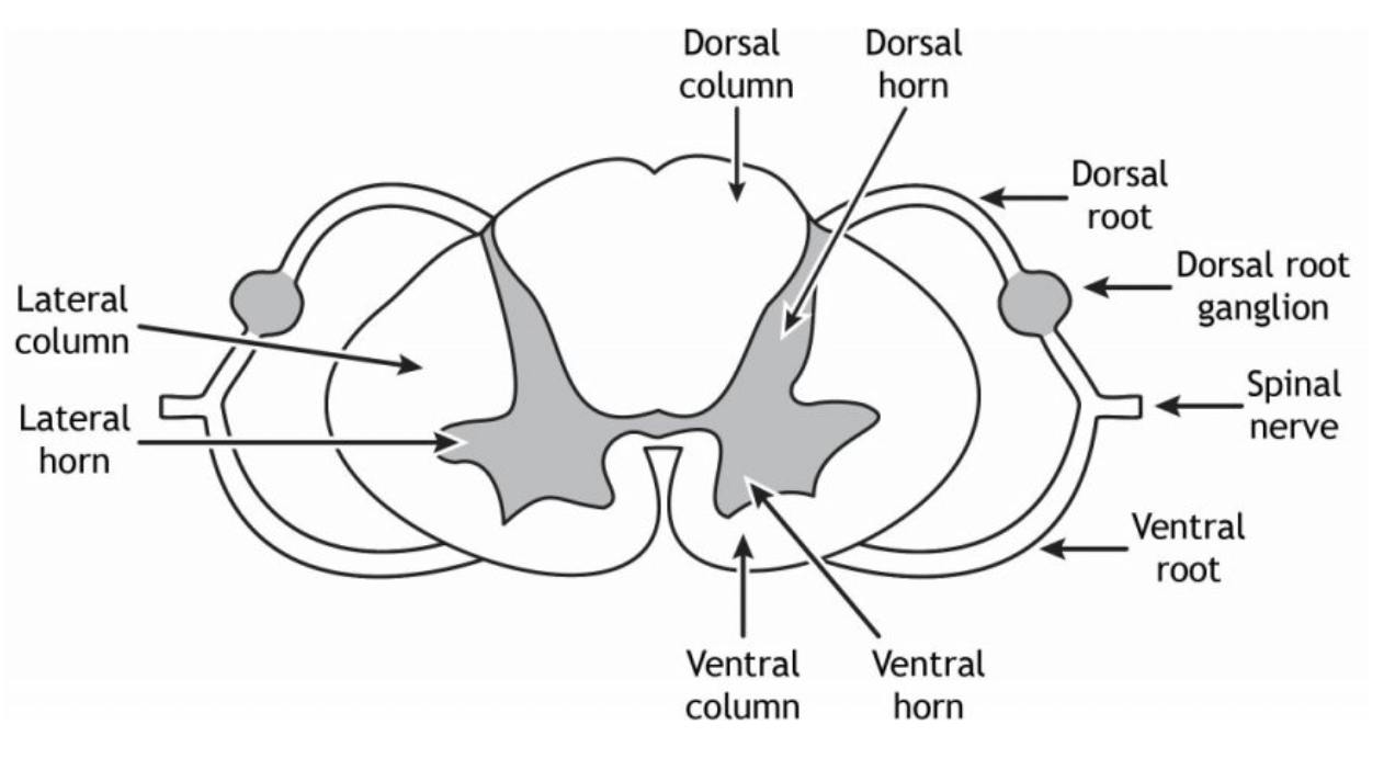
where do afferent signals enter?
dorsal root
where do motor/efferent signals exit?
ventral root
what is the dorsal root ganglion?
cluster of sensory neuron cell bodies
what is the pathway from stimulus to spinal cord?
Stimulus → Receptor potential → Action potential → Peripheral afferent fiber → Dorsal root ganglion (cell body) → dorsal root
what are the two types of somatosensory pathways? how do they differ?
ascending anterolateral pathway: immediately synapse in spinal cord → crossover in spinal cord → ascend to brain through anterolateral column (side) → ascends to thalamus → reaches somatosensory cortex
dorsal column pathway: ascend to brain through dorsal column (back) → synapse in brain → crossover in brainstem → ascends to thalamus → reaches somatosensory cortex
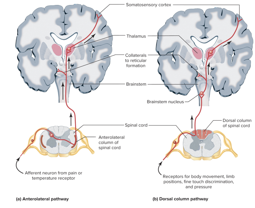
what is the full somatosensory pathway, from sensory to motor command?
Receptor → sensory neuron (afferent pathway) → dorsal root ganglion → dorsal root → synapse at dorsal horn → ascends to brain → thalamus (relay center) → somatosensory cortex → motor cortex (generates motor response) → motor command travels down descending motor tract → motor neuron synapses in ventral horn → exits spinal cord via ventral root → muscle contracts
what is the somatosensory reflex arc pathway
*does not require brain (e.g., knee jerk reflex)
Receptor → sensory neuron → dorsal root → dorsal horn → interneuron → motor neuron (ventral horn) → ventral root → muscle
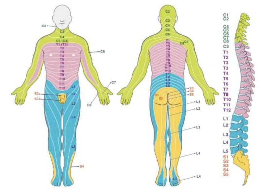
what are dermatomes? how do they relate to pathologies?
areas of skin innervated by nerves in a segmented fashion; location of sensation is where the spinal cord will receive the signal
ex: shingles—dormant virus in dorsal root ganglion; rash follows form of dermatome
ex: spinal cord injury—loss of sensation in body can indicate which vertebrae has been injured
how do mechanoreceptors work?
direct activation due to lipid tension (membrane bilayer pulling receptor)
direct linkage via. intracellular and extracellular proteins (proteins pulling receptor)
indirect activation via. a system that activates the channel
how does threshold stimuli relate to sensitivity to touch?
low threshold = require little mechanical force to open mechanically-gated ion channels → high sensitivity (detect extremely subtle touch)
how does receptive field size influence sensitivity and acuity?
small receptive field size → high sensitivity + acuity
detects fine detail
large receptive field size → broad detection + lower acuity
usually detects pressure or vibration
how does adaptation affect touch?
phasic (rapidly adapting) → movement, texture, vibration
tonic (slowly adapting) → grip control, maintain continuous awareness of objects in hand
what are nociceptors?
pain receptors that respond to tissue damage, chemical, mechanical or thermal stimulation
what are the types of nociceptor fibers?
Aδ (delta) fibers:
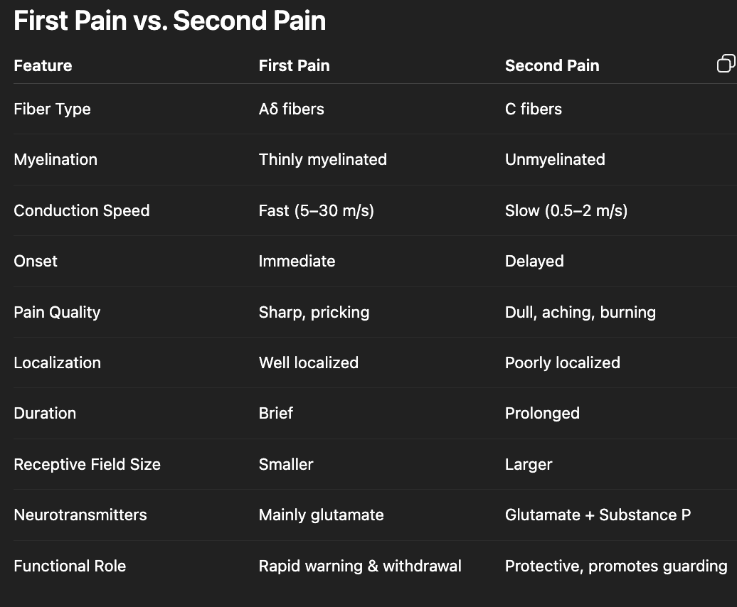
light myelinated, fast conducting →first pain
smaller receptive field size → increased acuity → sharp, localized pain
higher intensity pain for shorter period of time
polymodal nociceptors (C fibers):
unmyelinated, slow conducting → second pain (dull, aching, throbbing, nonlocalized pain)
lower intensity pain for prolonged time
what is unique about sleeping nociceptors?
activated by chemicals released during tissue damage
located on C fibers (slow, chronic, dull second pain)
ex: washing hand under water w/ cut
what neurotransmitters are involved in nociception?
glutamate: primary fast transmitter; released from Aδ and C fibers
substance P (neuropeptide): produce slower, prolonged excitation for chronic pain signaling; released mainly from C fibers
what are A-alpha fibers
associated with tonic (slow adaptation) in muscle
what are A-beta fibers?
associated with fast adaptation (phasic) in touch
what is the difference between mechanosensitive and mechanothermal receptors?
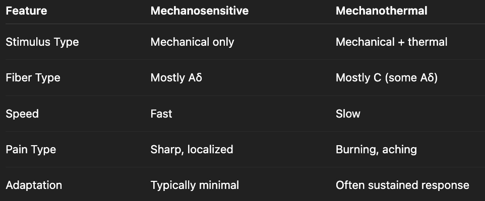
what is the difference between anesthesia and analgesics?
anesthesia: blocks all sensations
analgesic: blocks pain signaling
what are the subcategories of anesthesia?
general anesthesia: induces total unconcsciousness; affects entire body
local anesthesia: numbs specific small area
what are common forms of analgesics?
opiods/oppiates
cox inhibitors
what is hyperalgesia vs. hypoalgesia?
hyperalgesia: increases pain sensitivity
hypoalgesia: decreases pain sensitivity
what is the difference between first and second pain? how is it coded?
Describe the pain transduction pathway and how it relates to perception of pain.
Tissue injury → releases chemical signals (e.g., prostaglandins, histamine)
Nociceptor activation
Aδ and C fibers transmit signal
Synapse in dorsal horn
Cross in spinal cord
Ascend via anterolateral column
Thalamus relay
Cortex processes → conscious pain
How is hypoalgesia naturally activated?
stress → release serotonin + norepinephrine → releases opioids (e.g., beta-endorphins) → prevents release or binding of pain neurotransmitters from nociceptors (glutamate and substance P)
How is hyperalgesia naturally activated?
after tissue injury, inflammatory mediators are released (e.g., prostaglandins, histamine, cytokines) which lowers activation threshold of nociceptors → nociceptors fire more easily?
why does opioid addiction occur?
opioids bind to opiate receptors and inactivate GABA-ergic neuron → inhibits GABA release → allows dopamine release
what is the COX-1 pathway in nociception?
tissue injury releases inflammatory mediators (e.g., histamine, cytokines)
inflammatory mediators bind to GPCR on nociceptor
Gq activates PLC → cleaves PIP2 to IP3 + DAG → IP3 increases Ca2+, DAG activates PKC
activation of PLA2 causes release of arachidonic acid
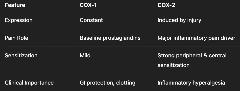
COX-1 converts arachidonic acid into prostaglandins & thromboxanes
sensitizes nociceptors & lowers activation threshold, leading to increased excitability and hyperalgesia
Compare COX-1 and COX-2
how do NSAIDs work?
COX-1 inhibitor
block COX pathway (*mainly prostaglandins) → reduced inflammation
can cause stomach ulcers & kidney issues w/ prolonged use (bc COX-1 enzymes found in gastro + renal tract)
What are the parts of the brain that influence pain perception?
reticular alertness, arousal
thalamus: dull, poorly localized (chronic pain); main relay station that sends signals to cortex
cerebrum (cortex): localizes + characterizes pain
amygdala: emotions/memory, experience, suggestions
what is the cross-bridge cycle? List out the steps
describes how myosin interacts with actin to generate contraction, with ATP, ADP, and Pi controlling each step
Ca2+ binds to troponin → induces conformational change
shifts tropomyosin on actin → exposes myosin-binding sites on actin
myosin (ADP + Pi bound—cocked state) binds to actin → forms cross-bridge
release of Pi leads to power stroke
ADP release leads to rigor state
ATP binds to myosin → detaches myosin from actin
ATP hydrolysis (ATP → ADP + Pi) leads to cocking (high energy state)
how is Ca2+ released into muscle cells, starting from motor neuron?
acetylcholine released from motor neuron
synapse on motor end plate
bind to acetylcholine receptors on sarcolemma of muscle cell and trigger action potential via. voltage-gated Na+ and K+ channels
action potential arrives at T-tubules and reaches DHP
DHP receptor (mechanical voltage receptor) detects voltage change and undergoes conformational change
DHP mechanically opens ryanodine receptor on sarcoplasmic membrane reticulum
Ca2+ release from sarcoplasmic reticulum
Ca2+ binds to troponin → shifts tropomyosin → muscle contraction
how does curare work?
plant-derived toxin
competitive antagonist of acetylcholine receptors → acetylcholine is released but does not bind to receptor → no end plate potential → no muscle action potential → no DHP activation → no Ca2+ release → no contraction → muscle weakness/paralysis
how does sarin (nerve gas) work?
inhibit acetylcholinesterase (AChE) → acetylcholine not broken down → continuous stimulation of Ach receptors
early phase: constant depolarization + Ca2+ release + sustained contraction → muscle spasms
later phase: depolarization block, voltage gated Na+ channels inactivated, Ca2+ regulation disrupted → paralysis
what are the types of muscle contractions?
isometric contraction: muscle tension increases but muscle length does not change (e.g., carrying groceries)
isotonic contraction: muscle tension increases and muscle length changes
what are the two types of isotonic contractions?
concentric contraction: muscle shortens while producing force; muscle force > external load (e.g., lifting dumbbells)
eccentric contraction: muscle lengthens; muscle force < external load (e.g., lower dumbbells)
what is the difference between twitch and tetanus?
twitch: single contraction in response to one action potential
tetanus: repeated stimulation prevents Ca²⁺ from being fully removed, leading to summation of force and sustained contraction (e.g., maintaining posture, lifting weights)
what is the difference between unfused and fused tetanus?
unfused tetanus: incomplete; minimal relaxation; fluctuating elevated Ca2+
fused tetanus: complete; no relaxation; sustained high Ca2+

what factors can increase muscle tension?
stimulate more cross bridges by recruiting more sarcomeres + frequent stimulation (summation)
optimal degree of overlap to maximize cross bridge formation
larger diameter of muscle fibers → increase formation of cross bridges
what is a motor unit? how does it regulate muscle tension?
single motor neuron and all the muscle fibers it innervates
determines how much force is produced
regulation: recruit motor unit by size & increase firing frequency for temporal summation
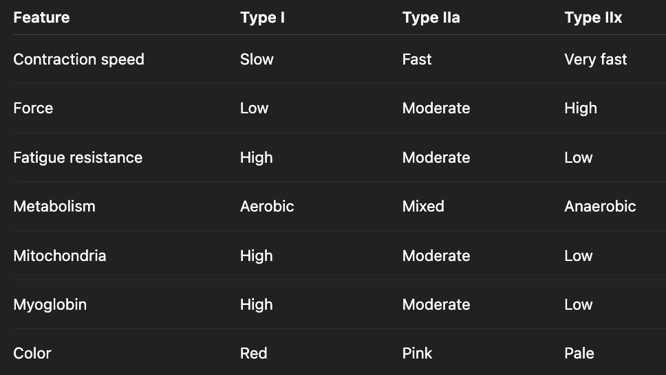
what are the three types of muscle fibers?
type I: slow-oxidative; postural
type IIa: faster-oxidative-glycolytic; endurance
type IIb: fastest-glycolytic; speed
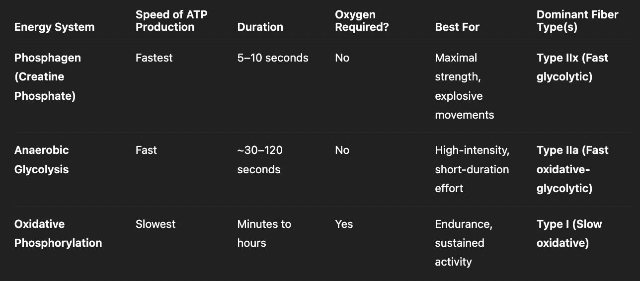
what are the pathways that muscles use to get ATP?
creatine phosphate + ADP → creatine + ATP (very fast ATP production)
glycolysis (generates atp fastest, anaerobic, amount of atp per waste product)
oxidative phosphorylation (produces most atp but also most time consuming; requires lots of O2—aerobic)
what are the causes of muscle fatigue?
reduced motor neuron firing: K+ saturated T tubules causes hypoerpolarization → conduction failure of action potential
lactic acid buildup → lower pH → troponin malfunction
accumulation of ADP + Pi: inhibit cross bridge cycle & release of myosin from actin
what is the motor end plate?
specialized region of the sarcolemma where motor neuron communicates with muscle cell at the neuromuscular junction
results in Ca2+ release from sarcoplasmic reticulum & muscle contraction
what is the difference between central and peripheral fatigue?
central fatigue (brain-level): reduced motor neuron firing + reduced recruitment of motor units
peripheral fatigue (muscle-level): accumulation of ADP+Pi, troponin malfunction
what are key structural features of smooth muscle?
gap junction (electrical coupling) vs. desmosomes (mechanical attachment)
no sarcomeres → actin + myosin organized in lattice network instead of parallel bundles
no troponin → use MLCK and MLCP instead
actin + myosin filaments
unique proteins: tropomyosin (different from skeletal), calponin, caldesmon
dense bodies: actin (thin filaments) is anchored to dense bodies
desmosomes: where two adjacent smooth muscle cells are physically attached to each other
what’s the difference between single-unit and multi-unit smooth muscle?
single unit smooth muscle
structure: cells connected by gap junctions; few cells innervated
activation: myogenic (self-activation) triggered by stretch and local signals
ex: GI tract
multi-unit smooth muscle
structure: little/no gap junction coupling; each cell acts independently + directly innervated
activation: neurogenic (nerve-controlled), allows for finer control
eyes adjusting light
what are the enzymes important for smooth muscle contractions?
MLCK (myosin light-chain kinase): phosphorylates myosin to allow myosin and actin to interact
MLCP (myosin light-chain phosphatase): removes phosphate from myosin
Rho-kinase: phosphorylates and inactivates MLCP so myosin stays phosphorylated
what are the steps of smooth muscle contraction?
agonist binds to GPCR on smooth muscle cells
activates PLC → IP3 binds to receptors on sarcoplasmic reticulum → Ca2+ release
Ca2+ enters smooth muscle cell (graded)
Ca2+ binds + activates calmodulin
Ca2+-calmodulin binds + activates MLCK (myosin light-chain kinase)
**dependent on ratio of MLCK and MLCP
contraction: MLCK phosphorylates MLC (uses ATP) to MLC-P → allows myosin and actin to bind → cross-bridge cycling (generates powerstroke) → smooth muscle contraction
relaxation: MLCP (myosin light-chain phosphatase) dephosphorylates MLC-P to MLC → myosin isn’t able to bind to actin → no cross-bridge cycling → smooth muscle relaxation
Rho-kinase phosphorylates + inactivates MLCP → MLC-P stays phosphorylated → cross-bridge cycling occurs → allows muscle contraction
why are smooth muscle myosin ATPase slower than in striated muscle cells?
provides a graded contraction
leads to slow, sustained contraction (maintains tension) and low fatigue
what is latch state?
when myosin stays latched into actin and can’t be released
maintain muscle tension w/ minimal ATP use + lack of fatigue over longer periods of time
when does latch state occur?
when MLCP dephosphorylates myosin while myosin and actin are attached → myosin stays latched onto actin and can’t be released → allows muscle to maintain tension with very little energy
what needs to happen to release from latch state?
decrease intracellular Ca2+ → stop cross-bridge cycling
increase MLCP activity → promotes detachment
what are the two types of smooth muscle contractions?
phasic contraction: contraction followed by relaxation
function: move things along
ex: peristalsis
tonic contraction: sustained contraction for long period of time; very energy-efficient (latch state); little to no relaxation
function: maintain pressure or resistance
ex: bladder holding urine
What factors influence blood flow?
directly proportional to pressure difference
indirectly proportional to resistance
what factors influence resistance? which factor has the greatest influence on resistance?
increase in viscosity (affected by water) → increase in resistance
decrease in vessel radius** (affected by temp) → increase in resistance
increase in length (affected by weight/height gain) → increase in resistance
what is the difference between increase vs. decrease vascular tone?
vascular tone: degree of smooth muscle contraction in blood vessels (diameter of vessel)
increase vascular tone = vasoconstriction (vessel narrowing, increase resistance, decrease bloodflow, increase blood pressure)
decrease vascular tone = vasodilation (vessel widening, decrease resistance, increase bloodflow, decrease blood pressure
what drugs decrease vascular tone?
decrease vascular tone (vasodilation) → treat hypertension
L-type Ca2+ channel blockers
K+ channel openers
nitrodilators
direct-acting vasodilators
angiotensin pathway
endothelin antagonists
a-adrenoreceptor antagonists
what are the different mechanism pathways to decrease vascular tone?
decrease vascular tone → muscle relaxation or decrease muscle contractions
decrease Ca2+
decrease MLCK activity
increase MLCP activity
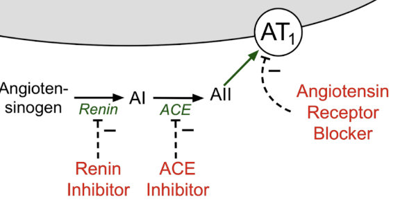
how does inhibiting the angiotensin pathway affect vascular tone?
inhibiting angiotensin pathway → decrease vascular tone
renin-inhibitor: block angiotensinogen → angiotensin I formation (earliest step)
ACE inhibitor: block conversion of angiotensin I → angiotensin II
angiotensin receptor blockers (ARBs): block angiotensin receptor directly → Ang II present but no Gq → decrease Ca2+ signaling → vasodilation
what are the different types of adrenoreceptors? where are they located and how they relate to affinity?
alpha adrenoreceptors: low affinity
a1: smooth muscle vasculature → cause vasoconstriction
a2: smooth muscle vasculature + neurons → decrease norepinephrine release → vasodilation
beta adrenoreceptors: high affinity
b1: heart → increase heart rate + myocardial contractions
b2: smooth muscle vasculature → cause vasodilation
how does the a1 adrenergic pathway work?
sympathetic nerve release epinephrine/norepinephrine
epinephrine/norepinephrine binds to a1 receptor (Gq) on smooth muscle
activates IP3-DAG pathway → releases Ca2+ → vasoconstriction