Chapter #10 Muscle Tissue
1/55
There's no tags or description
Looks like no tags are added yet.
Name | Mastery | Learn | Test | Matching | Spaced | Call with Kai |
|---|
No analytics yet
Send a link to your students to track their progress
56 Terms
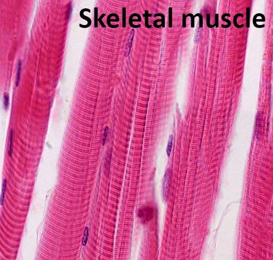
Skeletal Muscle
This tissue is packaged into skeletal muscles, organs that are attached to bones and skin.
Skeletal muscle fibers are the longest of all muscles and have striations (stripes)
Also called voluntary muscle: can be consciously controlled
Contract rapidly; tire easily; powerful
Keywords for skeletal muscle: skeletal, striated, and voluntary
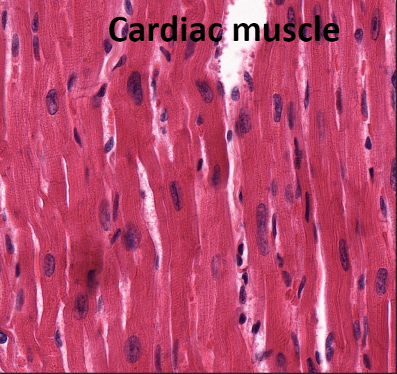
Cardiac Muscle
This tissue is found only in heart
Makes up bulk of heart walls
Striated
Involuntary: cannot be controlled consciously
Contracts at steady rate due to heart’s own pacemaker, but nervous system can increase rate
Key words for cardiac muscle: cardiac, striated, and involuntary
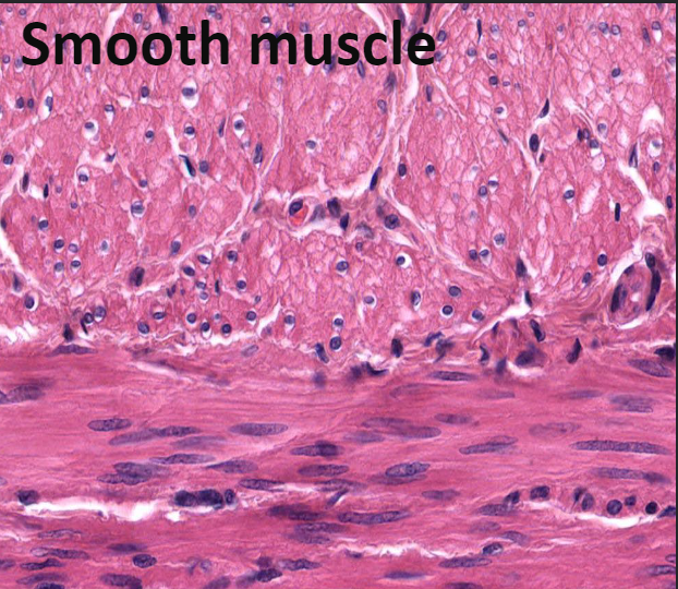
Smooth Muscle
This tissue is found in walls of hollow organs
Examples: stomach, urinary bladder, and airways
Not striated
Involuntary: cannot be controlled consciously
Key words for smooth muscle: visceral, nonstriated and involuntary
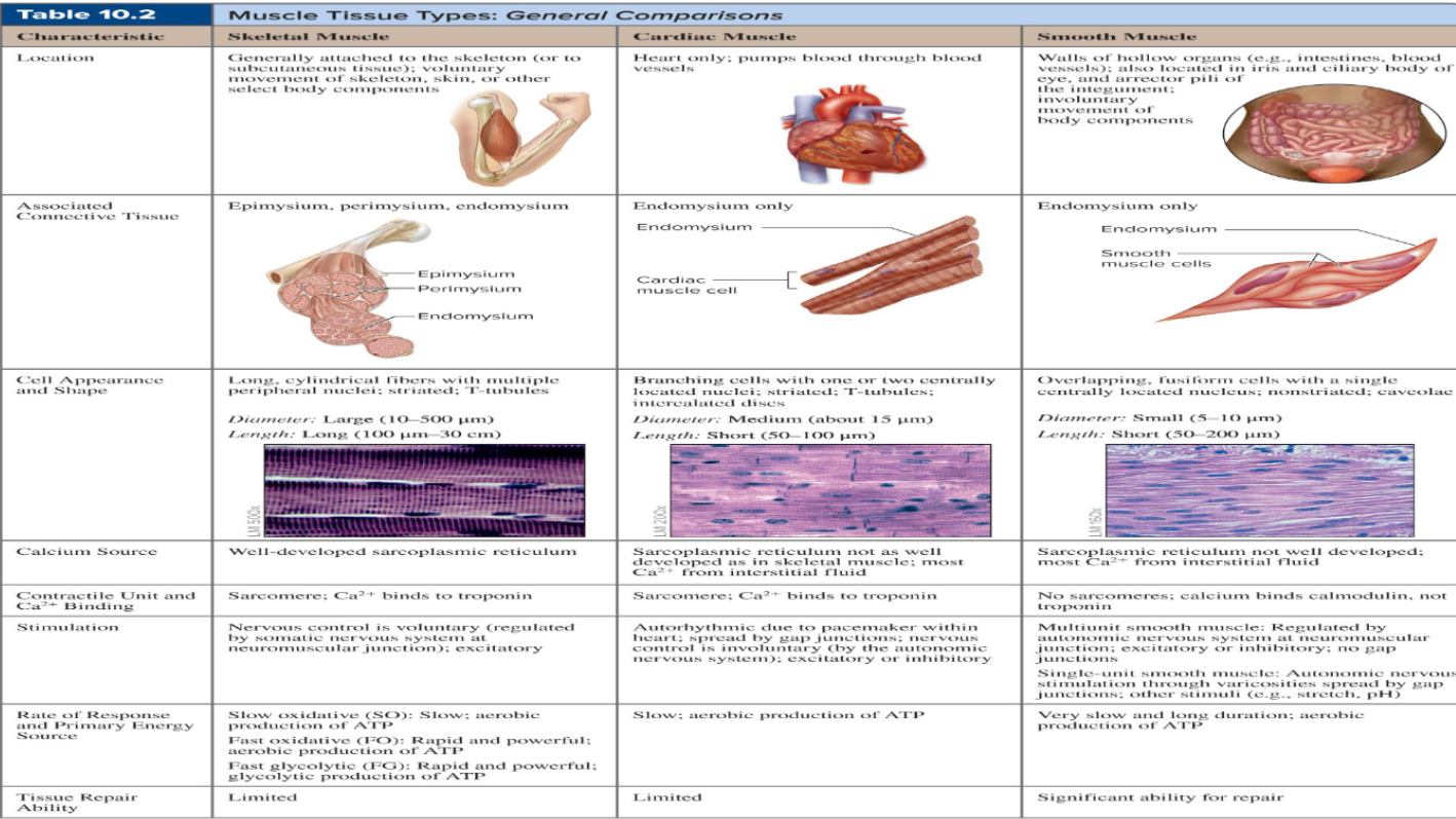
Muscle Tissue Comparisons
Skeletal Muscle ( Explained)
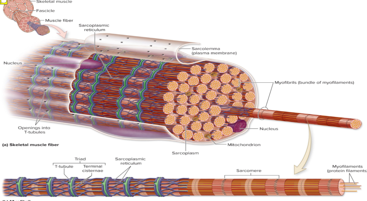
Skeletal muscle is an organ made up of different tissues with three features: nerve and blood supply, connective tissue sheaths, and attachments
Each muscle receives a nerve, artery, and veins
Consciously controlled skeletal muscle has nerves supplying every fiber to control activity
Contracting muscle fibers require huge amounts of oxygen and nutrients
Also need waste products removed quickly
Each skeletal muscle, as well as each muscle fiber, is covered in connective tissue
Support cells and reinforce whole muscle
Sheaths from external to internal:
Epimysium »Perimysium»Endomysium
Epimysium
dense irregular connective tissue surrounding the entire muscle; may blend with fascia
Perimysium
fibrous connective tissue surrounding fascicles (groups of muscle fibers)
Endomysium
fine areolar connective tissue surrounding each muscle fiber
Sarcolemma
muscle fiber plasma membrane

Sarcoplasm
: muscle fiber cytoplasm
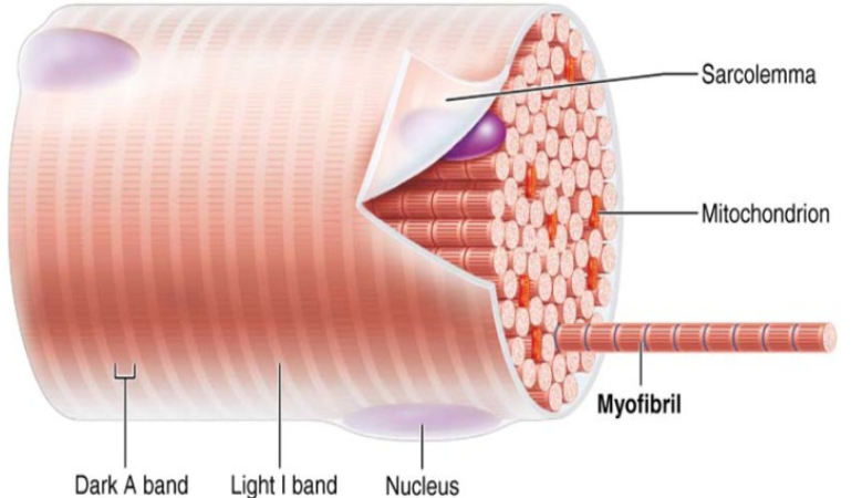
Myofibrils
Myofibrils are densely packed, rodlike elements
Single muscle fiber can contain 1000s
Accounts for ~80% of muscle cell volume
Myofibril features Striations, Sarcomeres, Myofilaments, Molecular composition of myofilaments

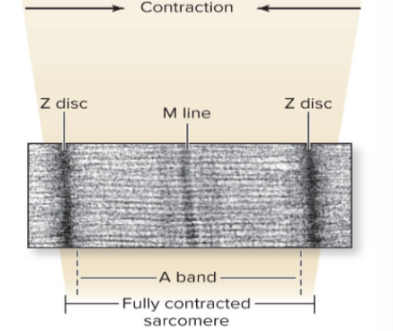
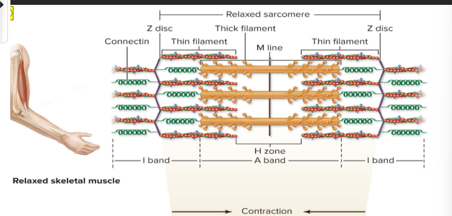
Sarcomere
Smallest contractile unit (functional unit) of muscle fiber
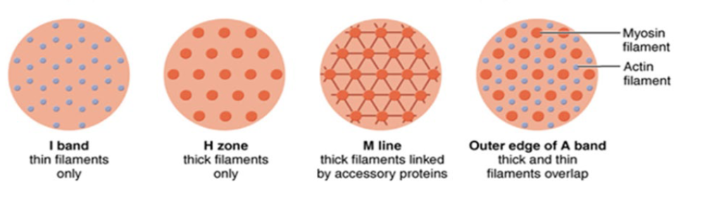
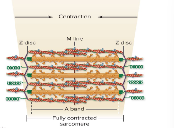
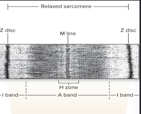
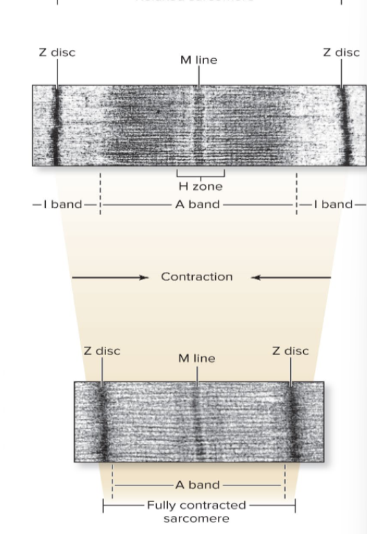
A bands: dark regions
H zone: lighter region in middle of dark A band
M line: line of protein (myomesin) that bisects H zone vertically
I bands: lighter regions
Z disc (line): coin-shaped sheet of proteins on midline of light I band
Contains A band with half of an I band at each end
Consists of area between Z discs
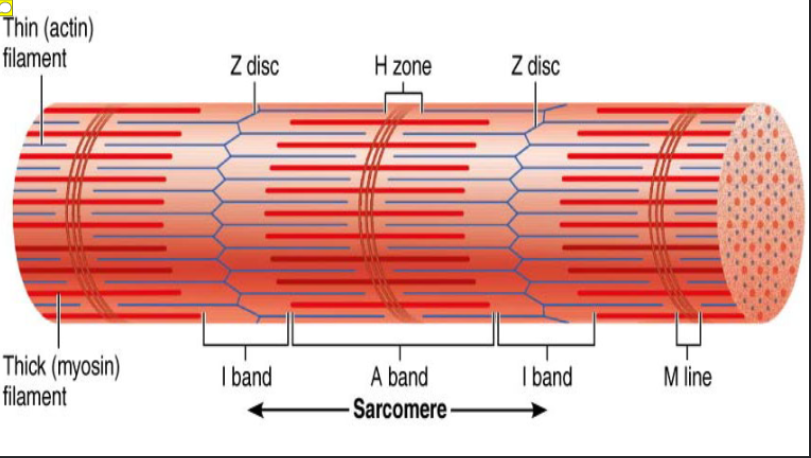
Striations
: stripes formed from repeating series of dark and light bands along length of each myofibril
Actin myofilaments
: thin filaments Extend across I band and partway in A band and Anchored to Z discs
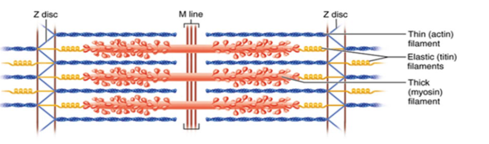
Myosin myofilaments
: thick filaments Extend length of A band Connected at M line
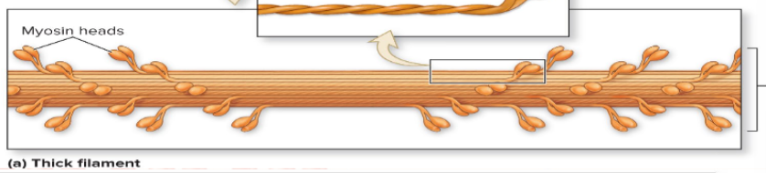
Thick filaments
: composed of protein myosin that contains two heavy and four light polypeptide chains
Heavy chains intertwine to form myosin tail
Light chains form myosin globular head
Myosins are offset from each other, resulting in staggered array of heads at different points along thick filament
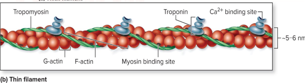
Thin filaments
: composed of fibrous protein actin
Actin is polypeptide made up of kidney-shaped G actin (globular) subunits
G actin subunits bears active sites for myosin head attachment during contraction
G actin subunits link together to form long, fibrous F actin (filamentous)
Two F actin strands twist together to form a thin filament
Tropomyosin and troponin: regulatory proteins bound to actin
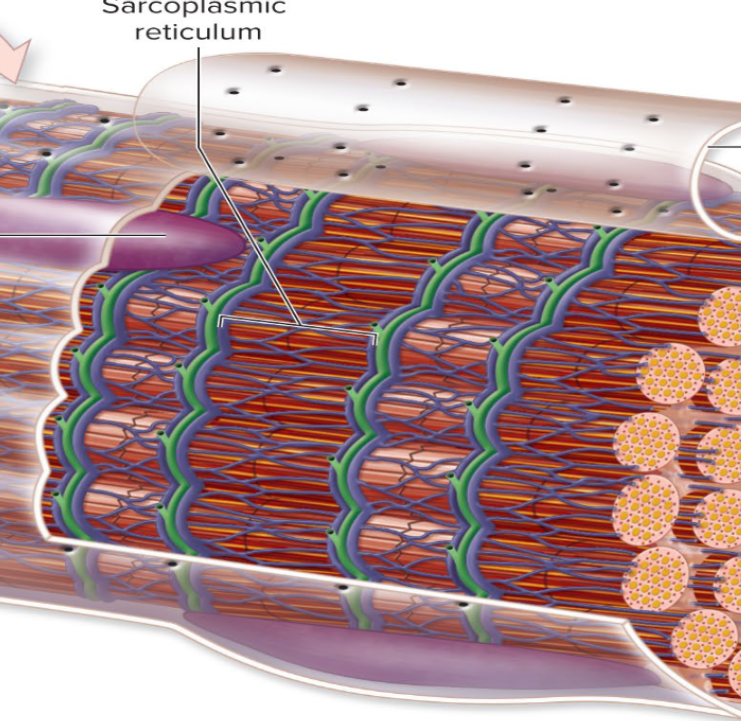
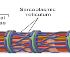
Sarcoplasmic reticulum
: network of smooth endoplasmic reticulum tubules surrounding each myofibril and Most run longitudinally
Sacroplasmic Reticulum (Explained)
Terminal cisterns form perpendicular cross channels at the A–I band junction
SR functions in regulation of intracellular Ca2+ levels and Stores and releases Ca2+
SR cistern membranes also have integral membrane proteins that protrude into intermembrane space
SR integral proteins control opening of calcium channels in SR cisterns
When an electrical impulse passes by, T tubule proteins change shape, causing SR proteins to change shape, causing release of calcium into cytoplasm
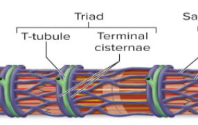
Triad
: area formed from terminal cistern of one sarcomere, T tubule, and terminal cistern of neighboring sarcomere
Triad relationships T tubule contains integral membrane proteins that protrude into intermembrane space (space between tubule and muscle fiber sarcolemma)

T tubules
Tube formed by protrusion of sarcolemma deep into cell interior
Increase muscle fiber’s surface area greatly
Lumen continuous with extracellular space
Allow electrical nerve transmissions to reach deep into interior of each muscle fiber
Tubules penetrate cell’s interior at each A–I band junction between terminal cisterns
Tubule proteins act as voltage sensors that change shape in response to an electrical current
Excitable cells
are capable of changing resting membrane potential voltages
Neurons and muscle cells are excitable cells capable of action potentials
Contraction
: the activation of cross bridges to generate force
Shortening occurs when tension generated by cross bridges on thin filaments exceeds forces opposing shortening
Contraction ends when cross bridges become inactive
Extension/Relaxed State of Muscle
In the relaxed state, thin and thick filaments overlap only slightly at ends of A band
Sliding filament model of contraction states that during contraction, thin filaments slide past thick filaments, causing actin and myosin to overlap more
Neither thick nor thin filaments change length, just overlap more
Muscle Fiber Stimulation
When nervous system stimulates muscle fiber, myosin heads are allowed to bind to actin, forming cross bridges, which cause sliding (contraction) process to begin
Cross bridge attachments form and break several times, each time pulling thin filaments a little closer toward center of sarcome in a ratcheting action
Causes shortening of muscle fiber
Z discs are pulled toward M line I bands shorten Z discs become closer H zones disappear and A bands move closer to each other
Ion Channels
Play a major role in changing of membrane potentials
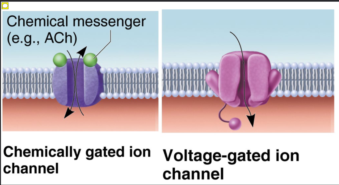
Two classes of ion channels:
Chemically gated ion channels – opened by chemical messengers such as neurotransmitters
Example: ACh receptors on muscle cells
Voltage-gated ion channels – open or close in response to voltage changes in membrane potential

Chemically gated ion channels
– opened by chemical messengers such as neurotransmitters
Example: ACh receptors on muscle cells

Voltage-gated ion channels
– open or close in response to voltage changes in membrane potential
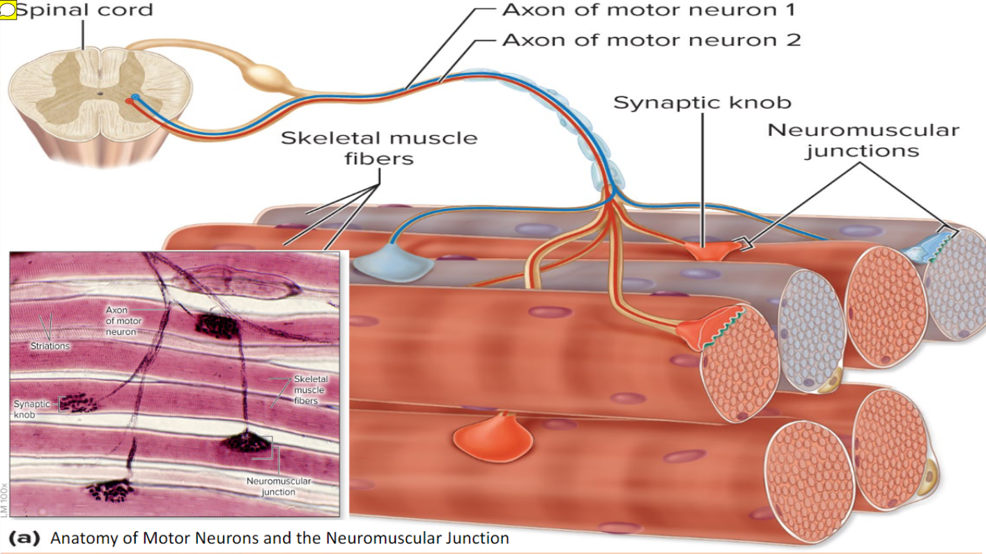
Anatomy of Motor Neurons and Neuromuscular Junction
Skeletal muscles are stimulated by somatic motor neurons
Axons (long, threadlike extensions of motor neurons) travel from central nervous system to skeletal muscle
Each axon divides into many branches as it enters muscle
Axon branches end on muscle fiber, forming neuromuscular junction or motor end plate
Each muscle fiber has one neuromuscular junction with one motor neuron
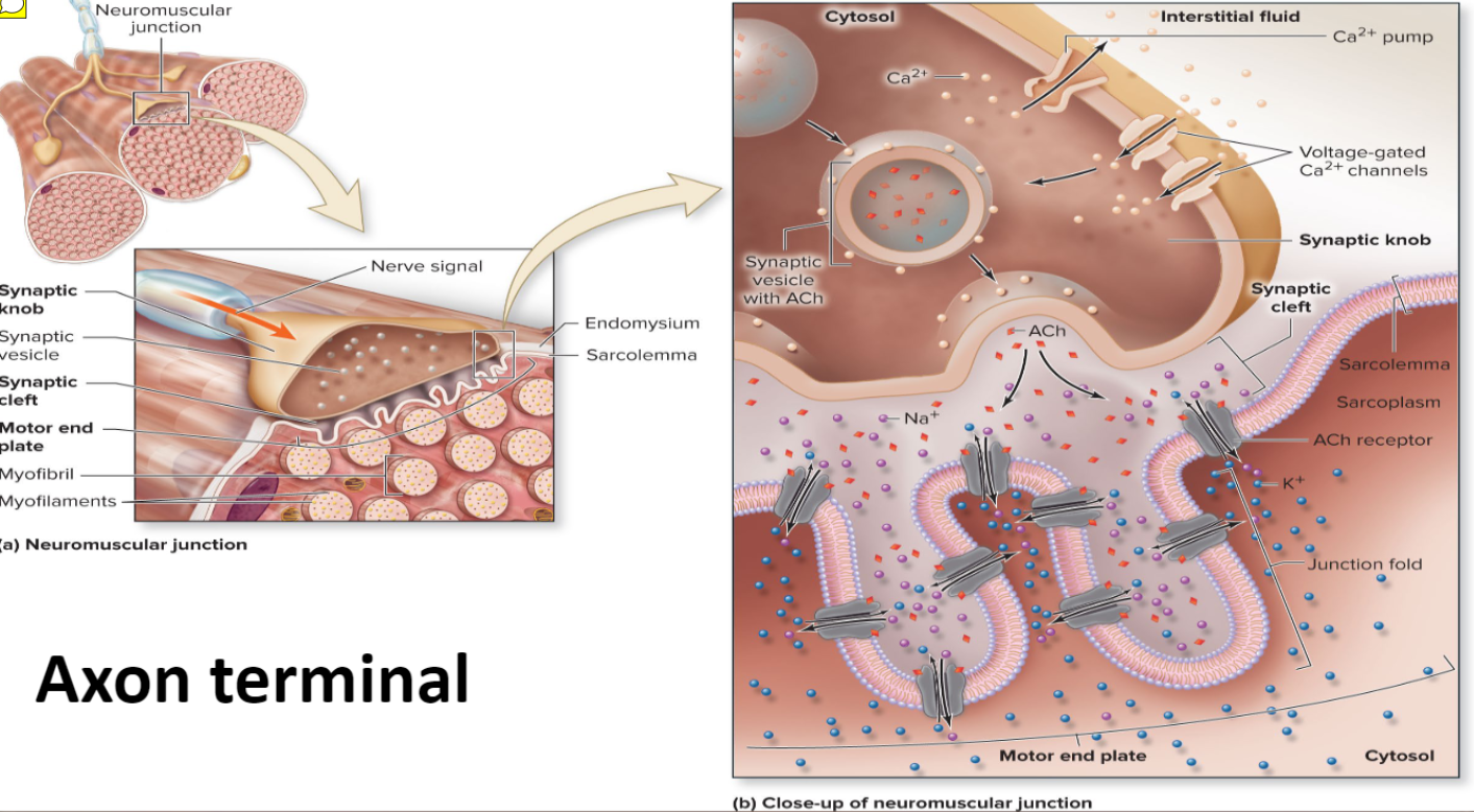
Axon Terminal
Axon terminal (end of axon) and muscle fiber are separated by gel-filled space called synaptic cleft
Stored within axon terminals are membrane-bound synaptic vesicles
Synaptic vesicles contain neurotransmitter acetylcholine (ACh)
Infoldings of sarcolemma, called junctional folds, contain millions of ACh receptors
NMJ consists of axon terminals, synaptic cleft, and junctional folds
Action potential
is caused by changes in electrical charges
Resting sarcolemma is polarized, meaning a voltage exists across membrane
Inside of cell is negative compared to outside
Neuromuscular Junction of Skeletal Muscle Fiber

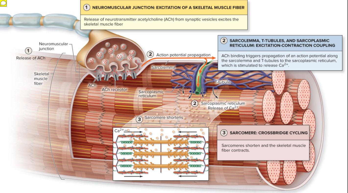
Neuromuscular Junction of Skeletal Muscle Fiber ( Explained)
Occurs in three steps
End plate potential
ACh released from motor neuron binds to ACh receptors on sarcolemma
Causes chemically gated ion channels (ligands) on sarcolemma to open
Na+ diffuses into muscle fiber Some K+ diffuses outward, but not much
Because Na+ diffuses in, interior of sarcolemma becomes less negative (more positive)
Results in local depolarization called end plate potential
Depolarization: generation and propagation of an action potential (AP)
If end plate potential causes enough change in membrane voltage to reach critical level called threshold, voltage-gated Na+ channels in membrane will open
Large influx of Na+ through channels into cell triggers AP that is unstoppable and will lead to muscle fiber contraction
AP spreads across sarcolemma from one voltage-gated Na+ channel to next one in adjacent areas, causing that area to depolarize
Repolarization: restoration of resting conditions
Na+ voltage-gated channels close, and voltage-gated K+ channels open
K+ efflux out of cell rapidly brings cell back to initial resting membrane voltage
Refractory period: muscle fiber cannot be stimulated for a specific amount of time, until repolarization is complete
Ionic conditions of resting state are restored by Na+-K+ pump
Na+ that came into cell is pumped back out, and K+ that flowed outside is pumped back into cell
Refractory period: muscle fiber cannot be stimulated for a specific amount of time, until repolarization is complete
Depolarization: generation and propagation of an action potential (AP)