Nerve Impulses and Synaptic Transmission
1/39
There's no tags or description
Looks like no tags are added yet.
Name | Mastery | Learn | Test | Matching | Spaced | Call with Kai |
|---|
No analytics yet
Send a link to your students to track their progress
40 Terms
Central Nervous System (CNS)
Brain and spinal cord of dorsal body cavity
Integration and control center
Interprets sensory input an dictates motor output
Peripheral Nervous System (PNS)
The portion of nervous system outside CNS
Consists mainly of nerves that extend from brain and spinal cord
Spinal nerves to and from spinal cord
Cranial nerves to and from brain
Walls of gastrointestinal tract also contain neurons called the enteric nervous system
Sensory (afferent) division
Somatic sensory fibers: convey impulses from skin, skeletal muscles, and joints to CNS
Visceral sensory fibers: convey impulses from visceral organs to CNS
Motor (efferent) division
Transmits impulses from CNS to effectors
Muscles and glands
Two divisions
Somatic nervous system
Autonomic nervous system
Cells of the Nervous System
Nervous tissue consists of two principal cell types
Neuroglia (glial cells): small cells that surround and wrap delicate neurons
Neurons (nerve cells): excitable cells that transmit electrical signals
Neuroglia of the CNS
astrocytes
microglial cells
ependymal cells
oligodendrocytes
Astrocytes
Most abundant, versatile, and highly branched of glial cells
Cling to neurons, synaptic endings, and capillaries
Functions:
Support and brace neurons
Play role in exchanges between capillaries and neurons
Guide migration of young neurons
Control chemical environment around neurons
Respond to nerve impulses and neurotransmitters
Participate in information processing in brain
Microglial Cells
Small, ovoid cells with thorny processes that touch and monitor neurons
Migrate toward injured neurons
Can transform to phagocytize microorganisms and neuronal debris
Ependymal Cells
Range in shape from squamous to columnar
May be ciliated
Cilia beat to circulate CSF
Line the central cavities of the brain and spinal column
Form permeable barrier between cerebrospinal fluid (CSF) in cavities and tissue fluid bathing CNS cells
Oligodendrocytes
Branched cells
Processes wrap CNS nerve fibers, forming insulating myelin sheaths in thicker nerve fibers
Satellite cells (Neuroglia of PNS)
Surround neuron cell bodies in PNS
Function similar to astrocytes of CNS
Schwann cells (neurolemmocytes) (Neuroglia of PNS)
Surround all peripheral nerve fibers and form myelin sheaths in thicker nerve fibers
Similar function as oligodendrocytes
Vital to regeneration of damaged peripheral nerve fibers
Neurons
Neurons (nerve cells) are structural units of nervous system
Large, highly specialized cells that conduct impulses
All have cell body and one or more processes
Special characteristics
Extreme longevity (lasts a person’s lifetime)
Amitotic, with few exceptioins
High metabolic rate: requires continuous supply of oxygen and glucose
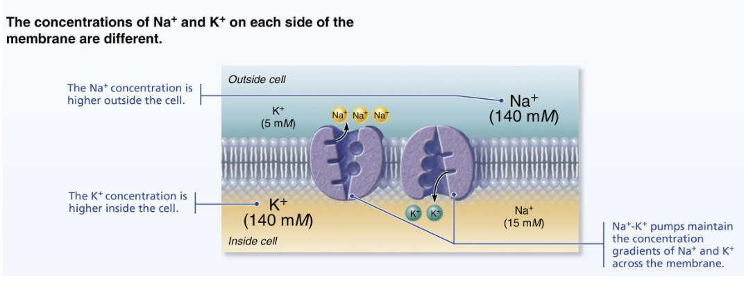
Resting Membrane Potential
Generating a resting membrane potential depends on differences in
K and Na concentrations inside and outside cells
in the permeability of the plasma membrane to these ions
Generating an Action Potential
Four main steps
Resting state: all voltage-gated Na and K channel are closed
only leakage channels Na and K are open for maintaining the resting membrane potential
Each Na channel has two voltage-sensitive gates
Activation gates: closed at rest; open with depolarization allowing Na to enter cell
Inactivation gates: open at rest; block channel once it is open to prevent Na from entering cell
Each K channel has one voltage-sensitive gate
Closed at rest
Opens slowly with depolarization
Depolarization: voltage-gated Na channels open
Depolarizing local currents open voltage-gated Na channels and Na rushes into the cell
Na activation and inactivation gates open
Na influx causes more depolarization, which opens Na channels more
As a result, ICF becomes less negative
At threshold (-55 to -50 mV), positive feedback causes the opening of all Na channels
Results in large action potential spike
Membrane polarity jumps to +30 mV
Repolarization: Na channels are inactivating and voltage-gated K channels open
Na channel inactivation gates close
Membrane permeability Na declines to resting state
AP spike stopss rising
Voltage-gated K channels open
K exits cells down its electrochemical gradient
Repolarization: membrane returns to resting membrane potential
Hyperpolarization: K channels remain open, Na channels reset
Some K channels remain open, allowing excessive K efflux
inside of membrane becomes more negative than in resting state
this causes hyperpolarization of the membrane (slight dip below resting voltage)
Na channels also begin to reset
Threshold and the All-or-None Phenomenon
Not all depolarization events produce APs
For an axon to “fire”, depolarization must reach threshold voltage to trigger AP
At threshold:
Membrane is depolarized by 15 to 20 mV
Na permeability increases
Na influx exceeds K efflux
The positive feedback cycles begins
All-or-none phenomenon: AP either happens completely, or does not happen at all
Neurotransmitter Receptors
Ionotropic: Ion channels
Metabotropic: G protein-coupled, enzymatic cascade
Neurotrnasmitters
Excitatory
Inhibitory
Ionotropic Receptors
Rapid synaptic transmission
Sensitive to molecules and sometimes, membrane potential
Mediates significant membrane currents
Selective for specific ions
Metabotropic receptors
G - protein coupled receptor
Structure of metabotropic receptors
A single polypeptide with 7 transmembrane alpha helix domains
Neurotransmitters that bind to metabotropic receptors
Amines (eg. dopamine, serotonin, noradrenalin)
Peptides
Amino acids have few metabotropic receptors
G protein linked receptors (neurotransmitter receptors)
Responses are indirect, complex, slow and often prolonged
Involves transmembrane protein complexes
Cause widespread metabolic changes
Examples
Muscarinic ACh receptors
Receptors that bind biogenic amines
Receptors that bind neuropeptides
Mechanism:
Neurotransmitter binds to G protein-linked receptor, activating G protein
Activated G protein controls production of second messengers such as cyclic AMP, cyclic GMP, diacylglycerol or Ca
Second messengers can then:
Open or close ion channels
Activate kinase enzymes
Phosphorylate channel proteins
Activate genes and induce protein synthesis
G protein-coupled receptors cause the formation of intracellular second messengers
Neurotransmitter (1st messenger) binds and activates receptor
Receptor activates G protein
G protein activates adenylate cyclase
Adenylate cyclase converts ATP to cAMP (2nd messenger)
cAMP changes membrane permeability by opening or closing ion channels
cAMP activates enzymes
cAMP activates specific genes
Neurotransmitters
Neurotransmitters, along with electrical signals, are the language of nervous system
50 or more neurotransmitters have been identified
Classified chemically and functionally
Acetylcholine (ACh)
first identified and best understood
Released at neuromuscular junctions
also used by many ANS neurons and some CNS neurons
Synthesized from acetic acid and choline by enzyme choline acetyltransferase
Degraded bu enzyme acetylcholinesterasse (AChE)
Catecholamines (biogenic amines)
Dopamine, norepinephrine (NE) and epinephrine: made from the amino acid tyrosine
Indolamines (biogenic amines)
Serotonin: made from the amino acid tryptophan
Histamine: made from the amino acid histidine
All widely used in brain: play roles in emotional behaviours and biological clock
Used by some ANS motor neurons, especially NE
Imbalances are associated with mental illness
Amino Acids
Amino acids make up all proteins: therefore, it is difficult to prove which are neurotransmitters
Amino acids that are proven neurotransmitters
Glutamate
Aspartate
Glycine
GABA
Peptides (neuropeptides)
Strings of amino acids that have diverse functions
Substance P
Mediator of pain signals
Endorphines (beta endorphin, dynorphin and enkephalins)
Act a natural opiates; reduce pain perception
Gut-brain peptides
Somatostatin and cholecystokinin (CCK) play role in regulating digestion
Glutamatergic receptors
EGluR=0mV
• AMPAR and KainateR
are rapid
Inital phase of glu EPSP
3 types of receptors
AMPA
Kainate
NMDA
The 3 types can be found at the same synapse
GABAeric receptors
GABA is responsible for most inhibitory transmission
Glycine is responsible for non-GABAergic inhibitory transmission
GABARs bind ethanol, benzodiazepine, barbiturate
GABA-A: Ionotropic
GABA-B: Metabotropic
Neural circuit
Pattern of synaptic connections between neuronw
Types of circuits
diverging
converging
reverberating
parallel after-discharge
Neural plasticity
Strength of a circuit or of pathways within a circuit change when new synapses form and old synapses removed
The basis of memory and learning
Peak during childhood and diminishes with age
Diverging circuit
One input, many outputs
An amplifying circuit
Example: A single neuron in the brain can activate 100 or more motor neurons in the spinal cord and thousands of skeletal muscle fibers
Converging circuit
Many inputs, one output
A concentrating circuit
Example: different sensory stimuli can all elicit the same memory
Reverberating circuit
Signal travels through a chain of neurons, each feeding back to previous neurons
An oscillating circuit
Controls thythmic activity
Example: involved in breathing, sleep-wake cycles and repetitive motor activities such as walking
Parallel after-discharge circuit
Signal stimulates neurons arranged in parallel arrays that eventually converge on a single output cell
Impulses reach output cell at different times, causing a burst of impulses called an after-dischagre
Example: May be involved in exacting mental processes such as math calculations