Cardiac Muscle + Circulatory System
 Call Kai
 Learn
Learn Practice Test
 Spaced Repetition
 Match
Match Flashcards
 Knowt Play
1/84

Earn XP
Description and Tags
A&P 335 Exam 3
Name | Mastery | Learn | Test | Matching | Spaced | Call with Kai | Chat |
|---|
No analytics yet
Send a link to your students to track their progress
85 Terms
Cardiac muscle structure
Striated and uni-nucleated
Components in cardiac muscle
Intercalated disks - provide mechanical and electrical connections
Desmosomes - keeps cells together as organ changes dimension
Gap junctions - protein channels linking cytosols of adjacent cells, allows small molecules to pass quickly from cell-to-cell
Functional syncytium
All cells in the cardiac muscle operate together
Explain contractile cell AP
Very negative resting membrane from lots of K+ leak
Rapid opening of voltage-gated Na+ channels
Prolonged “plateau” due to slow opening of voltage-gated Ca2= channels, L-type channels
Slower opening of v-g K+ channels back to repolarization
Long in duration to allow for refilling of ventricles
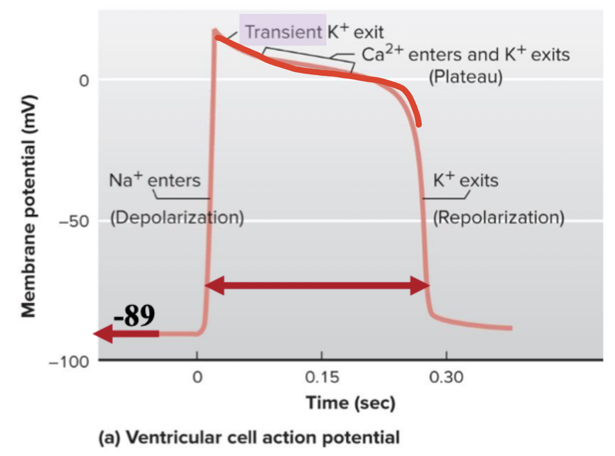
L-type Ca2+ channels
Long-lasting Ca2+ channels that are responsible for plateaus in APs after depolarization
The same thing as DHPRs
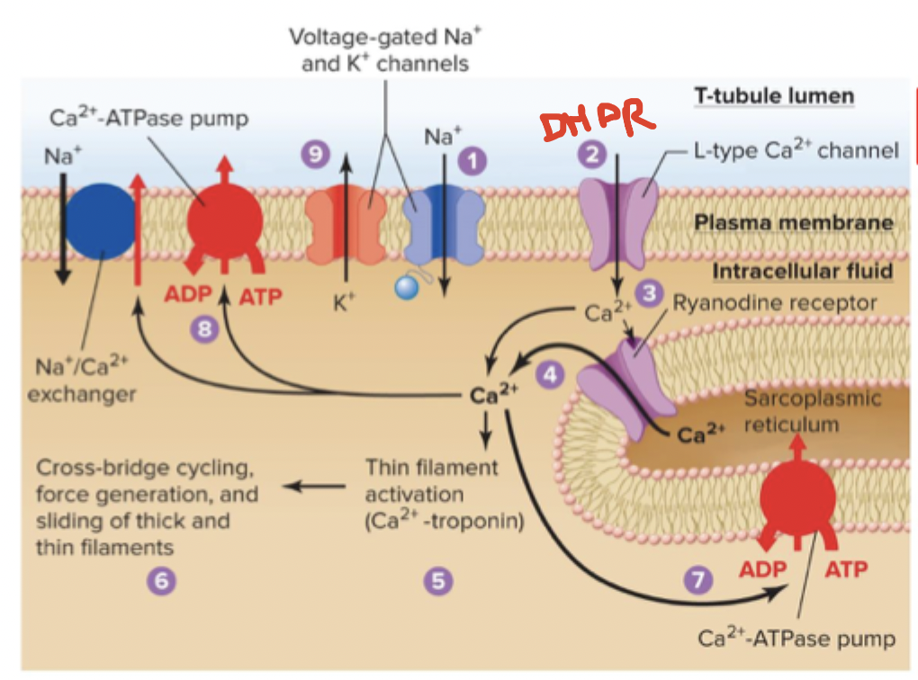
Walk through cardiac EC-coupling
Excitation
Membrane is depolarized by Na+ entry as AP begins
Depolarization opens DHPR (L-type Ca2+ channels) in T-tubules
CICR (calcium-induced calcium release) - entry of trigger Ca causes Ca to bind to and open Ryr —> Ca goes into the cytosol
Contraction
Binding of Ca exposes cross-bridge binding sites on thin filaments
Cross bridge cycling generates force, filaments slide
Relaxation
SERCA pump returns Ca to SR
Na/Ca exchangers
Ca ATPase pump
Membrane repolarizes
Cardiac muscles and tetanus
Tetanus is a sustained contraction. Can’t occur in cardiac muscles due to the prolonged refractory period and lack of summation in cardiac muscle
Occurs to allow ventricles to refill
All muscle cells…
Have myosin thick and actin thin filaments
Have a cross-bridge cycle
Have sliding filament mechanism
Use ATP to generate force
Include elevated cytosolic Ca to initiate contraction
These muscle cells are small and uninucleated
Smooth and cardiac
These muscle cells have troponin and tropomyosin
Skeletal and cardiac
What role does the cardiovascular system have on homeostasis
It’s the main transport system
Average total blood volume
5.5 L
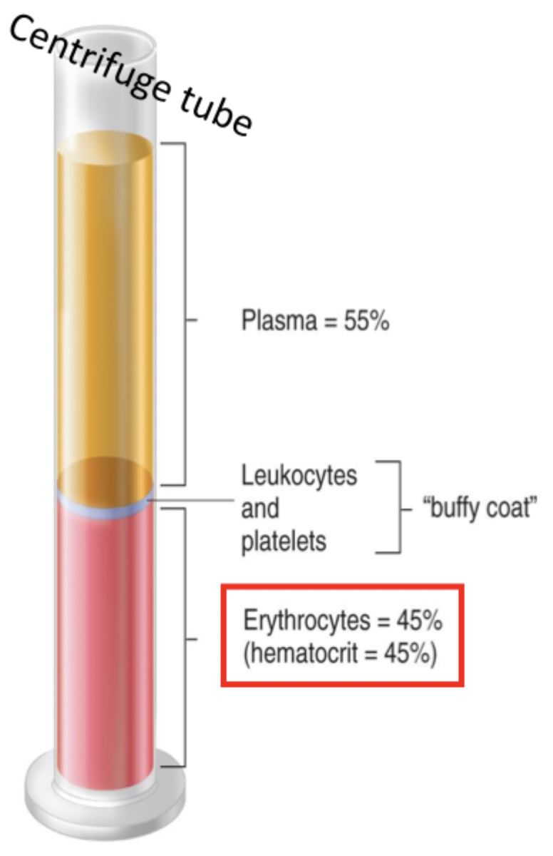
Plasma (centrifuge)
Averages 3 L or 55-58%
Is like the ISF but has plasma proteins
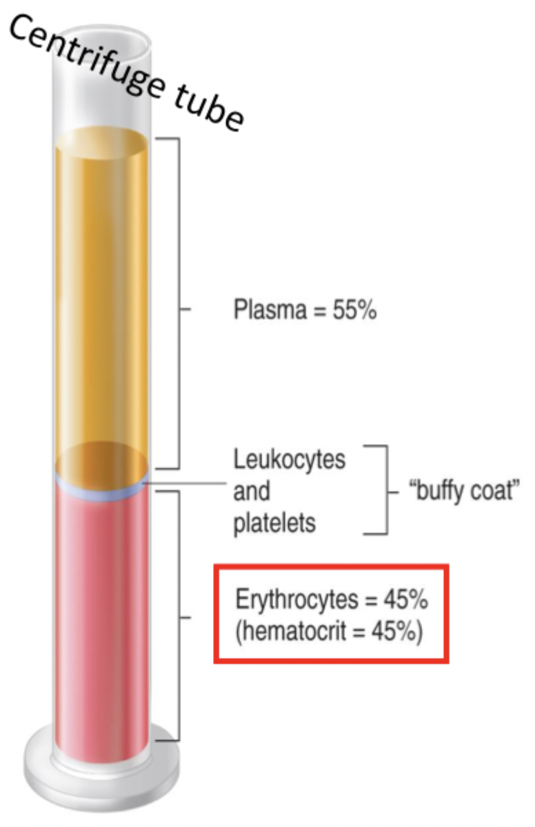
Buffy coat (centrifuge)
Insignificant volume
Contains leukocytes and platelets
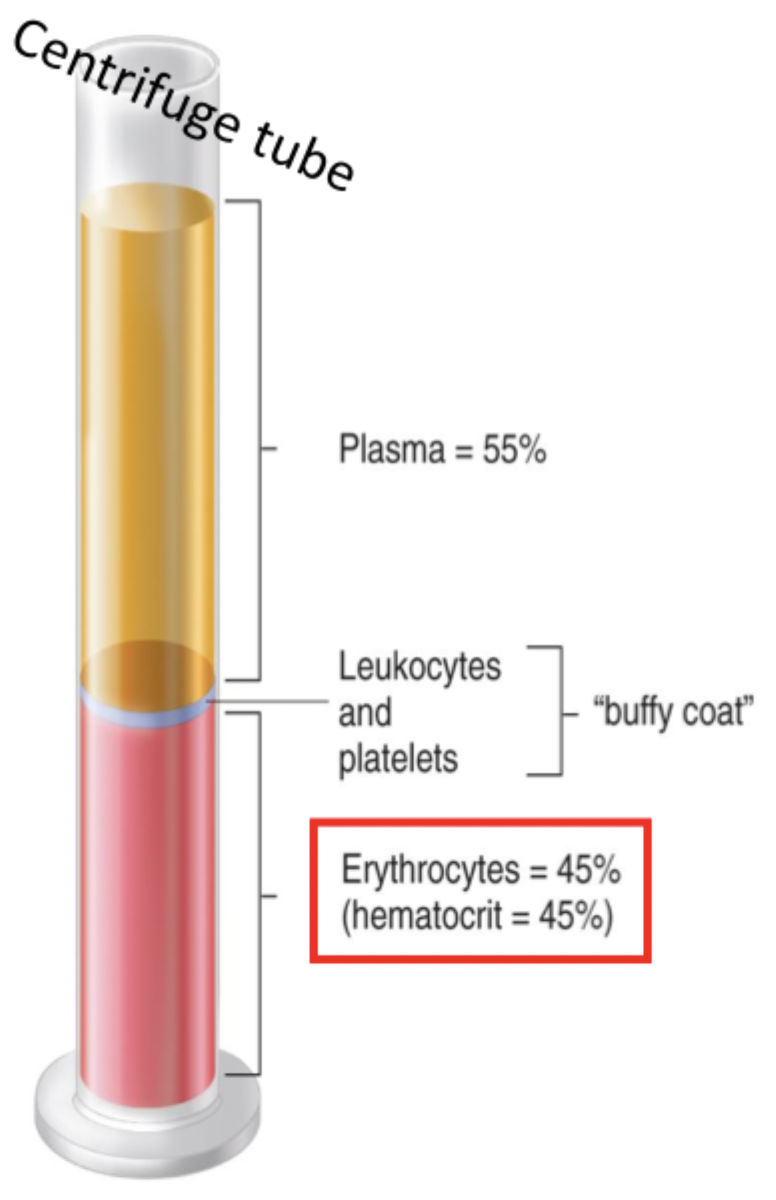
Hematocrit (centrifuge)
Averages 2.5 L or 42-45%
Contains erythrocytes/RBCs
Explain erythrocytes
Biconcave discs with a large surface area and small volume for rapid diffusion
Carries hemoglobin
Doesn’t contain DNA
4 commitments of blood cells
Reticulocyte —> RBC —> O2 transport
Megakaryocyte —> platelets —> clotting
Monocyte, neutrophil, eosinophil, basophil —> WBC —> immunity defense
B, T-lymphocytes —> immunity defense
Bone marrow
Produces erythrocytes
Define arteries
Carries blood away from heart
Define veins
carries blood toward the heart
Define perfusion
Passage of blood through a vascular bed, moves by bulk flow from high to low pressures
Why are vascular beds parallel and not in a series?
Blood quality - ensures every organ gets the same amount of O2, nutrients, CO2 waste
Flow regulation - reducing blood flow in one organ will still have lots of flow to other organs
Initial pressure - pressure is lost through each organ, but being parallel allows for 110/70 pressure for all organs
Follow the flow of blood through the pulmonary and systemic circulation (start at vena cava)
Vena cava —> right atrium —> right AV (tricuspid) valve —> right ventricle —> pulmonary semilunar valve —> pulmonary arteries —> arterioles —> lung capillaries —> pulmonary veins —> venules —> left atrium (pulmonary)
Left atrium —> left AV (bicuspid/mitral) valve —> left ventricle —> aortic semilunar valve —> aorta —> systemic arteries —> arterioles —> capillary beds —> systemic veins —> venules —> vena cava (systemic)
List the 3 types of cardiac muscle cells
Pacemaker, conducting, and contractile cells
Describe pacemaker cells
Has automaticity, SA node usually determines the heart rate as 100-120 APs/min
Uses nodal cell APs
Describe conducting cells
All are conducting (due to gap junctions) but few are specialized to rapidly spread electrical stimulus through chambers
Includes the bundle of his, bundle branches, and Purkinje fibers
Use conducting APs
Describe contractile cells
99% of cardiac muscle cells whose activity allows blood to be pumped out of the heart
Use fast-conducting APs
Describe coronary circulation
Blood supply to the heart through coronary arteries that branch off the aorta and coronary veins that return to the right atrium
Pressure, flow, and resistance equations
n = viscosity
Flow and resistance are inversely related
Radius and resistance are inversely related
Radius and flow are directly related

Relationship between resistance and radius
The smaller the radius (vasoconstriction), ↑ resistance, ↓ flow
The bigger the radius (vasodilation), ↓ resistance, ↑ flow
Purpose of heart valves
To promote a one-way direction of blood flow
How do pressure gradients affect valves
If the pressure is greater in 1 than in 2, blood will flow from 1 to 2. If pressure is greater in 2 than in 1, the valve will be closed and blood from 1 and 2 will bounce off the valve
Define prolapse
Heart valve flops backward onto the chamber behind it when it should be closed
Where are the chordae tendineae and papillary muscles located
Ventricular chambers
How do the chordae tendineae and papillary muscles prevent prolapse?
Ventricles get filled —> valves want to flip the other way —> papillary muscles contract and pull downward, pulling the chordae tendineae to keep the cusps closed against ventricular pressure
Define stenoic valve
A valve that doesn’t open all the way, turbulent flow sounds like a whistle and affects the lub/dub after it
Define insufficient valve
A valve that leaks backward due to poor closing, turbulent flow sounds like a gurgle and affects the lub/dub before it
Explain the “lub” and “dub”
Lub - AV valve closing
Dub - SL valve closing
Define sinoatrial (SA) node
A few cells collected in the right atrium that reach threshold first and are the true pacemaker
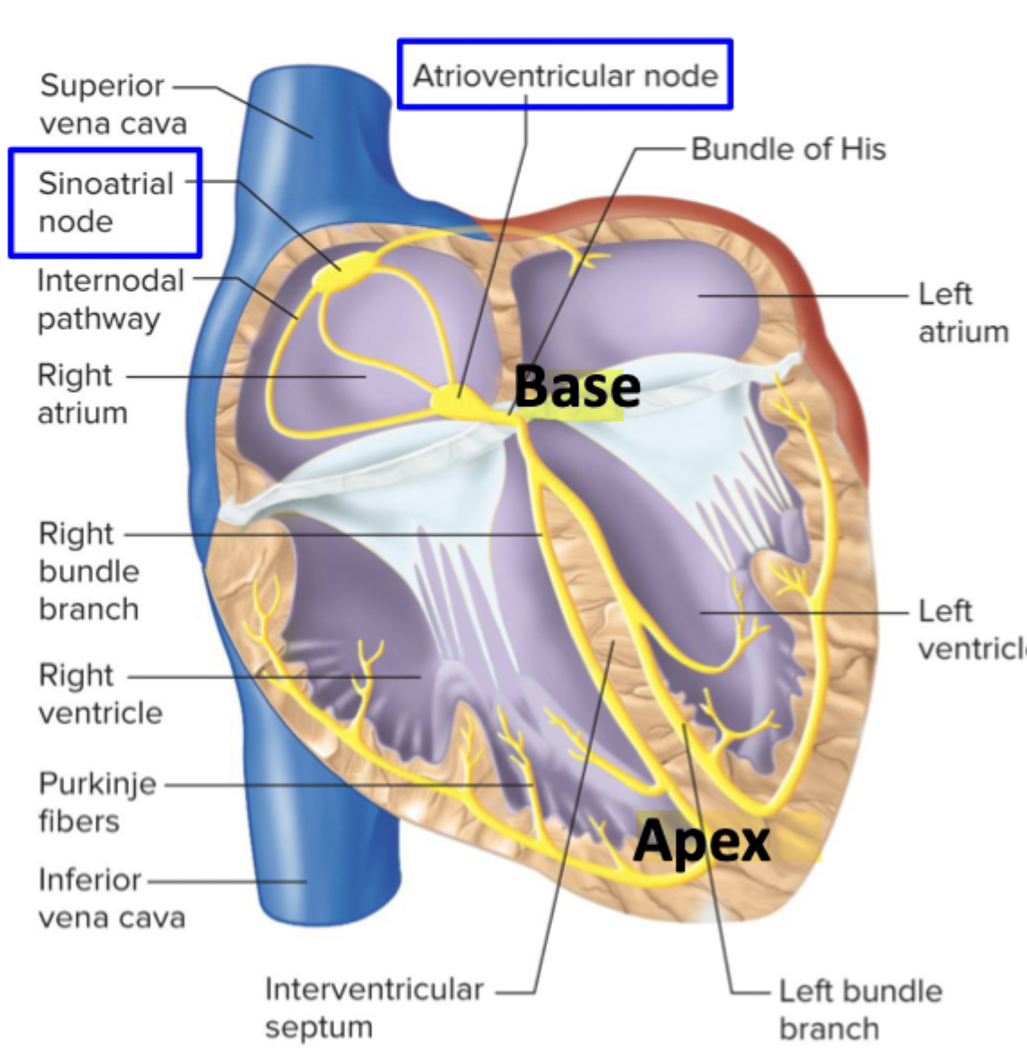
Define atrioventricular (AV) node
Electrical connection between atria and ventricles, slows AP propagation
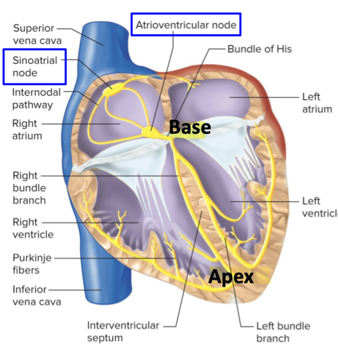
Define bundle of his, bundle branches, Purkinje fibers
All made of conducting cells so AP propagation is fastest, all in the ventricle
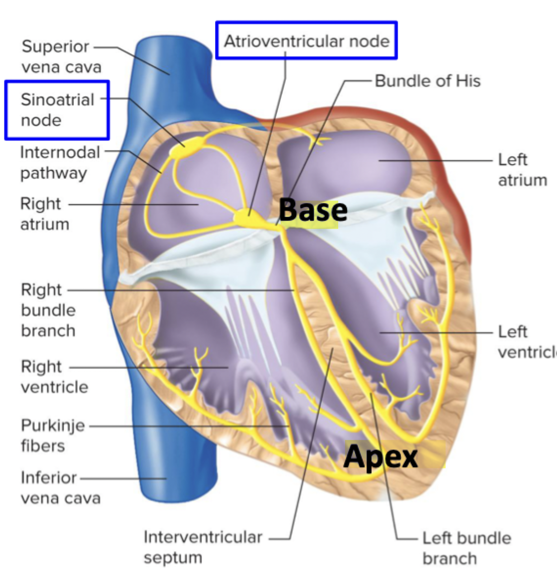
Implications of electrical syncytium
One cell starts AP
One diseased cell can cause fatal arrhythmia
Can install artificial pacemakers
No recruitment
Explain the Lead I ECG trace
P wave - atrial depolarization of both SA and AV nodes
AV node is too small, is a flat line after P wave
QRS - ventricular depolarization/systole
Atrial repolarization still occurs, isn’t shown
Q - septum, R - ventricular mass, S - base
T wave - ventricular repolarization/diastole
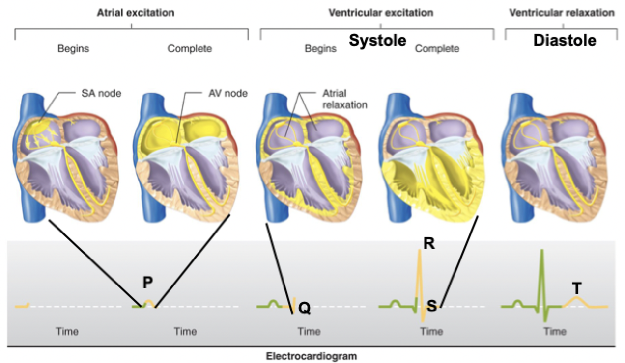
Define systole
Excitation/contraction of ventricles
Define diastole
Ventricular relaxation
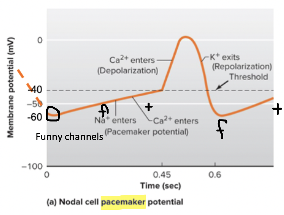
Explain the nodal cell AP
Starts at -60 mV
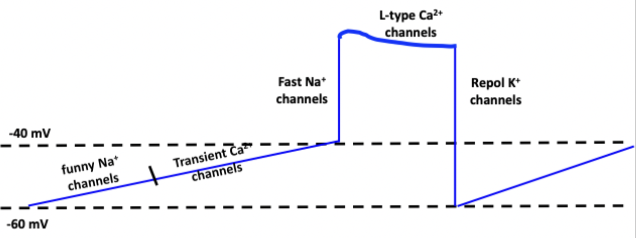
Na enters through funny Na channels, then Ca entry through T (transient) type channels, bringing membrane to threshold (-40mV)
Depolarization upstroke due to v-g L-type Ca channels
Repolarization by v-g K+ channels
Causes pacemaker potential
Explain fast-conducting cell APs
A mix of contractile and nodal APs
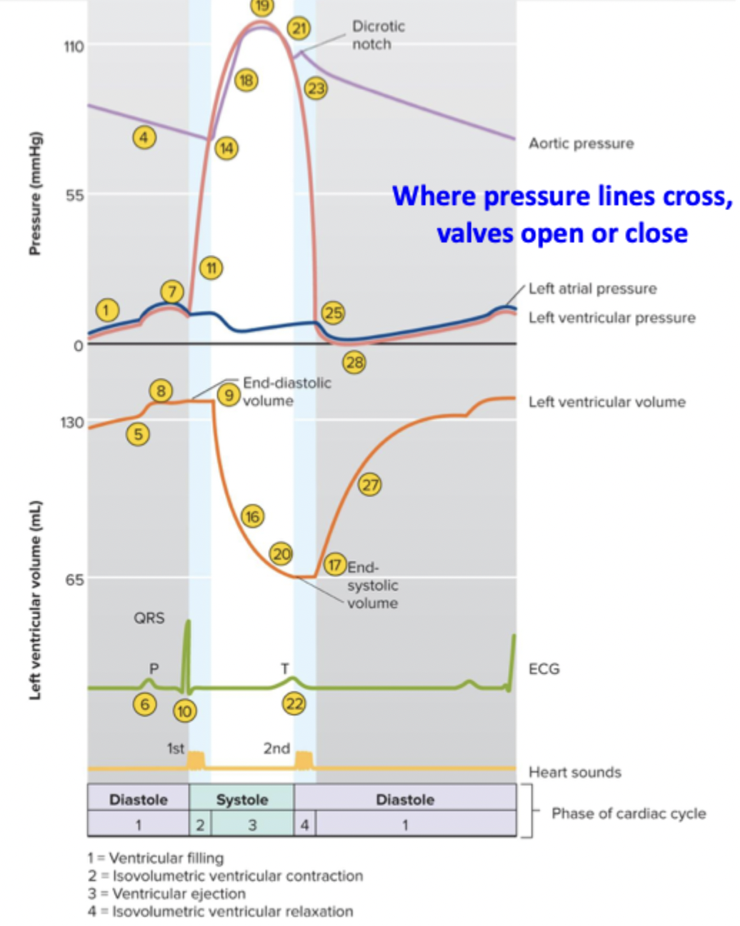
List the 4 periods in mechanical pumping
Ventricular filling (diastole), isovolumetric contraction (systole), ejection (systole), isovolumetric relaxation (diastole)
How much blood is in the left ventricle on average
135 L
Explain ventricular filling
There is an atrial kick where the atria first fill the ventricle with blood passively, then actively through contraction
Reaches end-diastolic volume
Diastole
End-diastolic volume
The final volume in the ventricle after filling, at least 135 mLs
Explain isovolumetric ventricular contraction
Ventricles contract
Hear “lub” (AV valves close), all valves are closed
Systole
Explain ventricular ejection
There is a stroke volume
Reach end-systolic volume
ESV = EDV - SV
SL valve is open
Systole
Stroke volume
The volume of blood ejected from each ventricle at rest, ~70mLs
End-systolic volume
The volume of blood remaining in ventricle after ejection
Explain isovolumetric ventricular relaxation
Hear the “dup”, SL valves close
Volume is constant at ESV
Diastole
Understand the Wigger’s diagram
Shows electrical events, pressures, volumes, valve events, and sounds of the heart
Understand Einthoven’s triangle
(+) is the recording electrode
(-) is the reference electrode
Explain the QRS axis and its shifts
QRS axis depicts a net direction of depolarization towards the left apex.
Left shift - directed left but up towards the base
Right shift- directed right
Define sinus rhythm
Typical cardiac excitation-contraction sequence beginning at SA node
Define latent pacemaker
Not active potential pacemakers, such as AV node and purkinje fibers
Define ectopic pacemaker
Abnormal, any site driving ventricular EC that isn’t the SA node. “If… then… has become…”
Define tachycardia, bradycardia, and fibrillation
Tachycardia - HR greater than 100 bpm
Bradycardia - HR slower than 60 bpm
Fibrillation - totally irregular and chaotic AP propagation
Define cardiac output
The volume of blood coming out of each ventricle per min. Is 5 L/min at rest
At rest, which autonomic nervous system dominates
Parasympathetic
How does increased physical activity affect autonomics?
↓ Parasympathetic output, ↑ sympathetic output
Cardiac output formula
= Heart rate x stroke volume
Intrinsic rate of pacemaker cells normal + rest (APs/min)
SA node - 100 → 50-70
AV node - 70 → 40-50
His/Purkinje - 30 → 30
Define chronotropic effects
Factors that affect heart rate
What 3 ion channels affect pacemaker potential (sympathetic)?
↑ Funny Na current, ↑ T-type Ca current, same K current
Gets potential to threshold sooner
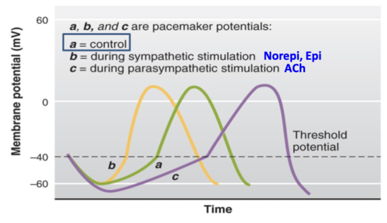
What 3 ion channels affect pacemaker potential (parasympathetic)?
↑ v-g K current, ↓ funny Na current, ↓ T-type Ca current
Slows down potential
Define dromotropic effects
Change in conduction velocity, specifically, the upstroke of the AP curve
What ion channel affects conduction velocity in nodal cells
L-type Ca current. ↓ in parasympathetic, ↑ in sympathetic
Stroke volume equation
End-diastolic volume - end-systolic volume
Explain the Frank-Starling mechanism / “preload”
If ventricle fills to a larger volume, improving overlap of actin and myosin crossbridging → next beat ejects more blood
An intrinsic mechanism of regulating SV by changing ventricular EDV (↑ EDV → ↑ length of cells → ↑ tension → ↑ SV)
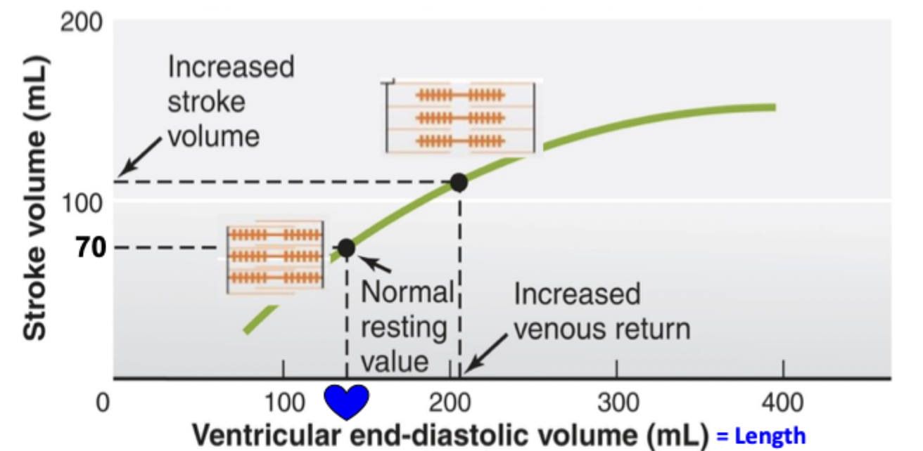
Implications of Frank-Starling mechanism
Prevents rise in ESV, preventing clotting
Prevents rise in venous pressure, forcing out blood back ups that can cause edema
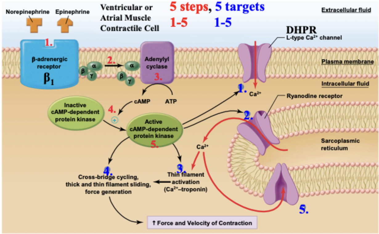
Explain sympathetic regulation of SV
Sympathetic nervous system releases NE + E, which can bind to a b1 receptor → G proteins → adenylyl cyclase —> activates protein kinase A —>
5 targets of phosphorylation for…
Faster and more Ca release (DHPR/L-type + RyR)
Stronger, briefer contraction (thin and thick filaments)
Faster Ca removal (SERCA pump)
Effects of sympathetic regulation of SV
Increased contractility by increasing Ca release at any given EDV, makes new Frank-Starling curve
Increased L-Ca current, more Ca in cytosol, more troponin saturation
Faster reuptake
Increased ejection fraction
Is an ionotropic effect
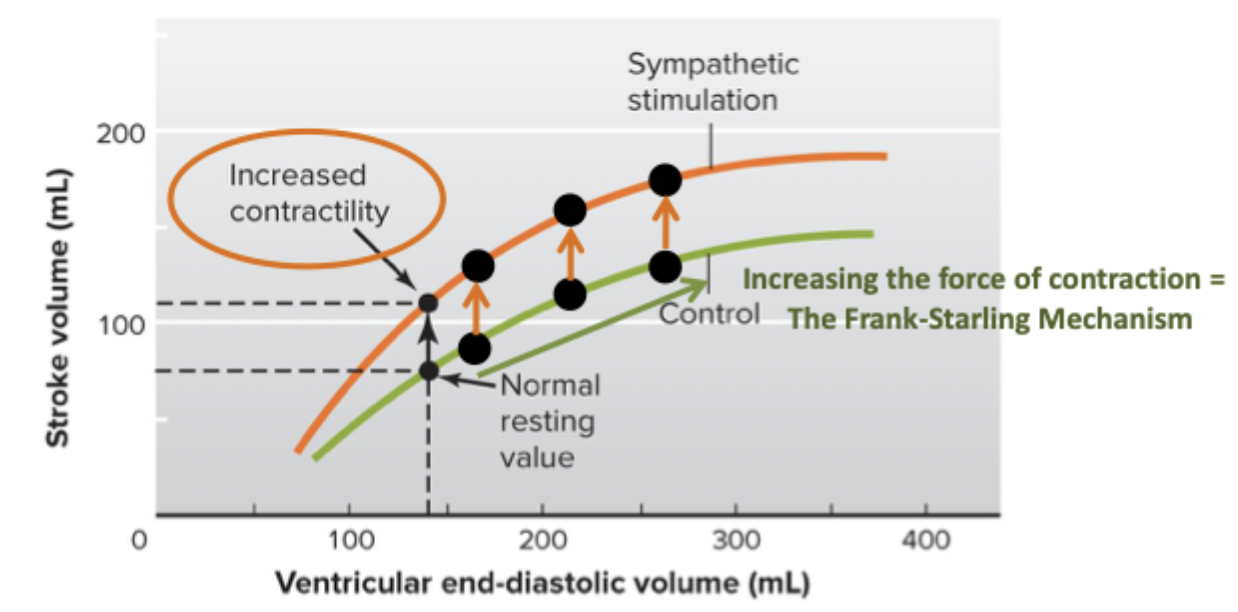
Ionotropic effect
Changing contractility that is resulting from any given EDV
Define ejection fraction + formula
Clinical measurement of contractility
EF = SV/EDV
Where are parasympathetic neuronal signals sent from
The vagus nerve
Explain parasympathetic ganglion anatomy
Sends signals through long pre-ganglionic axons, short post-ganglionic
Ganglion is near where heart is
Where are sympathetic neuronal signals sent from
Thoracic spinal nerves
Explain sympathetic ganglion anatomy
Sends signals through short pre-ganglionic axons, long post-ganglionic axons
What organ secretes epinephrine
Adrenal medulla