NOD 1 Afferent System and Neuroimaging Review
 Call Kai
 Learn
Learn Practice Test
 Spaced Repetition
 Match
Match Flashcards
 Knowt Play
1/120

There's no tags or description
Looks like no tags are added yet.
Name | Mastery | Learn | Test | Matching | Spaced | Call with Kai | Chat |
|---|
No analytics yet
Send a link to your students to track their progress
121 Terms
anterior to the chiasm
Where is the lesion/problem if there is unilateral vision loss?
amblyopia
What is the one exception to the localization of a lesion/problem causing unilateral vision loss?
between chiasm and occipital cortex
Where does the lesion localize if there is a homonymous hemianopia?
4
How many HPIs must be recorded?
foveolar vision, estimates central scotoma size, screens for hemianopic defects and metamorphopsia, functional visual loss and suggests anatomical localization
What does Snellen acuity measure and screen for?
foveal disease
What does the presence of metamorphopsia indicate?
any patient that isn't 20/20
When should pinhole be done?
central media opacity or scotoma, amblyopia, non-organic vision loss, pathology
What is indicated by no improvement even with pinhole?
40'
What should stereopsis be in a patient with 20/20?
20/400
What acuity is the number on a dollar bill?
20/70
What acuity are the serial numbers on a dollar bill?
20/40 - 20/50
What acuity of the numbers on a wristwatch?
5 degrees
What would the estimated size of a central scotoma be on a patient with 20/50 acuity?
20
What would the estimated size of a central scotoma be on a patient with 20/200 acuity?
20/70
What would the estimated acuity be on a patient with a 7 degree central scotoma?
red
What color is optic nerve function most sensitive to?
red desaturation (stationary red target) and confrontation fields with red target moved throughout field
What tests can be used to compare optic nerve function between both eyes?
brachium of the superior colliculus to get the pretectal nucleus
What does the light reflex pathway use?
no
Is the presence of an RAPD related to anisocoria?
optic neuropathy
What does the presence of an RAPD indicate?
anterior the LGN
Where would a lesion causing an RAPD localize to?
compression, glaucoma, inflammation, infarction, demyelination, trauma, papilledema
What are optic neuropathies that can cause an RAPD?
CRAO, CRVO, RD w/ macular involvement, macular disease
What are retinal causes of an RAPD?
early release
What does a 1+ RAPD grade indicate?
no initial movement followed by early release
What does a 2+ RAPD grade indicate?
immediate release
What does a 3+ RAPD grade indicate?
amaurotic pupil associated w/ no light perception
What does a 4+ RAPD grade indicate?
the good eye
Which eye is the neutral density filter placed over?
0.3 log filter over an eye
How is a neutral density filter used to tease out a subtle RAPD?
reverse RAPD testing, compare direct and consensual response to light
What is the next step if 1 pupil doesn't respond to light during RAPD testing?
outer retina or choroid
Where does a lesion resulting in a defect that fails to respect both horizontal and vertical meridians localize to?
arcutes
Where does a lesion resulting in a defect that respects the horizontal meridian localize to?
chiasm or retrochiasm
Where does a lesion resulting in a defect that respects the vertical meridian localize to?
occipital cortex
Where does a lesion resulting in a defect that respects both the horizontal and vertical meridians localize to?
No
Does the papillomacular bundle respect the horizontal raphe?
arcuate bundles
What retinal nerve fiber bundles respects the horizontal raphe?
temporal defect or enlarged blind spot
Which defects are not consistent with glaucoma?
nasal step, bjerrum scotoma, arcuate defect
What are typical glaucomatous defects?
No
Is optic disc pallor associated with glaucoma?
the ON appearance must match the VF and glaucoma is usually bilateral
If the visual field defect is consistent with glaucoma, what else must be present to be able to officially diagnose it as glaucoma?
enlarged blind spot, arcuate
What visual field defect is seen in a patient with papilledema?
central/cecocentral
What visual field defect is seen in a patient with optic neuritis?
altitudinal
What visual field defect is seen in a patient with NAAION?
devastating field loss
What visual field defect is seen in a patient with AAION?
central/cecocentral
What visual field defect is seen in a patient with toxic/nutritional optic neuropathy?
incongruous
The more anterior the lesion, the more _____________ the defect.
congruous
The more posterior the lesion, the more _____________ the defect.
total or complete hemianopia
When is it not possible to assess congruity of a defect?
RNFL thickness
What part of the OCT report must be reviewed when assessing for papilledema?
No
Is elevation of the NRR without RNFL thickness elevation indicative of papilledema?
optic nerve disease/damage
What is ganglion cell loss concerning for?
loss, pallor, edema
What must you look for when assessing the health of the neuro retinal rim/optic disc?
thickest regions swell first
Which regions is elevation first seen in edema?
ischemic optic neuropathy, compression, inflammation, trauma, hereditary/mitochondrial
What are non-glaucomatous causes of cupping?
1 month
How long after damage to the ganglion cells or their axons does pallor develop?
LGN and forward
Where would a lesion have to be to cause pallor?
compression, infection, inflammation
What does diffuse neuro retinal rim pallor indicate?
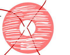
toxic/nutritional, demyelinating, compression, infection, inflammation
What does temporal neuro retinal rim pallor indicate?

optic tract compression
What does bow tie or band atrophy neuro retinal rim pallor indicate?
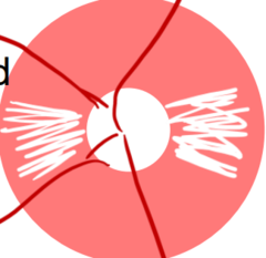
ischemic/NAION, infection, inflammation
What does altitudinal neuro retinal rim pallor indicate?

glaucoma
What does neuro retinal rim obliteration indicate?

glaucoma, compression
What does neuro retinal rim thinning without pallor indicate?

compression
What does neuro retinal rim thinning with pallor of the remaining neuro retinal rim indicate?

papilledema, optic neuritis, NAAION, AAION, Neuro-retinitis, disc drusen, anomalous disc
What are causes of indistinct optic disc margins?
1 mm
What is the length of the papilla optic nerve segment?
30 mm
What is the length of the intra-orbital optic nerve segment?
6 mm
What is the length of the intra-osseous optic nerve segment?
10 mm
What is the length of the intracranial optic nerve segment?
falx cerebri
What divides the cerebral hemispheres?
tentorium cerebelli
What divides the occipital lobe from the cerebellum?
septum pellucidum
What divides the lateral ventricles?
CT or X-ray
What imaging is used for bone?
2 layers of grey and white matter
What does the septum pellucidum consist of?
meninges
What is the falx cerebri derived from?
tells you the location/plane of the section you are viewing
What is a SCOUT scan?
bright
How do EOMs appear in an MRI with contrast?
grey
How do EOMs appear in an MRI without contrast?
temporalis muscle
What is being marked by the yellow star?

masseter
What is being marked by the pink star?

lateral pterygoid muscle
What is being marked by the blue star?

medial pterygoid muscle
What is being marked by the orange star?

paranasal sinuses
What drains mucus into the nasal cavity?
eustachian tube in the nasopharynx
What connects the paranasal sinuses to the middle ear?
close proximity (referred pain / trauma)
What is the relationship between the orbital cavity and the paranasal sinuses?
10 mm
How much space should be between the chiasm and pituitary in suprasellar cistern?
tissue density
What does a CT focus on?
tissue signal intensity
What does a MRI focus on?
CSF, fat, air
What is considered hypodense on a CT scan?
calcium, hemorrhage, metal, bone, iodine
What is considered hyperdense on a CT scan?
brain
What is considered isodense on a CT scan?
bone and blood
What is a CT scan useful in looking for?
CT
What scan would be helpful to detect a nerve sheath meningioma?
axial and coronal only
What sections are possible using a CT scan?
ionizing radiation, limited by head movement, more allergic rxns to its contrast
What are disadvantages of a CT scan?
sella turcica and suprasellar cistern
What cannot be viewed using a CT scan?
shellfish allergies, asthma, DM, kidney dysfunction
Who is CT contrast contraindicated in?
BUN/creatinine levels
What must be check prior to administering CT contrast to a patient?
CTA
What is the study of choice to look for aneurysms?
cervico-medullary junction, pituitary gland, pineal region, corpus callosum
What is the sagittal section of an MRI useful for viewing?
No
Is there radiation exposure in an MRI?