intro to rad positioning 8/31
 Call Kai
 Learn
Learn Practice Test
 Spaced Repetition
 Match
Match Flashcards
 Knowt Play
1/114

Earn XP
Description and Tags
merrill ch 2
Name | Mastery | Learn | Test | Matching | Spaced | Call with Kai | Chat |
|---|
No analytics yet
Send a link to your students to track their progress
115 Terms
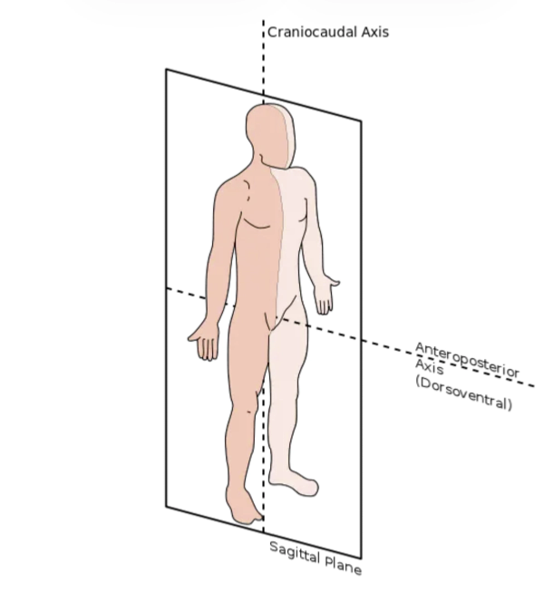
sagittal plane
divides the body or a body part into left and right segments vertically
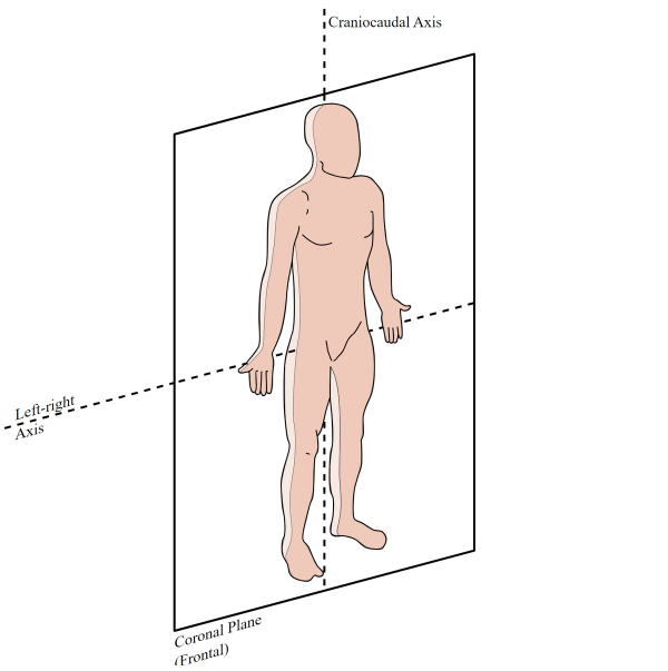
coronal plane
divides the body or a body part into anterior and posterior segments vertically
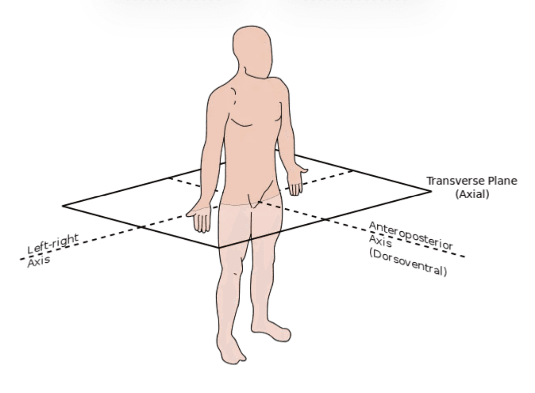
transverse plane
divides the body into superior and inferior segments horizontally (crosswise). also referred to as axial, transverse, or cross-sectional plane.
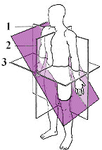
oblique plane
passes through the body or a body part at an angle
interiliac plane
special horizontal body plane that cuts across the pelvis at the highest points of the hip bones (iliac crests), level with the fourth lumbar (L4) vertebra

the interiliac plane serves as a surface landmark for which anatomical structures?
helps in positioning the lumbar spine, sacrum, and coccyx
occlusal plane
imaginary surface of where the upper and lower teeth meet when biting down
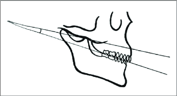
radiographic use of the occlusal plane
helps position the odontoid process and in some head projections
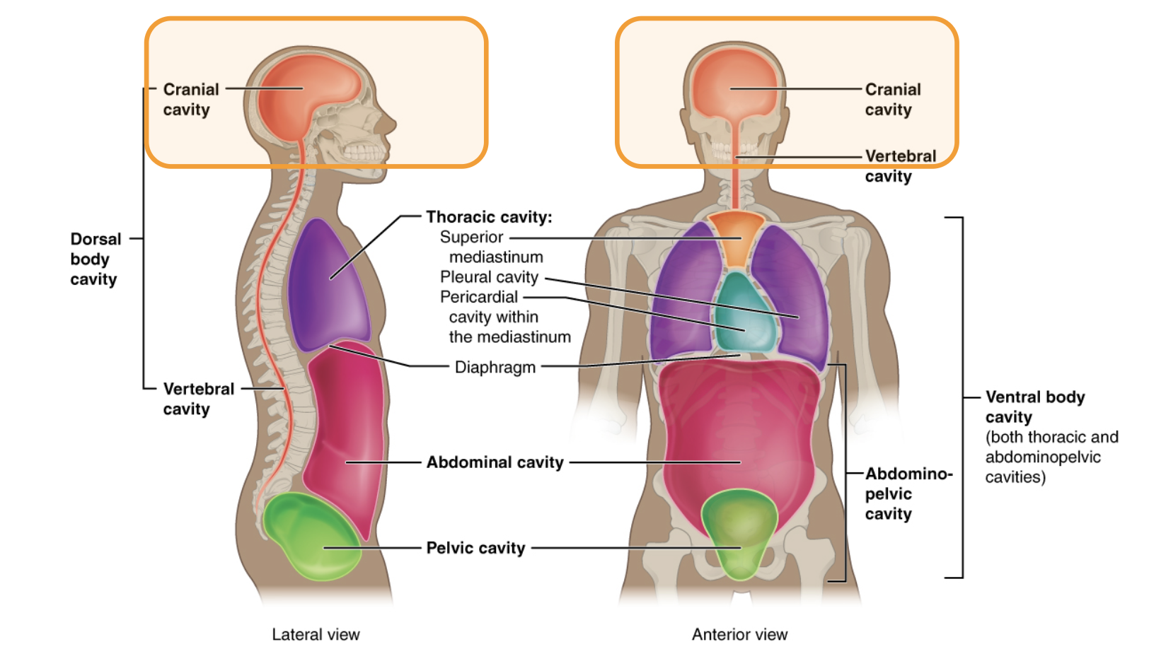
cranial cavity
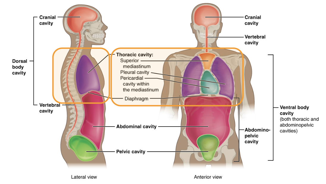
thoracic cavity
segments of the thoracic cavity
two pleural portions (left and right pleural cavity) and mediastinum

abdominal (abdominopelvic) cavity
what is body habitus?
refers to shape of the human body which determines the size, shape, and position of internal organs
how does body habitus affect radiography?
determines positioning, exposure factors, and IR size & placement
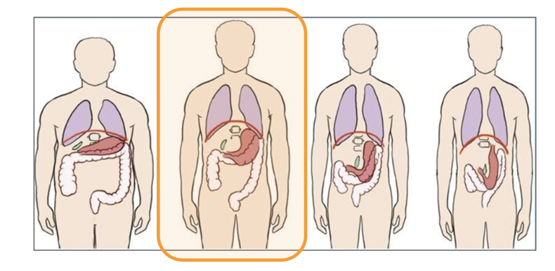
sthenic body habitus
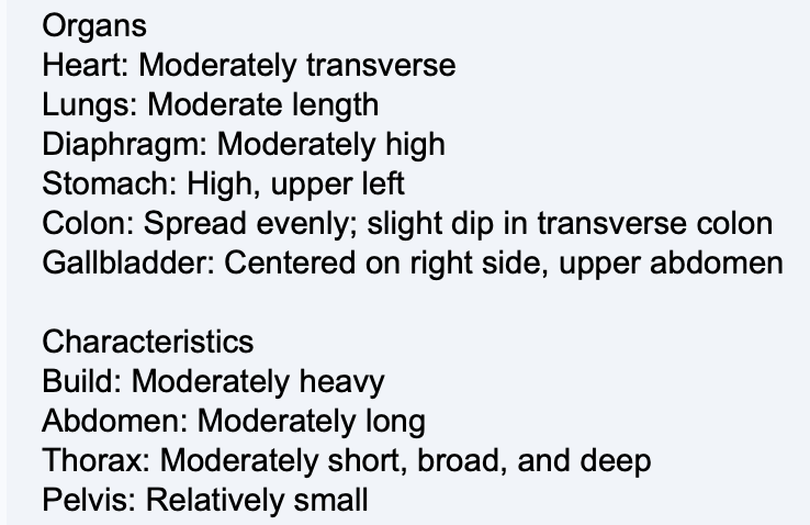
body habitus of 50% of human population
sthenic body habitus
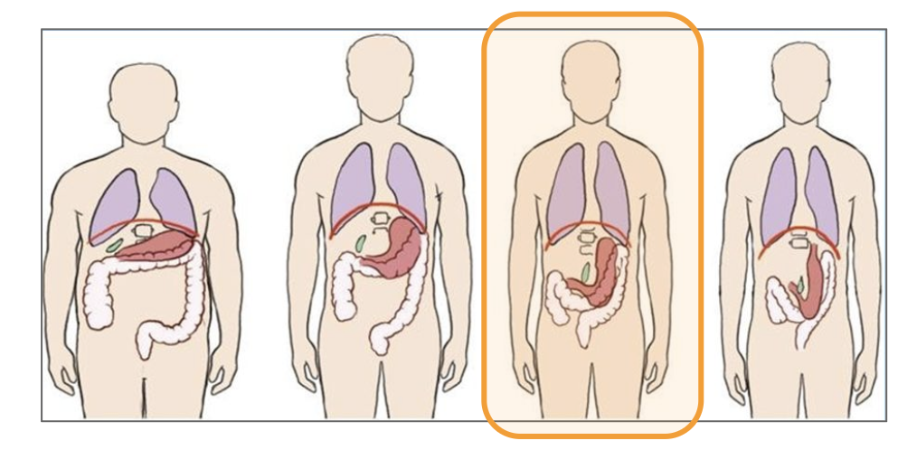
hyposthenic body habitus
characteristics of hyposthenic body habitus
most difficult to classify but it is intermediate between sthenic and asthenic. 35% of human population.
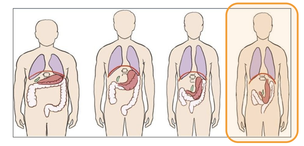
asthenic body habitus
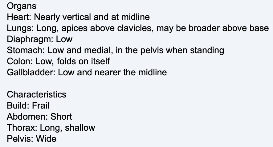
body habitus of 10% of human population
asthenic body habitus
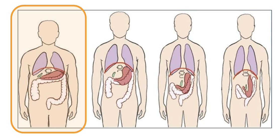
hypersthenic body habitus
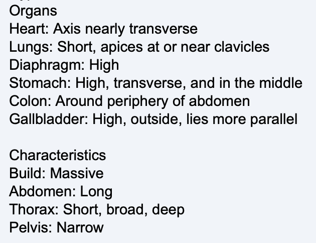
body habitus of 5% of human population
hypersthenic body habitus
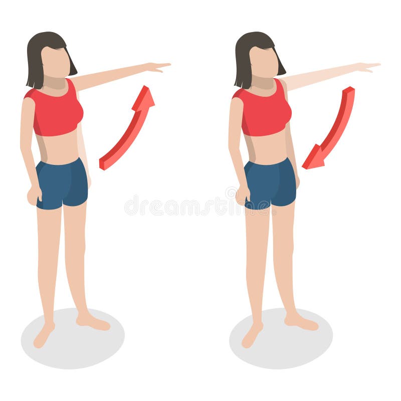
abduction/adduction

extension/flexion
eversion
outward turning of the foot at the ankle
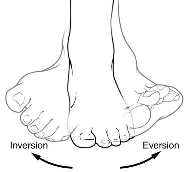
inversion
inward turning of the foot at the ankle

pronate
rotation of the forearm so that the palm is down

supinate
rotation of the forearm so that the palm is up (anatomic position)
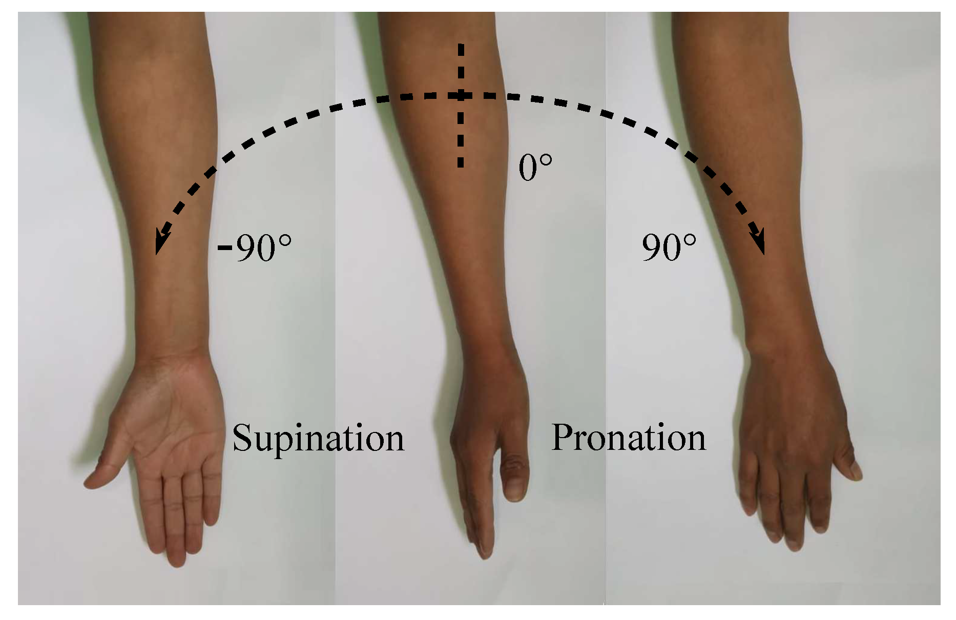
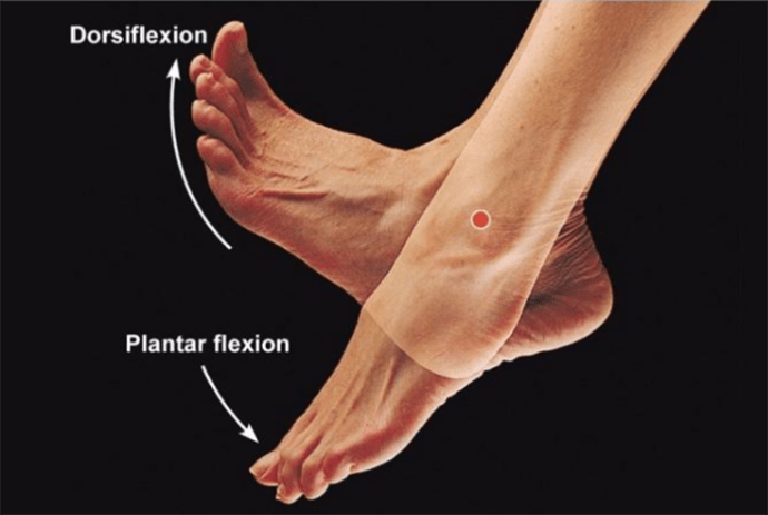
dorsiflexion
flexion or bending of the foot toward the leg
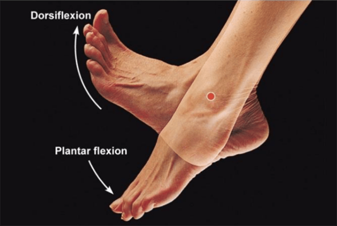
plantar flexion
flexion or bending of the foot downward toward the sole
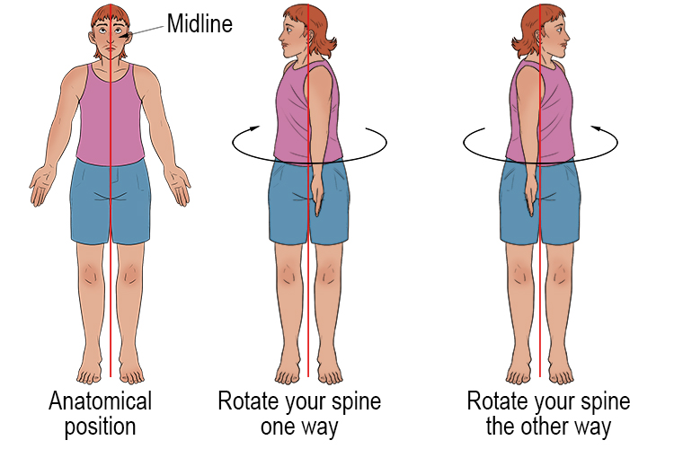
rotation
turning of a body or body part around its axis
circumduction
circular movement of limb

tilt
tipping or slanting a body part slightly
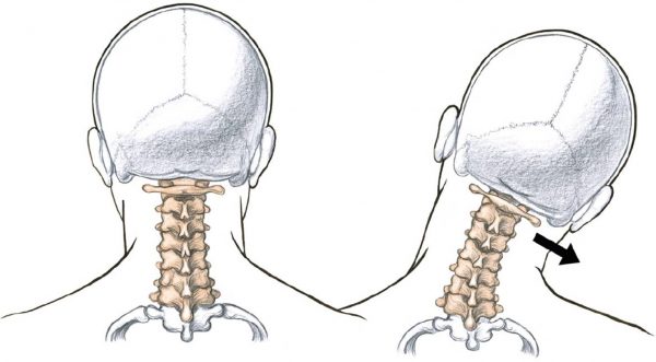
deviation
turning away from the regular standard or course
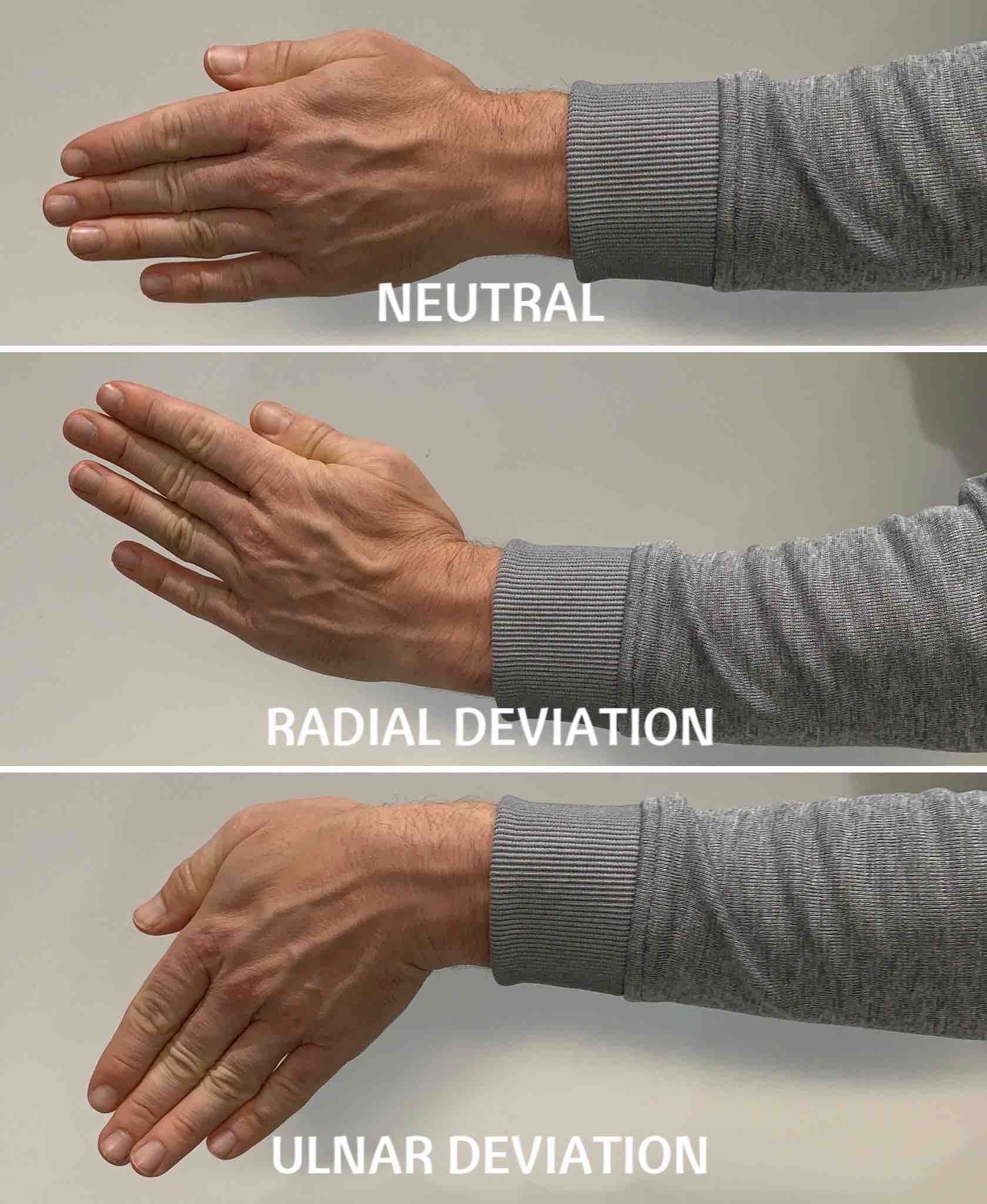
inferior
refers to nearer the feet or situated below
superior
refers to nearer the head or situated above
distal
refers to parts farthest from point of attachment
proximal
refers to parts nearest to point of attachment
distal
refers to parts farthest from point of attachment
superficial
parts near skin or surface
palmar
palm of hand
plantar
sole of foot
dorsum
top surface of foot or back surface of hand
peripheral
refers to parts at or near the surface, edge, or outside of another body part
ipsilateral
on the same side of the body
contralateral
on the opposite side of the body
parietal
refers to the wall or lining of a body cavity
visceral
refers to the covering of an organ
external
parts outside an organ
internal
parts within or inside of an organ
definition of osteology
detailed study of the body of knowledge related to bones
how many bones in the body?
206 primary bones
what are the main functions of bones?
• attachment for muscles
• mechanical basis for movement
• protection of internal organs
• frame to support the body
• storage for calcium, phosphorus, and other salts
• Production of red and white blood cells
division of the skeletal system
axial and appendicular skeleton
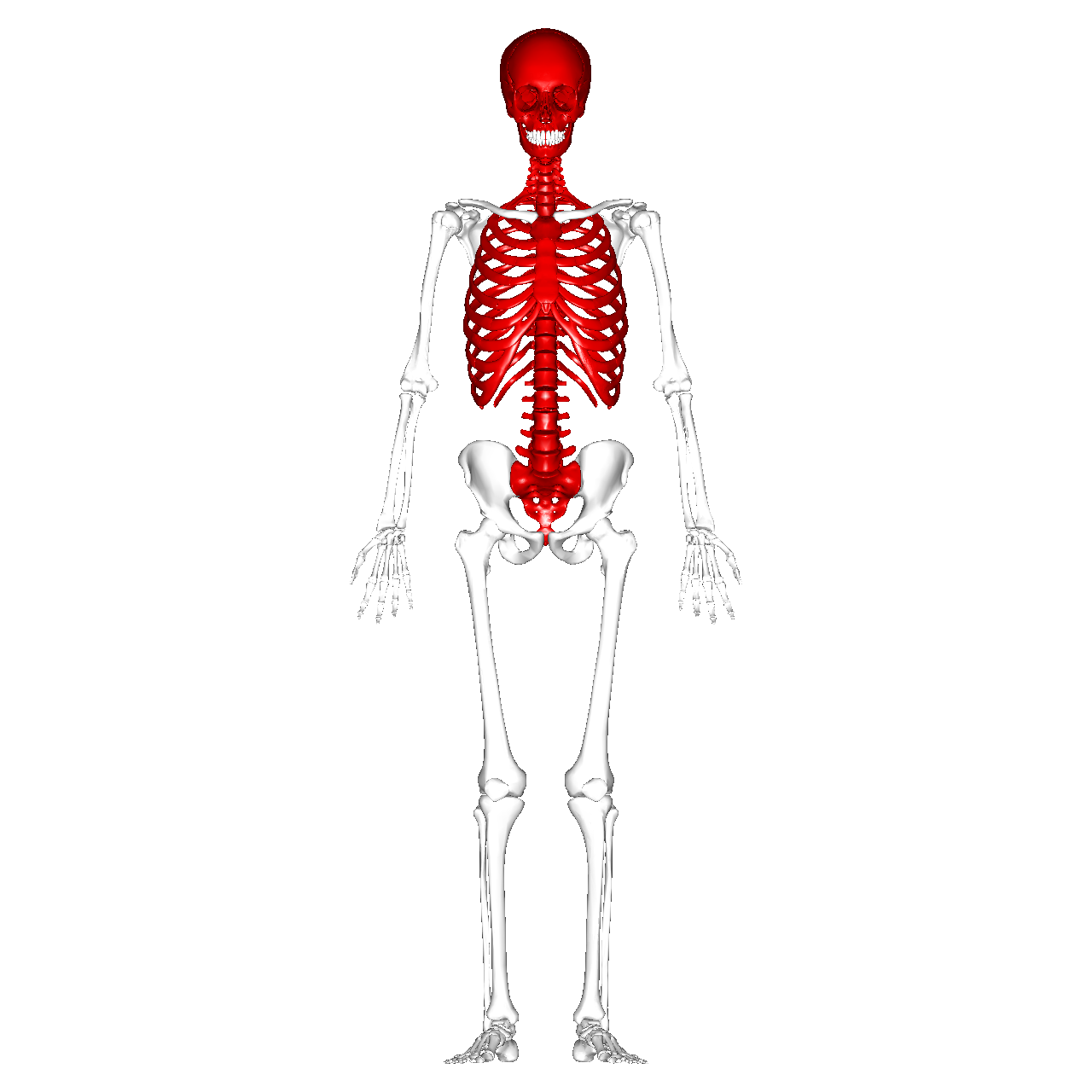
what makes up the axial skeleton?
80 bones that supports and protects the head and trunk
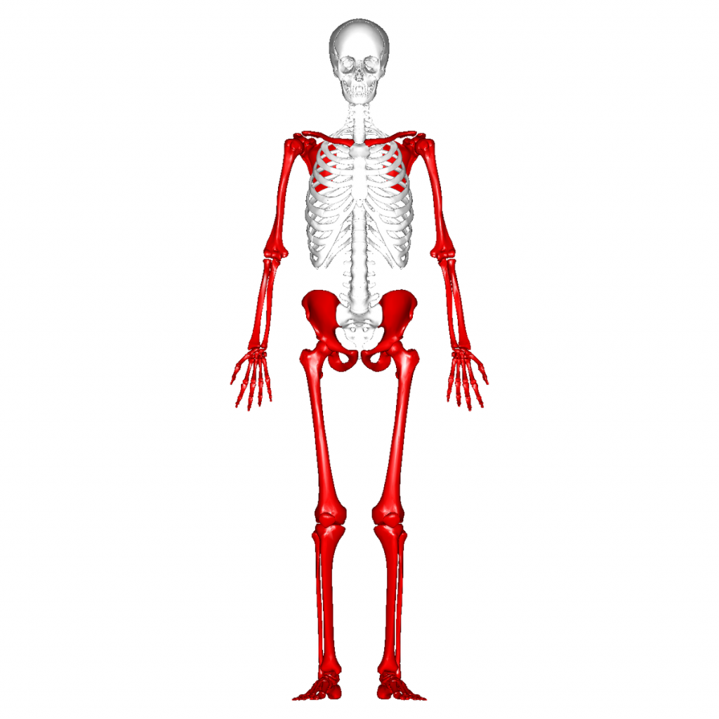
what makes up the appendicular bones?
126 bones that allows the body movement
compact bone
strong, dense outer layer of all bones
spongy bone
inner portion of bones that contains interconnecting spaces called trabeculae
red marrow
stored between the trabeculae
produces rbc and wbc
concentrated at the ends in long bones
yellow marrow
stores fat/adipose cells
medullary cavity
central cavity in long bones that are filled with yellow marrow
periosteum
tough, fibrous connective tissue covering all bone surfaces except articular surfaces
what are articular surfaces in bones?
specific parts of a bone that come into direct contact with another bone to form a joint
endosteum
tissue lining of the medullary cavity
foramina (openings in bones)
entrance and exit of blood vessels and nerves supplying the bones
ossification
development and formation of bones which begins during the second embryonic month
intermembranous ossification
bones that develop from fibrous membranes create flat bones
endochondral ossification
bones that develop from hyaline cartilage create short, irregular, and long bones
what are the two construction sites of endochondral ossification?
primary ossification begins before birth as diaphysis
secondary ossification begins after birth as epiphysis
what is the epiphyseal plate?
plate of cartilage between the diaphysis and epiphysis during growth
common site of pediatric fractures
fully ossifies and fuses into an epiphyseal line near age 21
classification of bones
long, short, flat, irregular, and sesamoid
long bones
found only in limbs
consists of body/shaft and two rounded articular ends

short bones
trabecular bone that varies in shape and allow minimal flexibility
carpal and tarsal bones only
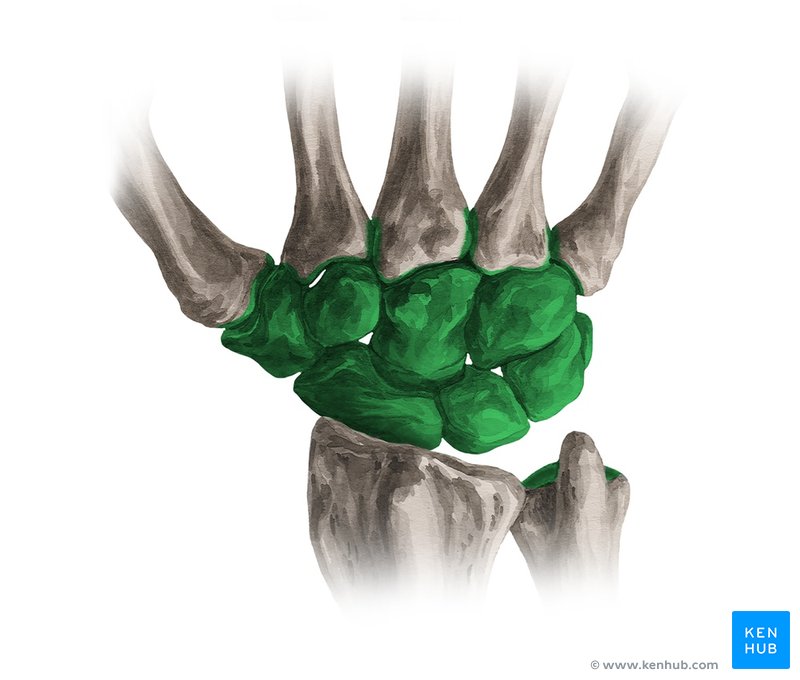
flat bone
consists of two tables of compact bone enclosing spongy bone

irregular bones
peculiar shapes that don’t fit with other categories
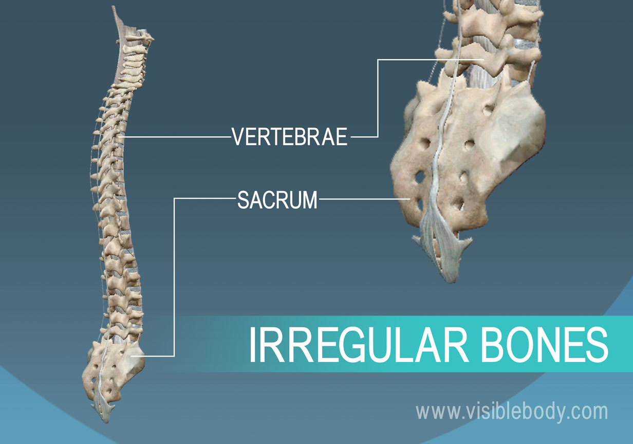
sesamoid bones
small, oval bones
develop in tendons to decrease friction

arthrology
study of joints (articulation between bones)
functional classification of joints
synarthroses = immoveable
amphiarthroses = slightly moveable
diarthroses = freely moveable
structural classification of joints
fibrous = immoveable
cartilaginous = virtually immoveable (intervertebral disc)
synovial = freely moveable (knee, elbow)
characteristics of fibrous joints
bound by dense connective tissue
lack a joint cavity
immoveable or slightly moveable
stronges joints
types of fibrous joint
syndesmosis (tibiofibular joint)
suture (skull)
gomphosis (tooth rooth)
closed fracture
fracture that does not break through skin
displaced fracture
fracture in which bones are not in anatomical alignment
nondisplaced fracture
fracture in which bone retains its normal alignment
open fracture
broken bone or bones project through the skin

compression fracture
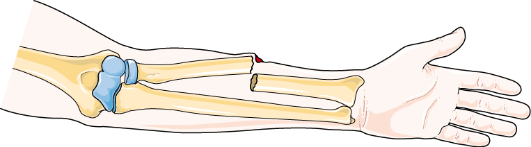
open/compound fracture
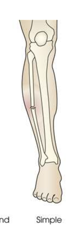
simple fracture
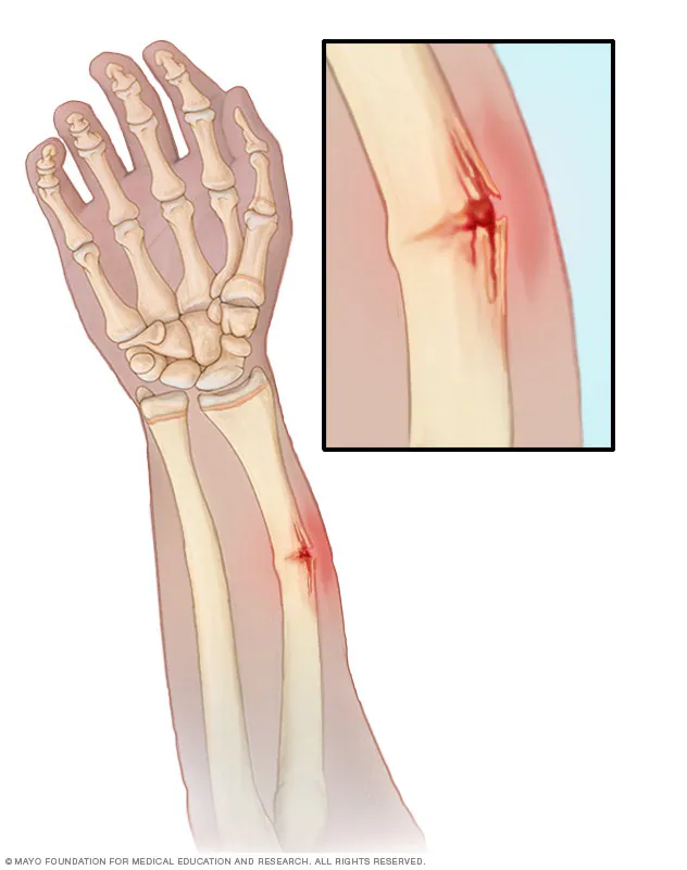
greenstick fracture
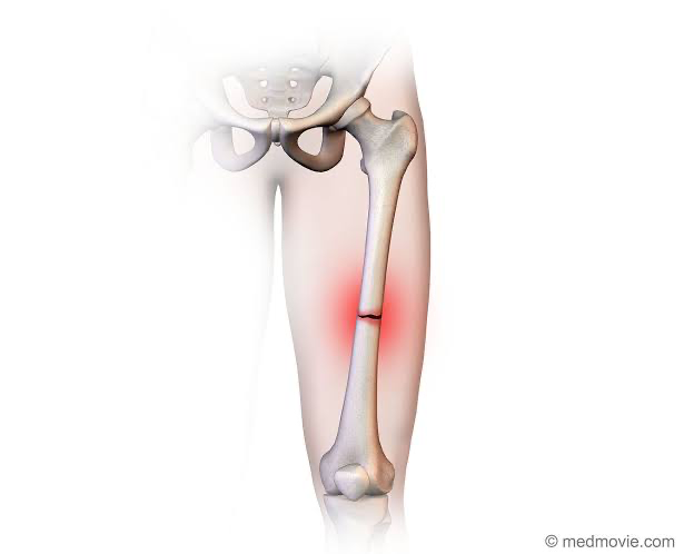
transverse fracture

oblique/spiral fracture
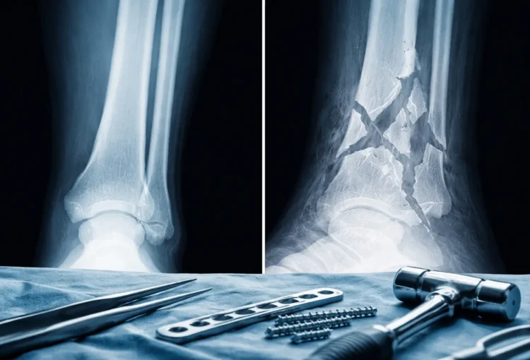
comminuted fracture
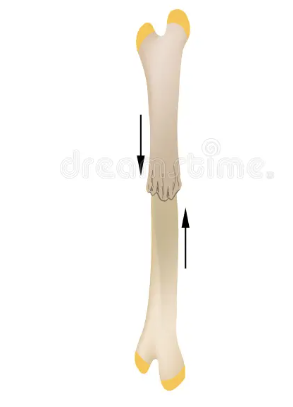
impacted fracture
processes or projections bone marking
bone markings that extend beyond from the main body of a bone
depressions bone marking
hollow or depressed areas
characteristics of cartilaginous joints
lack joint cavity and virtually immovable
united by cartilage
types of cartilaginous joints
symphysis (pubic symphysis, intervertebral joints)
synchondrosis (epiphyseal plate)
characteristics of synovial joints
synovial fluid-filled cavity that sits between connecting bones
meniscus: fibrocartilage pad as shock absorber
bursae: fluid filled sacs to reduce friction
6 types of synovial joints
gliding (carpals, tarsals)
hinge: extension/flexion (elbow, knees, fingers)
pivot: rotates around axis (neck, radius, ulna)
ellipsoid: slots into depression (wrist)
saddle (first carpometacarpal joint only)
ball and socket: multiaxial (hip, shoulder)
quadrants of the abdomen
RUQ, RLQ, LUQ, and LLQ
