RAD 1 unit 1
 Call Kai
 Learn
Learn Practice Test
 Spaced Repetition
 Match
Match Flashcards
 Knowt Play
1/76

Earn XP
Description and Tags
positions, projections, planes, xray exam details (SID, collimation, etc.)
Name | Mastery | Learn | Test | Matching | Spaced | Call with Kai | Chat |
|---|
No analytics yet
Send a link to your students to track their progress
77 Terms
what are projections?
Projections describe the path of the x-ray beam through the patient.
what are positions?
Overall posture of the patient or general body position.
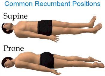
what is the supine position?
lying on the back.

what is the prone position?
lying on stomach, face down.

what is recumbent?
lying down in any position.
what is the upright position?
erect or vertical
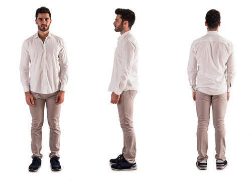
what is the seated position?
upright, but sitting on stool.


what plane is this?
sagittal, separates body into left and right halves
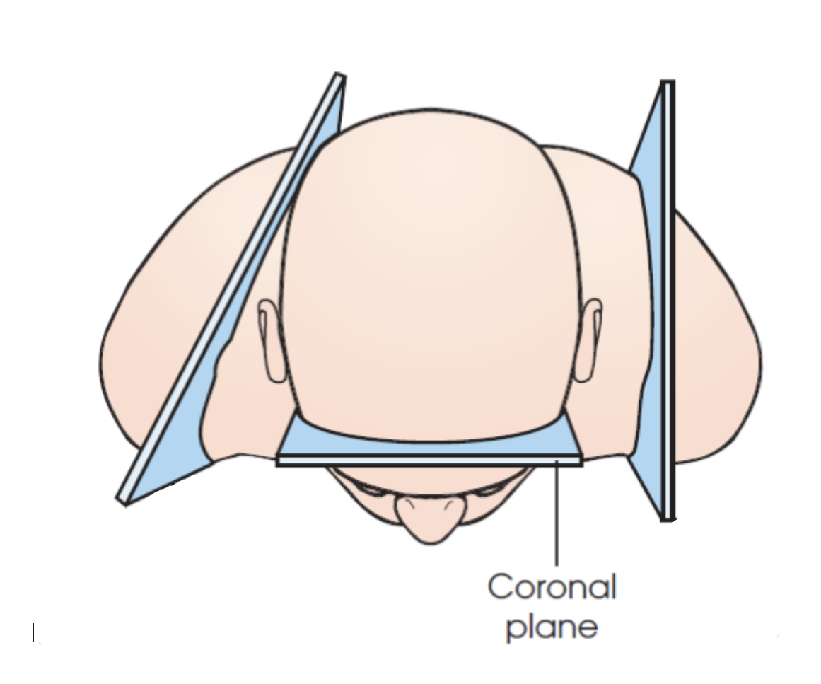
what plane is this?
coronal, separates body into anterior (ventral, front) and posterior (dorsal, back)
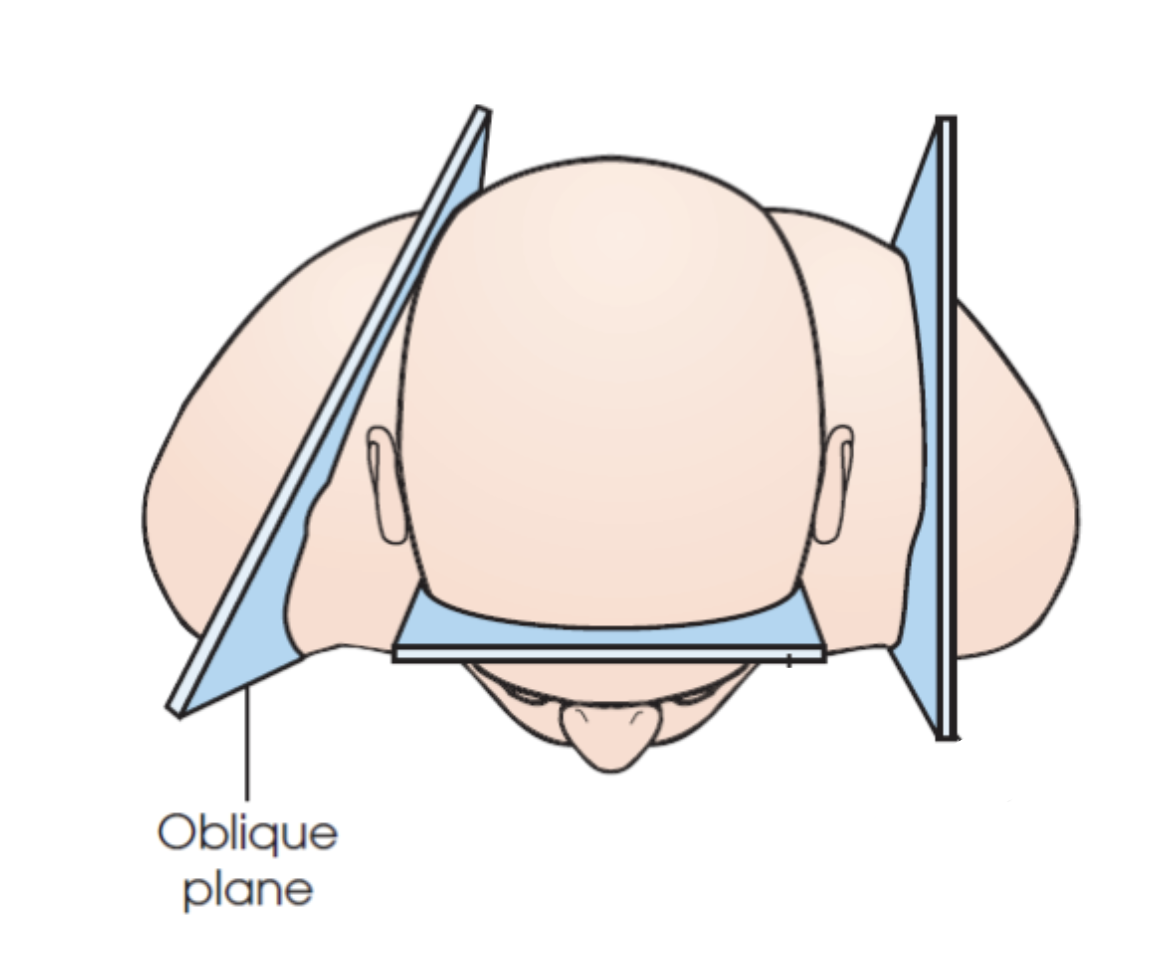
what plane is this?
oblique, at an angle
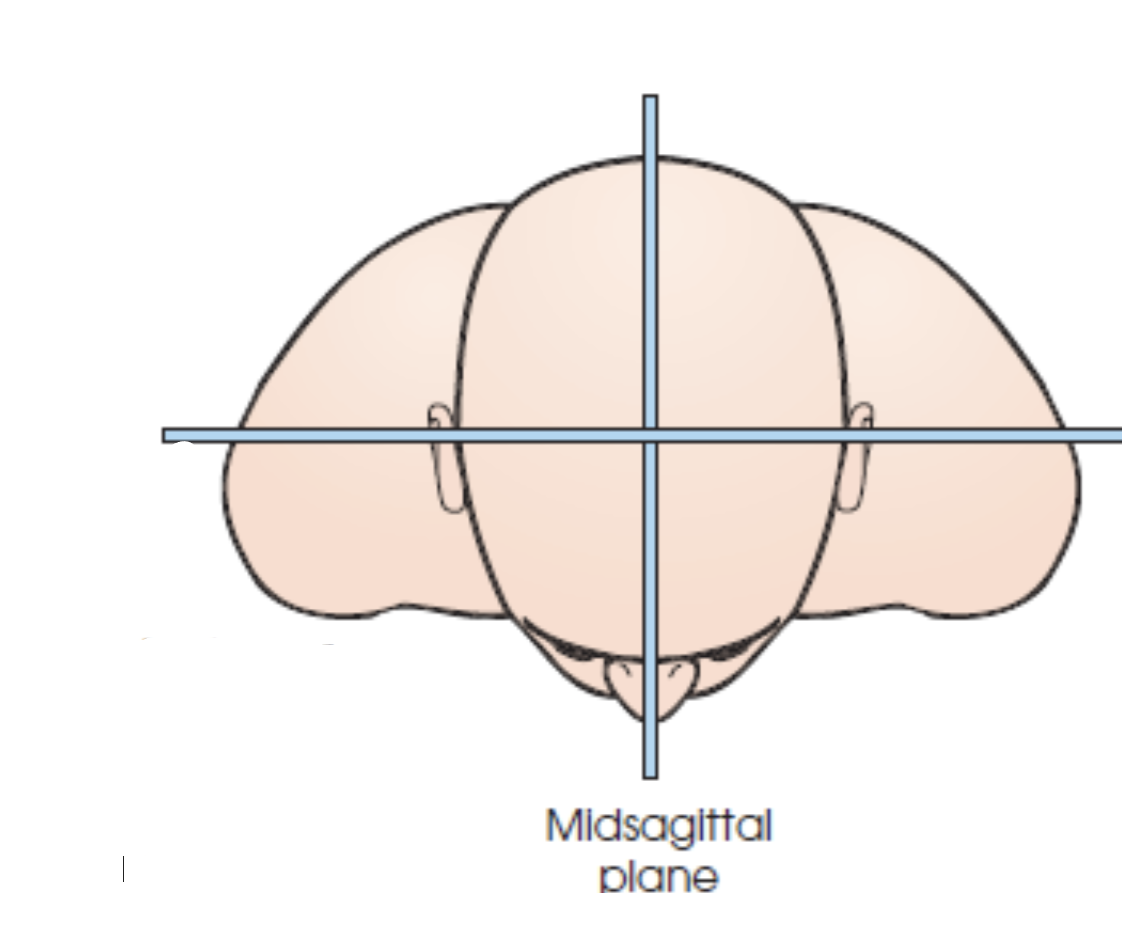
what plane is this?
midsagittal, separates body into EQUAL left and right halves.

what plane is this?
midcoronal, separates body into EQUAL anterior and posterior halves
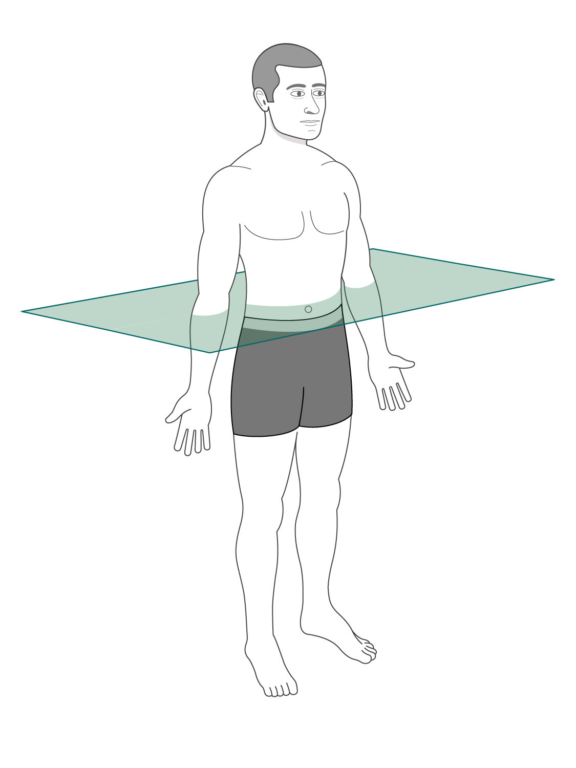
what plane is this?
horizontal (transverse), divides body into superior and inferior

what position is this?
Trendelenburg, supine with head lower than feet.
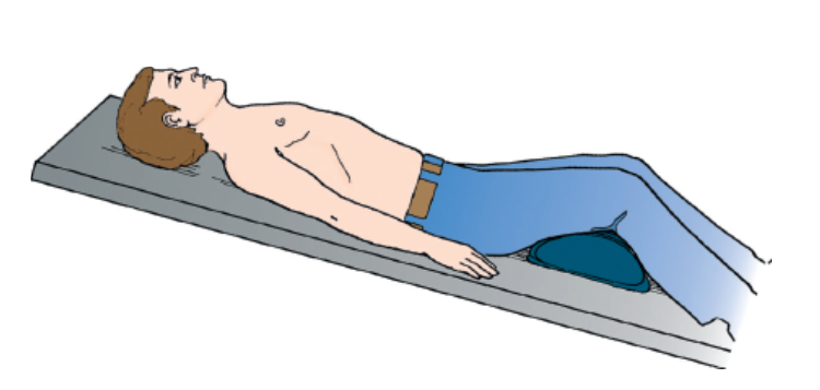
what position is this?
fowler, supine with feet lower than head. (FEET FIRST)

what position is this?
Sims’; recumbent, lying on left anterior side w/ left leg extended and right knee and thigh partially flexed
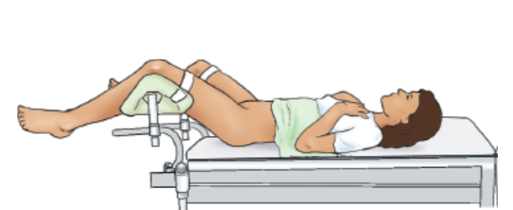
what position is this?
Lithotomy, supine with knees and hips flexed and thighs abducted and rotated externally, supported by ankle supports
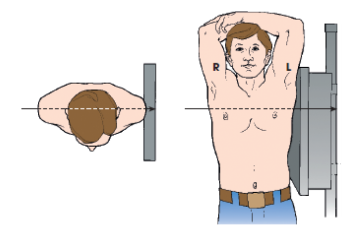
what position is this?
Lateral, named according to the side of the patient that is placed closer to the IR
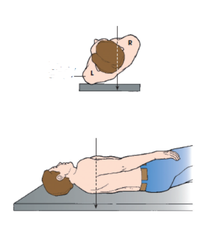
what position is this?
Oblique, Body is rotated so that the coronal plane is not parallel with the table or IR (Named according to side and surface of body closer to table or IR: “RPO, LPO, RAO, and LAO”)

what position is this?
Decubitus, Recumbent position with a horizontal CR
ØNamed according to the body surface on which the patient is lying
“RLD (right lateral), LLD, Dorsal Decub, Ventral Decub
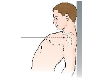
what position is this?
Lordotic, Upright position in which the patient is leaning backward
what is an AP projection?
CR enters the anterior surface and exits the posterior
what is a PA projection?
CR enters the posterior surface and exits the anterior
what is an AXIAL projection?
ØLongitudinal angle of the CR of 10 degrees or more
ØProduced by angling the central ray cephalad or caudad
ØAlso achieved by angling the entire body or body part while maintaining the central ray perpendicular to the IR.
what is a TANGENTIAL projection?
CR directed along the outer margin of a curved body surface
what is a LATERAL projection?
CR enters one side of the body, passing transversely along the coronal plane
what is an OBLIQUE projection?
ØCR enters from side angle
ØEntrance and exit surfaces still specified (e.g., AP oblique)
what are the 3 IR placements?
Lengthwise (portrait), Crosswise (horizontal), Diagonal
What is SID?
Source-to-image receptor distance. Defined as the distance from the anode inside the x-ray tube
what is SSD?
source-to-skin distance. Defined as the distance between anode inside the x-ray tube (source) to the patient’s skin
where to place a marker on a LATERAL projection?
Always mark the side closest to the IR; if the left side is closest, use the L marker, typically placed anterior to the anatomy.
where to place a marker on an AP or PA projection?
typically Use the R marker for the right side of the body.
where to place a marker for OBLIQUE projections?
Mark the side down or nearest the IR; for a right posterior oblique (RPO) position, use the R marker.
where to place marker on EXTREMITY projections?
Use appropriate R or L markers within the edge of the collimated x-ray beam.
how to place a marker on EXTREMITIES THAT ARE SIDE BY SIDE?
For extremity projections where R and L sides are imaged side by side on one IR (e.g., R and L, AP knees), R and L markers must be used to identify the two sides clearly.
where to place a marker for DECUBITUS projections?
For decubitus positions of the chest and abdomen, R or L marker should always be placed on the side up (opposite the side laid on) and away from the anatomy of interest
what is required information on a radiograph?
-Date
-Patient’s name or ID number
-Right or left side marker
-Institution identity
what are the 4 body habitus?
Sthenic, Hyposthenic, Asthenic, Hypersthenic
(sthenic, hyposthenic = average) (asthenic, hypersthenic = extreme)
how many bones in human body?
206
what is the axial skeleton, and how many bones are in it?
Bones in the head and trunk, 80
what is the appendicular skeleton, and how many bones are in it?
bones in the extremities, 126
what is caudad?
towards the feet.
what is chephalad?
towards the head.
what is central?
Mid area or main part of an organ
what is peripheral?
At or near the surface, edge, or outside of another body part
what is ipsilateral?
Parts on the same side of the body
what is contralateral
Parts on the opposite side of the body
what is plantar?
Sole of the foot
what is palmar?
Palm of the hand
What is dorsum?
Anterior, or top, of the foot or the back of the hand
How to conduct PA chest?
Patient stands facing IR, erect with hands on hips and shoulders rounded.
Align MSP (mid-sagittal plane) to IR. IR is placed CROSSWISE.
SID= 72. Align CR to MSP and T7 (palpate for vertebra prominens and extend pinky down).
Collimated to 14W×17L.
Hold breath after second inspiration.
how to conduct lateral chest?
Patient stands erect with side against IR (usually their left = heart closer to IR). IR PLACED LENGTHWISE
SID = 72, collimation = 14w×17L.
CR= centered to T7 (palpate for vertebra prominens, extend pinky and follow scapula edge to patients side) and midcoronal plane. Hold breath after second inspiration.
how to conduct KUB/AP supine abdomine
Patient lies in supine position. IR is placed LENGTHWISE.
SID= 40, collimation= 14Wx17L.
Palpate the iliac crest, center CR to the iliac crest level on the midsagittal plane.
Hold breath after first expiration.
How to conduct AP upright abdomen?
Patient stands erect with back to the IR. IR is placed LENGTHWISE
SID = 40, collimation = 14wx17L.
Palpate for iliac crest, center CR 2 inches ABOVE iliac crest on midsagittal plane.
Hold breath after first expiration.
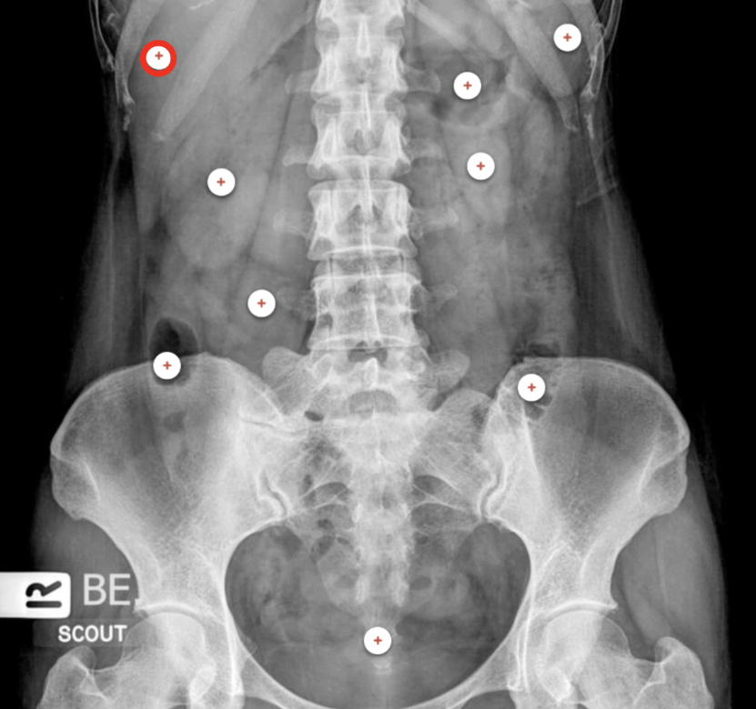
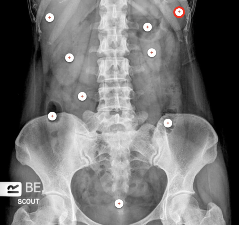
what structure is the red circle labeling?
Liver
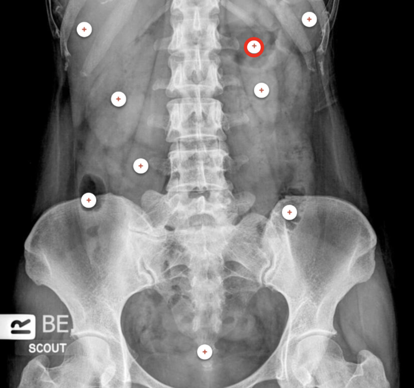
what structure is the red circle labeling?
Spleen
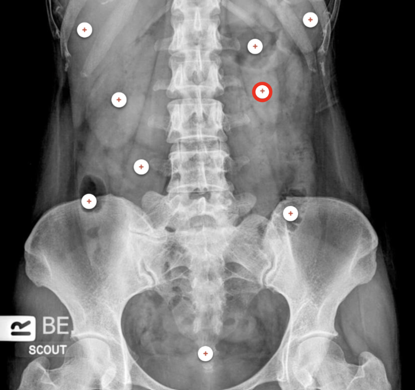
what structure is the red circle labeling?
Stomach
what structure is the red circle labeling?
Left kidney shadow

what structure is the red circle labeling?
Right kidney shadow

what structure is the red circle labeling?
Right psoas muscle
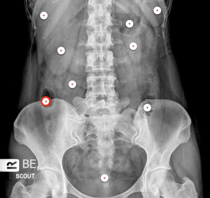
what structure is the red circle labeling?
Ascending colon

what structure is the red circle labeling?
Descending colon
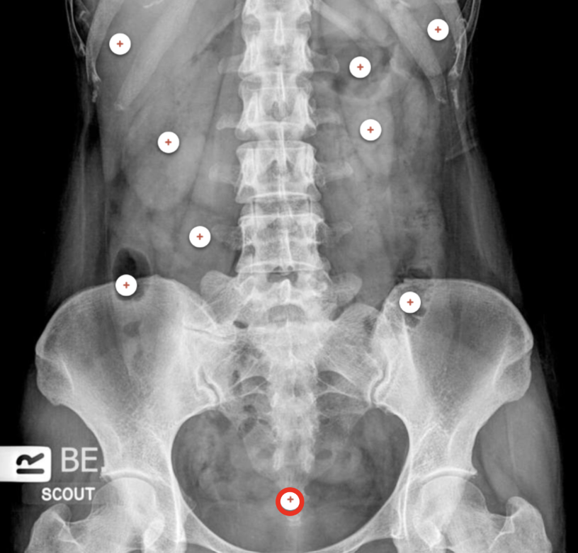
what structure is the red circle labeling?
Bladder

what structure is the red circle labeling?
air filled trachea
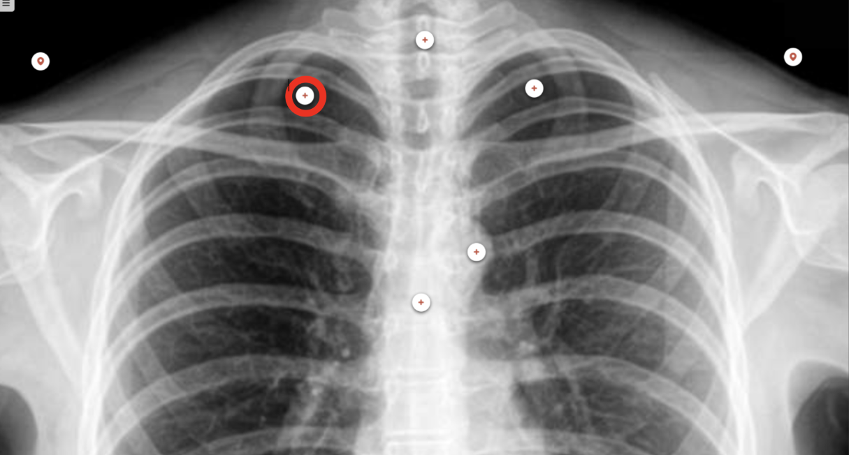
what structure is the red circle labeling?
right lung apex
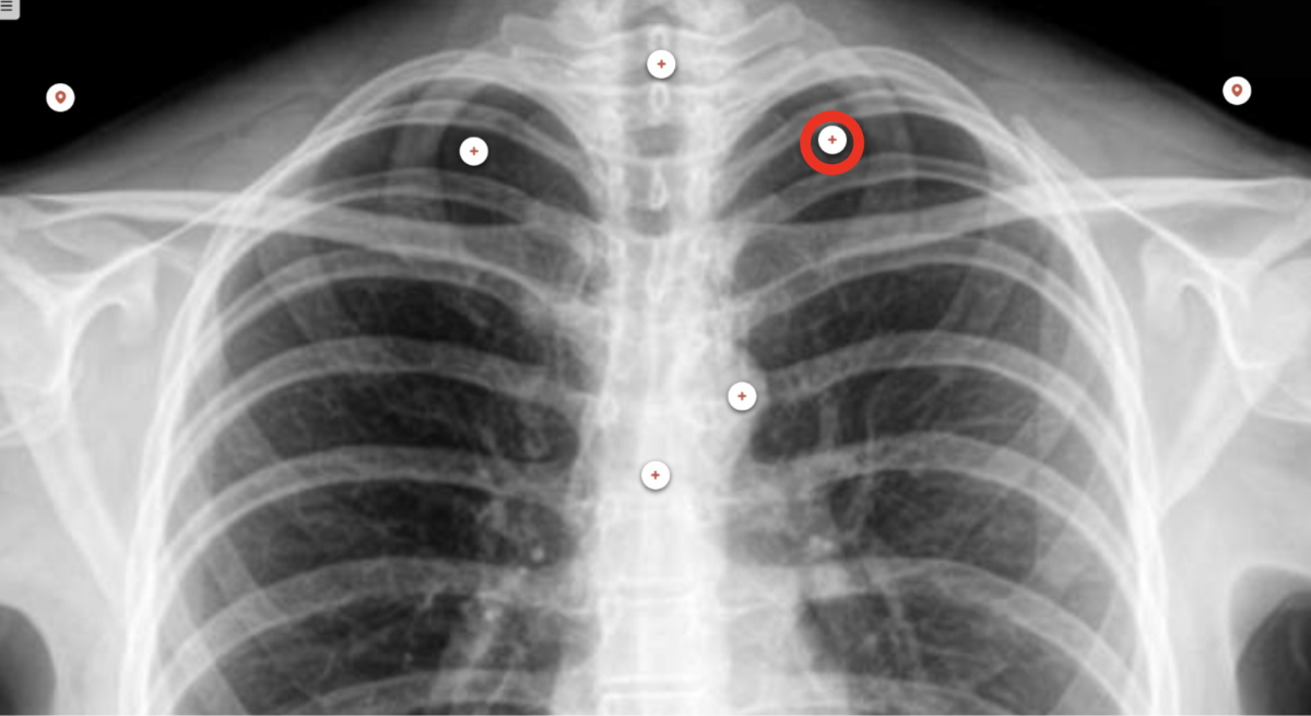
what structure is the red circle labeling?
left lung apex

what structure is the red circle labeling?
aortic knob
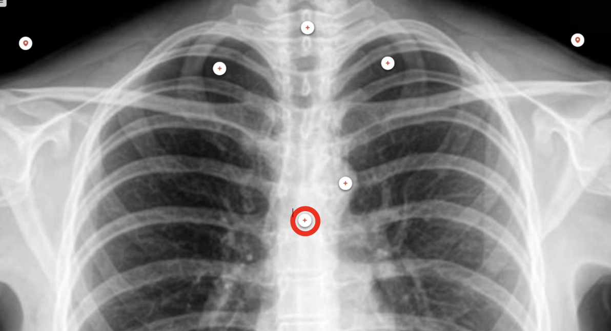
what structure is the red circle labeling?
carina (The carina is the area where the trachea bifurcates to the left and right main stem bronchus.)
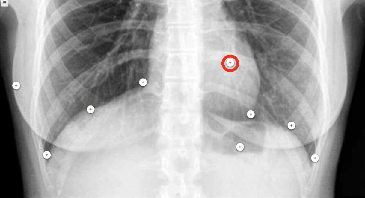
what structure is the red circle labeling?
heart shadow
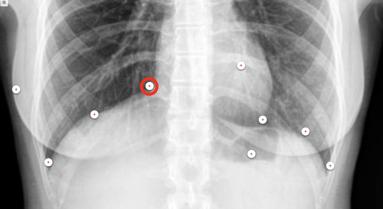
what structure is the red circle labeling?
right cardiophrenic angle (the junction point where the shadow of the heart meets the diaphragm on a chest X-ray or CT scan)

what structure is the red circle labeling?
left cardiophrenic angle (the junction point where the shadow of the heart meets the diaphragm on a chest X-ray or CT scan)

what structure is the red circle labeling?
Right Hemidiaphragm (Right hemidiaphragm is slightly higher than the left. The liver is below the diaphragm.)
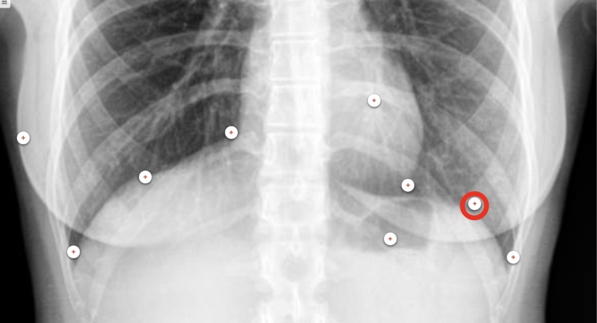
what structure is the red circle labeling?
Left hemidiaphragm
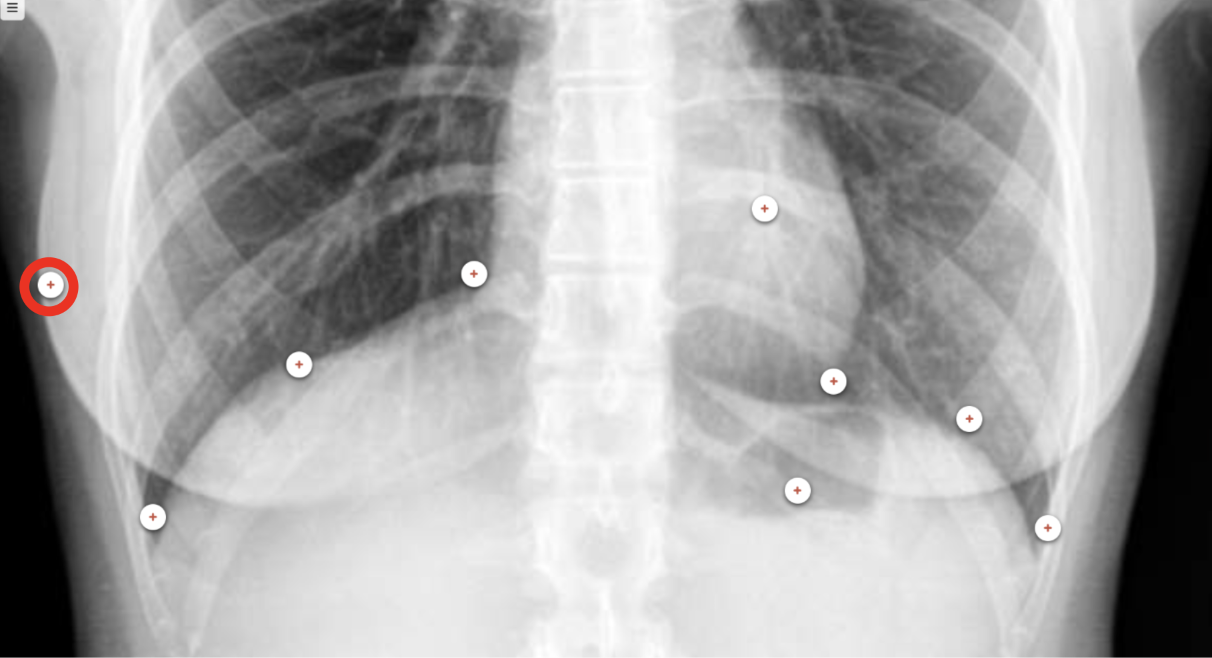
what structure is the red circle labeling?
right breast shadow
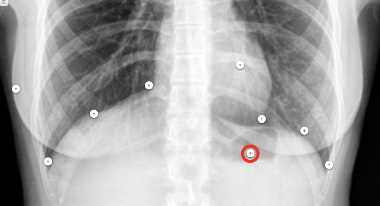
what structure is the red circle labeling?
air filled stomach

what structure is the red circle labeling?
right castophrenic angle
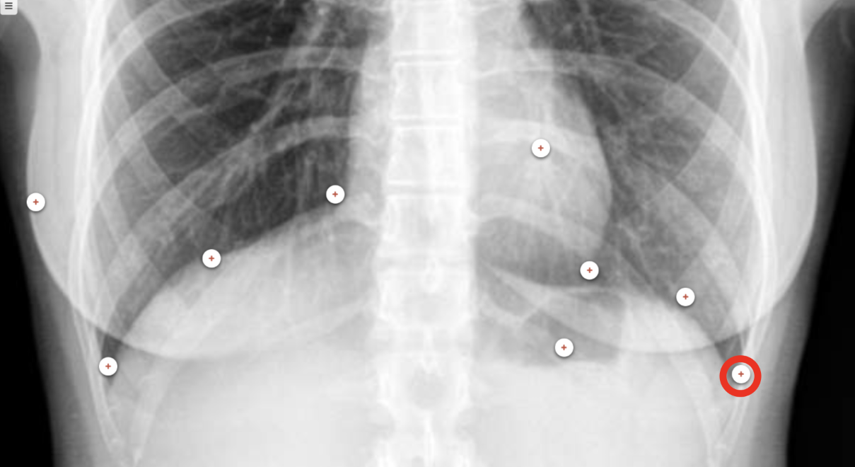
what structure is the red circle labeling?
left costophrenic angle