Comprehensive Guide to EKG Interpretation and Cardiac Pathophysiology
 View linked note
 Call Kai
 Learn
Learn Practice Test
 Spaced Repetition
 Match
Match Flashcards
 Knowt Play
1/221

There's no tags or description
Looks like no tags are added yet.
Name | Mastery | Learn | Test | Matching | Spaced | Call with Kai | Chat |
|---|
No analytics yet
Send a link to your students to track their progress
222 Terms
ECG/EKG
A recording of the electrical activity of the heart at the body surface. ECG and EKG refer to the same test.
Normal cardiac conduction sequence
SA node → atrial cells → AV node → Bundle of His → bundle branches → Purkinje fibers.
Narrow QRS association with normal ventricular conduction
Conduction slows through the AV node but travels rapidly through the bundle branches and Purkinje fibers, producing rapid coordinated ventricular depolarization.
Leads I, II, and III
Bipolar limb leads with positive and negative poles that view the heart in the frontal plane.
Leads aVR, aVL, and aVF
Augmented unipolar limb leads that view the heart in the frontal plane.
Leads V1–V6
Unipolar precordial/chest leads that view the heart in the horizontal plane.
10 electrodes producing a 12-lead ECG
4 limb electrodes and 6 precordial electrodes that generate 12 lead views.
Positive electrode of an ECG lead
The viewpoint from which the heart’s electrical activity is being recorded.
One small horizontal ECG box
0.04 seconds.
One large horizontal ECG box
0.20 seconds.
One small vertical ECG box
0.1 mV.
One large vertical ECG box
0.5 mV.
Standard ECG paper speed
25 mm/sec.
Large ECG boxes that equal 3 seconds
15 large boxes.
Difference between ECG interval and segment
An interval includes waves, whereas a segment is the time between waves.
P wave
Represents atrial depolarization.
Normal P-wave duration
Approximately 0.08–0.10 sec.
QRS complex
Represents ventricular depolarization.
Normal QRS duration
0.04–0.12 sec.
T wave
Represents ventricular repolarization.
PR interval
Represents atrial depolarization plus AV nodal/conduction delay.
Normal PR interval
0.12–0.20 sec = 3–5 small boxes.
ST segment
The isoelectric period during which the ventricles are depolarized.
QT interval
Represents ventricular depolarization + ventricular repolarization; corresponds approximately to ventricular action-potential duration.
QT cutoff for basic ECG interpretation
QT
Clinical importance of prolonged QT
It can occur from hypocalcemia or medications and predisposes to torsades de pointes.
Q wave
The first downward deflection of the QRS complex.
R wave
The first upward deflection of the QRS complex.
S wave
A downward deflection following an R wave.
R-prime (R′) wave
A subsequent upward deflection following the initial R wave.
Systematic approach on EVERY ECG
Rate, Rhythm, Axis, Examine P, PR, QRS, ST, T, QT.
5-step approach for rhythm strip
Rate, Regularity, P waves, PR interval, QRS duration/morphology.
Where to evaluate rate/rhythm, axis, structural/ischemic abnormalities
Rate and rhythm from the rhythm strip; axis from limb leads; all 12 leads for hypertrophy, ischemia, and infarction.
Heart-rate ranges in the lecture
Normal = 60–99 bpm; bradycardia = <60; tachycardia = ≥100.
Calculate heart rate from a 6-second rhythm strip
Count the R waves and multiply by 10.
Calculate heart rate from a 10-second rhythm strip
Count the R waves and multiply by 6.
Large-box method for calculating heart rate
Find an R wave on a bold line and count large boxes until the next R: 300 → 150 → 100 → 75 → 60 → 50.
Preferred method for very slow heart rates or irregular rhythms
The 6-second rate method.
Regular rhythm definition
Consistent R-R intervals.
Irregular rhythm definition
Variable R-R intervals.
P waves presence favoring rhythm origins
Sinus or atrial.
P waves absence favoring rhythm origins
Junctional or ventricular.
Identical P waves suggest what
One pacemaker focus.
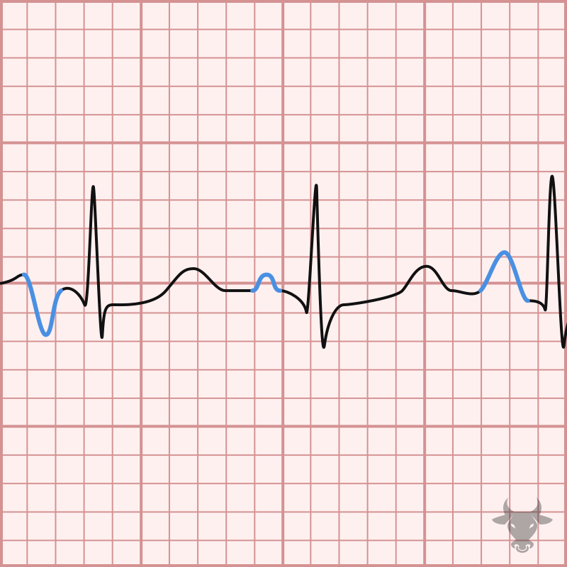
Varying P-wave morphology suggests what
Multiple atrial automaticity foci.
Every P wave is followed by a QRS suggests what
No second- or third-degree AV dissociation/block.
Some P waves not followed by QRS suggests what
AV block.
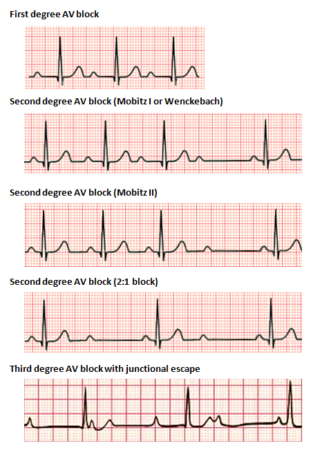
Consistently prolonged PR interval indicates what
First-degree AV block.
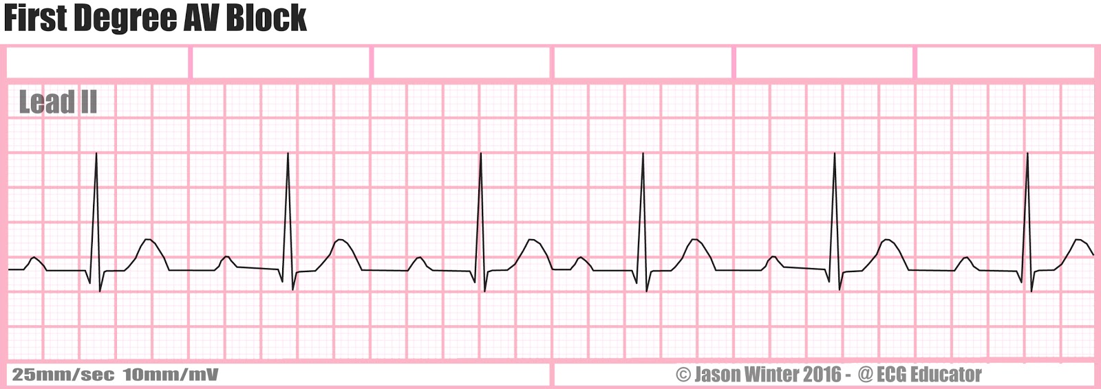
Progressively increasing PR duration indicates what
Second-degree AV block Mobitz I/Wenckebach.
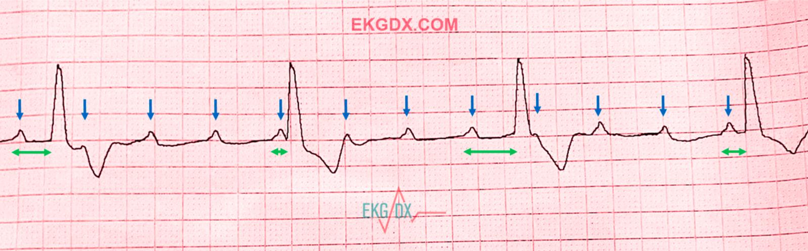
Completely variable PR interval suggests what
Third-degree AV block.
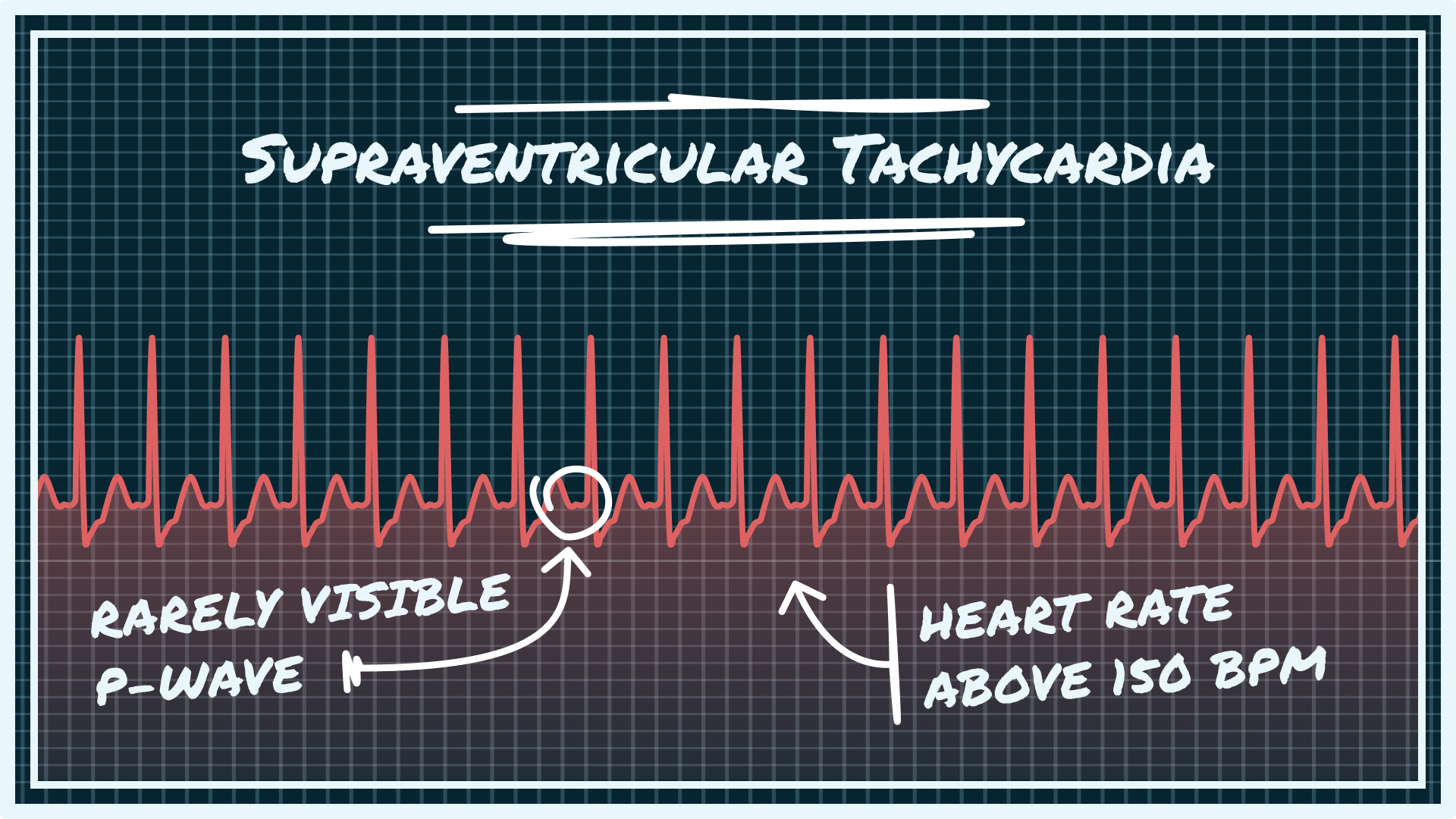
QRS <0.12 sec indicates what
A supraventricular impulse with normal ventricular conduction.
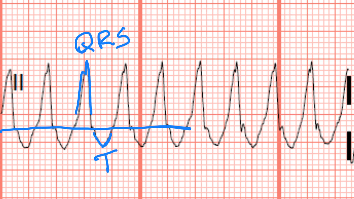
QRS >0.12 sec indicates what
Ventricular origin or delayed intraventricular conduction such as bundle branch block.
ECG features of normal sinus rhythm
Rate 60–100; regular; normal P waves; P before every QRS; PR 0.12–0.20; QRS 0.04–0.12 sec.
ECG features of sinus arrhythmia
Rate 60–100; irregular; normal P waves; normal PR; narrow QRS.
Physiology of sinus bradycardia
The SA node depolarizes more slowly than normal, but conduction through the rest of the system remains normal.
ECG features of sinus tachycardia
Rate >100; regular; normal P waves; normal PR; narrow QRS.
Notable aspect of sinus tachycardia
It is generally a response to physical or psychological stress rather than a primary arrhythmia.
Sinus arrest definition
The SA node stops firing.
Appearance of sinus arrest on ECG
An irregular pause interrupts an otherwise sinus rhythm.
Occurrence after sinus arrest
A lower automaticity focus can produce an escape beat or escape rhythm.
Prolonged electrical inactivity can lead to
Asystole.
Escape beat definition
A backup automaticity focus fires after a pause in normal pacing activity.
Escape rhythm definition
A lower automaticity focus takes over when the dominant pacemaker stops entirely.
Consecutive new beats required to call it an escape rhythm
At least 3 beats.
Intrinsic pacemaker hierarchy
SA node: 60–100, Atrial cells: 60–80, AV junction: ~40–80, Ventricular cells: 20–40 bpm.
Atrial escape beat appearance
Occurs after a pause; the P wave looks different; QRS remains narrow.
Atrial escape rhythm features
Rate 60–80; regular; P waves present but different morphology from sinus P waves; normal PR; narrow QRS.
Junctional escape beat appearance
Occurs after a pause; P wave absent or inverted; PR absent; QRS narrow.
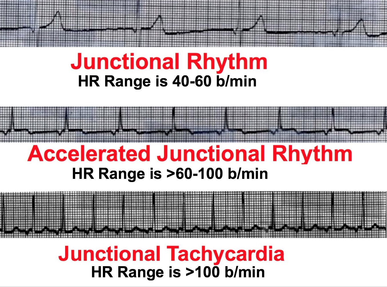
Junctional escape rhythm features
Rate ~40–60; regular; P waves absent or inverted; PR absent; QRS narrow.
Ventricular escape beat appearance
Occurs after a pause; no P wave; no PR interval; wide QRS >0.12 sec.
Ventricular escape rhythm features
Rate 20–40; regular; absent P waves; absent PR; wide QRS.
Key difference between premature beat and escape beat
Premature: abnormal beat comes early; Escape: pause occurs first, then the backup beat appears.
Causes of irritable ectopic focus
Caffeine, cocaine, methamphetamines, and medications.
PAC features
Premature altered P′ wave; altered timing/morphology; generally normal PR; narrow QRS.
Reason for QRS being narrow during a PAC
The premature impulse originates in the atrium but travels normally through the AV node.
Notable fact about PACs
PACs are the most common premature beat.
PJC features
Premature beat; P absent or inverted; PR absent; QRS narrow.
PVC features
Premature beat; no preceding P; absent PR; wide/bizarre QRS >0.12 sec.
Reason for PVC being wide and bizarre
The impulse begins within ventricular myocardium.
Monomorphic/uniform PVCs
PVCs with the same morphology, suggesting one ventricular focus.
Polymorphic/multiform PVCs
PVCs with different morphologies, suggesting multiple ventricular foci.
Wandering atrial pacemaker definition
The atrial pacemaker focus changes from beat to beat.
Wandering atrial pacemaker ECG features
Rate 60–100; irregular; P′ morphology varies; PR generally normal; narrow QRS.
Multifocal atrial tachycardia distinction from wandering atrial pacemaker
Rate >100 bpm.
MAT ECG features
Rate >100; irregular; varying P′ morphologies; PR may vary/shorten; narrow QRS.
Disease commonly associated with MAT
COPD.
Reason MAT is associated with COPD
Right atrial dilation, hypercarbia, and increased sympathetic activity can contribute.
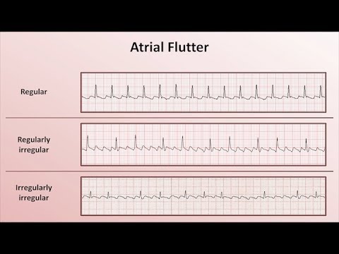
Atrial flutter ECG features
Rapid identical flutter waves producing a sawtooth baseline, with predictable AV conduction.
Mechanism of atrial flutter
Reentry.
Reason not all atrial flutter waves produce QRS complexes
The AV node blocks many of the rapid impulses.
Atrial fibrillation ECG features
No discernible P waves; fuzzy/chaotic baseline; narrow QRS; irregularly irregular rhythm.
Reason atrial fibrillation is irregularly irregular
Chaotic atrial impulses reach the AV node.
First-degree AV block features
PR >0.20 sec consistently; every P produces a QRS; regular rhythm.
Core concept of first-degree AV block
Every impulse conducts, but conduction is delayed.
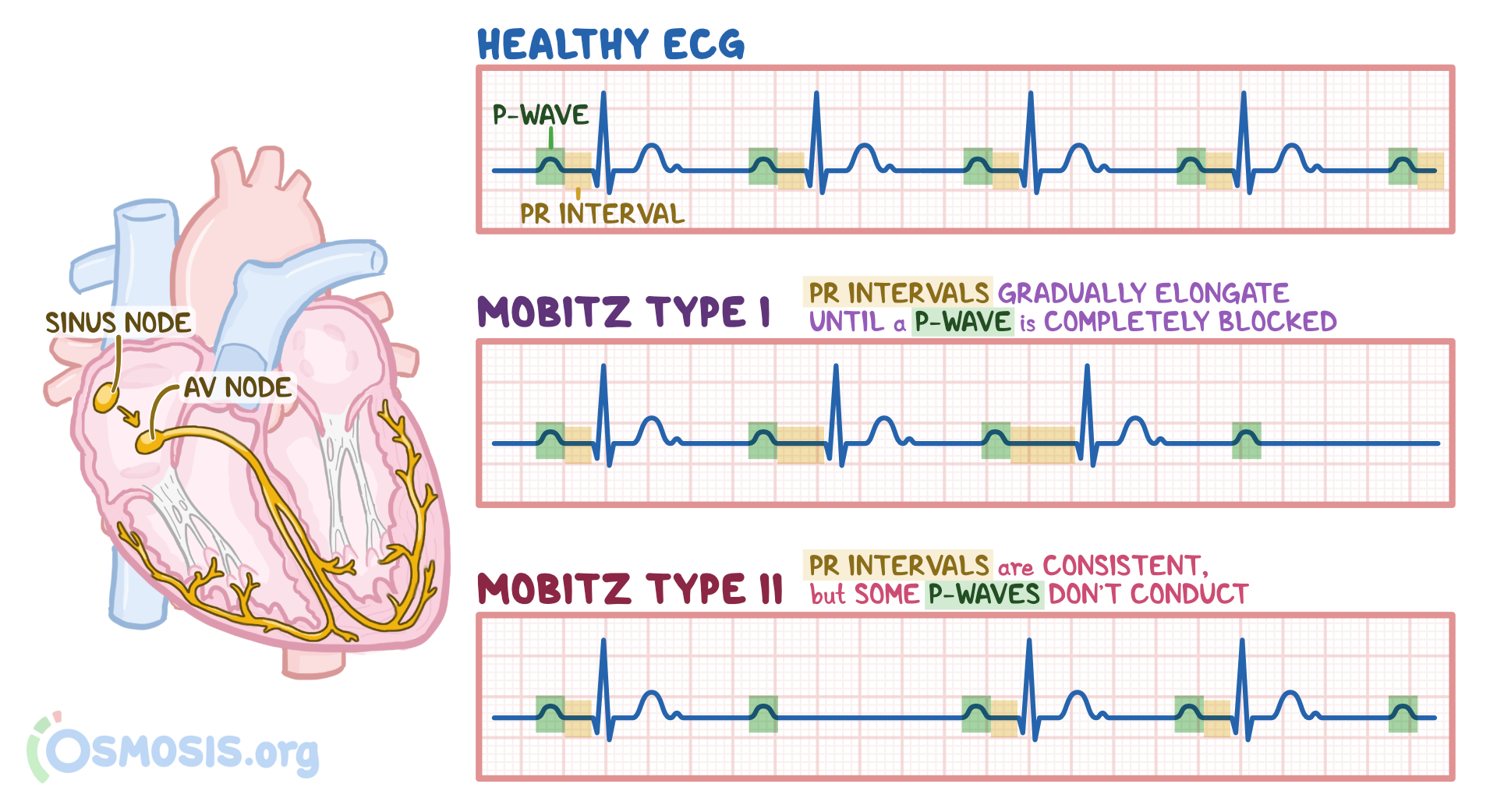
Mobitz I/Wenckebach features
PR progressively lengthens until a P wave is not followed by a QRS.
Mnemonic for identifying Wenckebach
“Long, longer, drop = Wen, kee, bach.”
Mobitz II features
Constant PR interval; some P waves fail to conduct and QRS complexes are dropped.
Mobitz II QRS width
May be narrow or wide depending on the level of the block.
Distinction between Mobitz I and Mobitz II
Mobitz I = PR progressively lengthens. Mobitz II = PR stays constant.
Third-degree AV block features
P waves march independently of QRS; completely variable PR intervals.
Another name for third-degree AV block
Complete heart block.