BMS1052 - W6: Motor Control - Peripheral Control of Movement
1/16
There's no tags or description
Looks like no tags are added yet.
Name | Mastery | Learn | Test | Matching | Spaced |
|---|
No study sessions yet.
17 Terms
Describe this image:
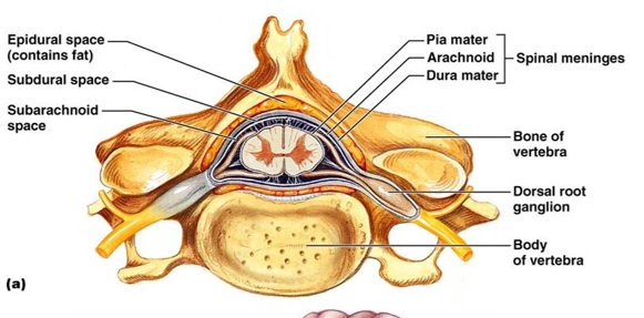
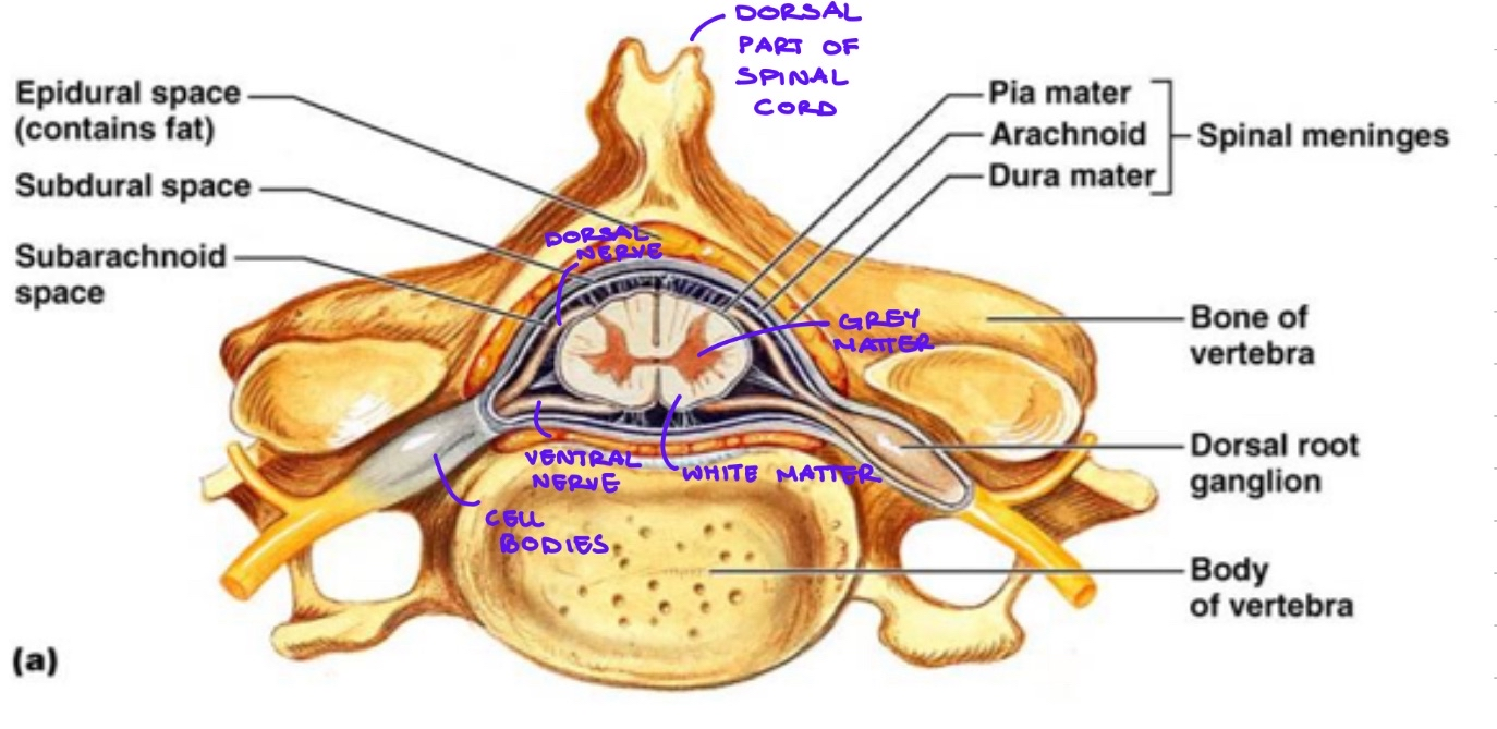
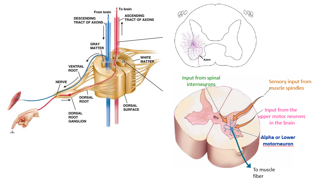
This is an image of a section of the spinal cord
dorsal part of spinal cord: boney thing you can feel in your back
white matter: axons of neurons going up and down spinal cord
grey matter: neuron cell bodies
Dorsal nerve root: sensory nerve coming in. Primarily somatosensory neurons bringing in sensory information from the periphery
Ventral nerve root: carrying motor information away from the spinal cord
(CHECK THE IMAGE HERE, I ADDED LABELS)
What is a lower motor neuron?
What are the two special properties of muscle fibers?
A neuron that innervates the muscle
also referred to as alpha motor neuron, motoneuron or final common pathway
they are the common mechanisms for causing muscle contractions
involved in all movements (voluntary and reflexive)
directly innervate muscle
cell bodies in spinal cord
Muscle fibers special properties:
can change length
can generate force
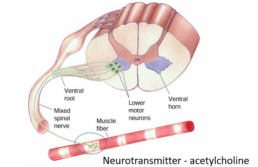
Where can neurons in the spinal cord collate information from?
Dendritic arbor - all the dendrites of the neuron where it can collate information
inputs from spinal interneurons (local circuitry within the spinal cord)
sensory inputs from muscle spindles (sensory receptors associated with the muscles)
descending inputs from the upper motor neurons in the brain (sent through the blue axons - the tube - in image)
What is a motor unit?
What is the motor neuron pool?
What is a motor unit?
1 alpha motor neuron (lower motor neuron) + the muscle fibers that it innervates
we can have different size motor units (and the size is primarily determined by the number of muscle fibers that are innervated by this motor neuron)
While a muscle contains multiple muscle fibers, each of the muscle fibers is innervated by only one motor neuron.
But a motor neuron can innervate multiple muscle fibres?
small motor units: involves <10 muscle fibres (generated with small amount of force or precise movements e.g. fingers/ eyes)
large motor units: involve >1000 muscle fibres (large force e.g. calf muscles)
What is a motor neuron pool?
Collection of all alpha motor neurons that innervate a single muscle
What is rate coding in the motor system?
Rate coding corresponds to intensity
higher rates of action potentials lead to higher amounts of force generation
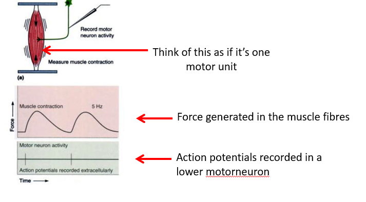
In the experiment depicted in the image:
we can see that each motor neuron activity is associated with a muscle contraction
the muscle contraction lasts quite a long time (~well over 100ms) compared to the motor neuron activity (~1 ms)
How can force generation be controlled?
rate of action potentials (force summation)
size principle
What is force summation?
What is unfused and fused tetanus?
A very different kind of summation compared to summation of action potentials in excitatory post synapses
Looking at the image
when we increase rate of action potentials to 10Hz, we get a phenomenon called force summation - where the generation of force within the motor unit rides upon the force that was already generated
further increasing results in smoother and smoother muscle contractions
Force summation: where the responses to individual action potentials summate (can be seen in the top right, bottom left and bottom right images)
Unfused tetanus: oscillatory force generation (top right and bottom left images)
Fused tetanus: smooth force summation - generates maximal force within an individual motor unit (bottom right image) - when all possible binding site within the motor unit are activated
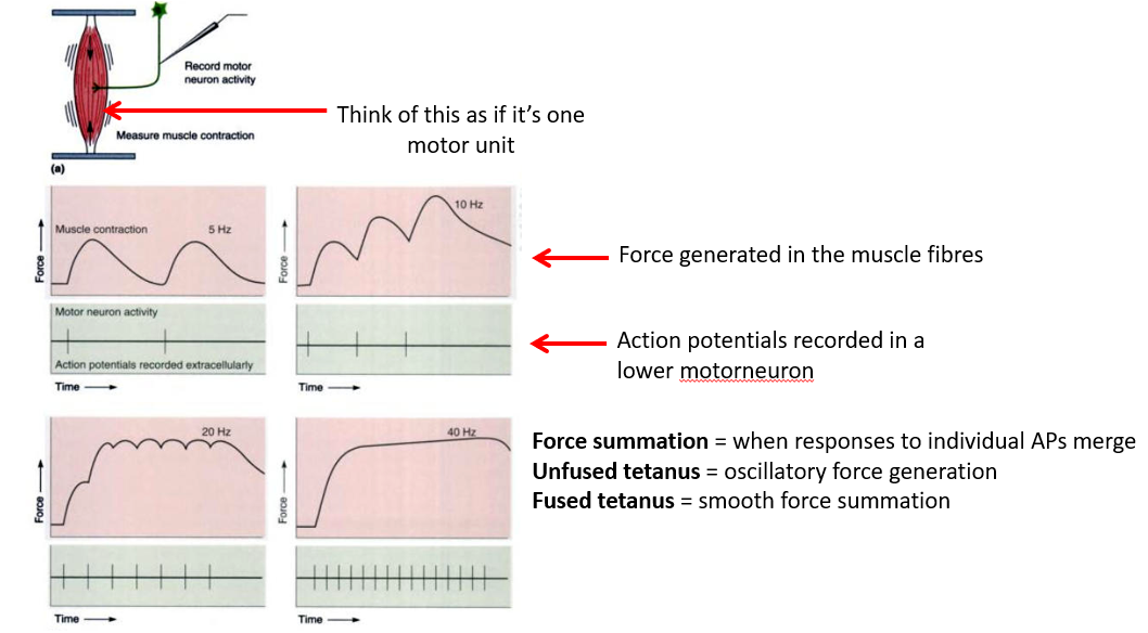
Usually we don’t make the jerky kind of movements. Usually our movements are quite smooth. Does this mean we are generating a fused tetanus within all our motor units?
No. Rather, it is because we have many, many motor units within a muscle and they will be activated at different times. So it is the summation of force that occurs across an entire muscle that then accounts for that smoothness of contraction rather than the smoothness generated by a single motor unit.
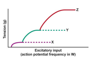
What is the size principle?
Why does it happen?
What is proportional control?
Differences in neural control (often referred to as the size principle)
From image:
can see that an upper motor neuron (W) innervates 3 separate lower motor neurons (in spinal cord)
they have different size cell bodies
they then innervate muscle fibres
to generate graded forces, first recruit small motor units, then progressively recruit larger motor units so we generate larger and larger amounts of force over time
This essentially happens automatically because of the size principle (cell body size takes care of the graded motor recruitment automatically)
Basically, because cell X is smaller, it is activated first causing it to generate action potentials within the muscle fibers
Neuron x fires action potentials before Y, and Y fires before Z even though they receive the same inputs from neuron W (ignore the difference in axon length)
Why does it happen?
small motor neurons have higher membrane resistance and reach AP threshold more easily
smaller motor neurons = smaller surface area = higher membrane resistance
Ohms Law: ΔVmembrane= I (current) * R (Resistance)
so larger resistance = larger change in membrane potential for a fixed input current
so the tension being generated is going to start out small as neuron x is recruited, then going to increase as y and z are recruited
so size of cell body determines when they are recruited, and that’s how we get graded generation of force across an entire muscle
Proportional control:
a feature of the size principle
inverse relationship between the number of motor units in a muscle and their force generating capacity
many small motor units; progressively fewer large motor units
initially not generating much force, then change the type of motor units being recruited and generate force more quickly, then rapidly scale up the amount of force generated as we recruit more motor units. We recruit more motor units for different activities (e.g. fewer motor units standing, more for walking and running)
What are the types of muscles?
Skeletal muscles - striated muscle under voluntary control
used for controlling body movements, speaking, etc.
Cardiac muscle - striated muscle involuntary control
found in heart
smooth muscle - involuntary control
primarily found in stomach and gut
A lil note: When muscle is activated by action potential inputs (from a lower motor neuron) it is referred to as a contraction or twitch contraction (if it is in response to a single action potential) - even if the muscle doesn’t change length or gets longer. Does not imply that the muscle is getting shorter despite the name.
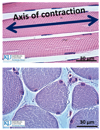
Skeletal muscle:
multi-nucleated (each cell has multiple nuclei) on periphery of cell body
has a single axis of contraction (when stimulated, will contract along that axis only)
Top image = longitudinal section of muscle
Bottom image = cross section of muscle
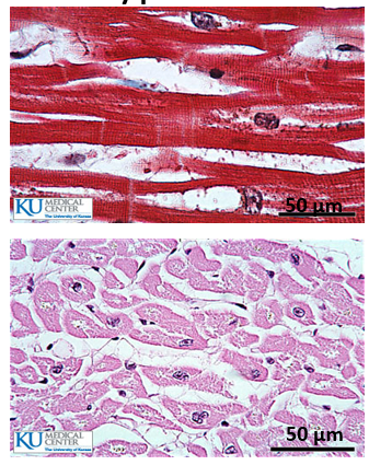
Cardiac muscle:
Contains a very branched structure
usually has a single central nucleus per fibre
due to branching, and unordered striations, there is no single direction of contraction
Top image = longitudinal section of muscle
Bottom image = cross section of muscle
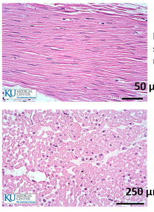
Smooth muscle:
no striations
has a single nucleus per fibre
this muscle type is not under voluntary control
Top image = longitudinal section of muscle
Bottom image = cross section of muscle
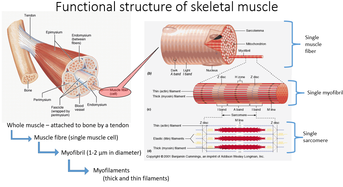
Structure of skeletal muscle:
Muscle is attached to bone via tendon
muscles apply force to bones via tendons
Fascicle: a collection of muscle fibers
Single muscle fiber = a single cell
Muscle fiber:
made up of many myofibrils
myofibrils have a regular repeated a appearance which gives rise to the striations/ stripy appearance of muscle fiber
myofibrils have 3 types of protein filament (myofilaments)
actin (thin) filaments (shown in blue)
myosin (thick) filament (shown in red)
titin (elastic) filaments (shown in yellow)
What allows for changing length of the muscle and build up of tension in the muscle?
The relative movement of the actin and myosin
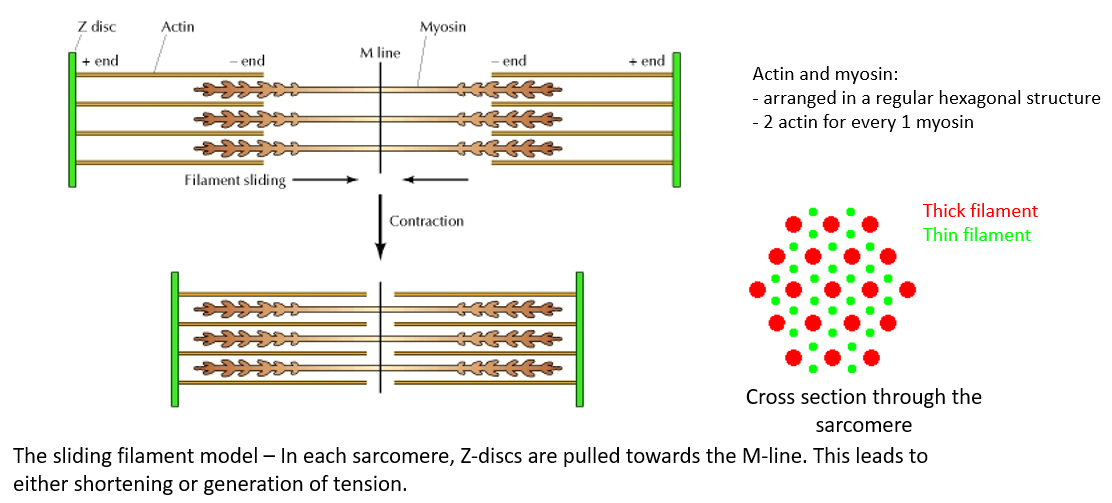
What is a sarcomere?
The region between two Z discs in myofibril
The sarcomere is the basic fucntional unit of a muscle fibre
actin filaments alternating with myosin filaments which attach to a separate structure (the M line)
myosin heads directly attach to the actin filaments