CO III Anesthetics and Tonometry
 Call Kai
 Learn
Learn Practice Test
 Spaced Repetition
 Match
Match Flashcards
 Knowt Play
1/48

There's no tags or description
Looks like no tags are added yet.
Name | Mastery | Learn | Test | Matching | Spaced | Call with Kai | Chat |
|---|
No analytics yet
Send a link to your students to track their progress
49 Terms
Topical anesthetic purpose
Temporarily anesthesize the cornea and/or conj to allow certain procedure to be done by blocking nerve conduction
Topical anesthetic actions
Increase corneal permeability to increase the clinical effectiveness of mydriatics and cycloplegics
Prevent stinging and/or burning of dilating agents, increasing pt comfort
Decrease systemic absorption due ot anesthetic-induced inhibition of tear flow
Topic anesthetic mechanism of action
Local anethestics stabilize neuronal membrane si the nuron is less permeable to Na+ ions by preventing initiation and transmission of nerve impulses, therevby producing the local anesthetic actions
Topical anesthetic procedures
ERG
Goldmann
Gonio
Forced duction
ERG purpose
Determines function of rods and cones
DTL electrode
Silver thread that touches conj/cornea to anesthesize pt
Goldmann tono
Touches cornea
Accurate
Vasovagal syncope
Handheld applanation tonometers examples
Tono pen
Anesthetic drop recommended
Gonio
Uses anesthetic drops
Uses 3-mirror and 4-mirror lens
Suctions eyeball and makes more contact with cornea so more anesthetic is needed
Measures ant. chamber angle
Forced duction testing
For pts with trapped/paretic muscles- can perform with cotton-tipped application
Can use topical lidocaine 4%, proparacaine HCl 0.5%, tetracaine HCl 0.5%, or TOOTHED FORCEPS
Toothed forceps
Used in forced duction testing
Can use topical cocaine 4%, lidocaine, proparacaine HCl 0.5%, or tetracaine HCl 0.5%
ASSESS CORNEA/CONJ OF FB
Schirmer No. 2 Test
Paper touching cornea to ASSESS THE TEAR FILM QUANTITY AND REFLEX TEARING
Anesthetic takes away the reflex tearing so that you can measure the true amount of tears
Tears collect on paper tab and blue dye drains down- measure in mm how much the blue dye has moved down to measure the quantity of the tear film
Topical anesthetic therapeutic provedures
Cornea/conj. FB removal
Lacrimal dilation and irrigation
Punctal occlusion
Periocular injections
Corneal FB removal
SLE: High mag of corneal surface
Check upper lid (lid eversion)
Lacrimal dilation and irrigation
Treats blocked tear ducts
Small metal rods widens cannulicula and injects saline- you can taste saltwater in the back of your throat
Blocked = you're meeting hard resistance
Punctal occlusion
For pts with dry eye
Insert pieces of collagen into puncta to create a clogged duct to keep tears on the eye
Periocular injections
i.e.: Avastin
Use speculum to keep eye open and inject needle
When to NOT use topical anesthetics
Pts have:
Hypersensitivity
Known allergic reaction
Cultures (preservative-free)
Cannot exceed recommended dose because they can prevent healing and cause total breakdown of the epi, stromal edema, and severe pain
NEVER GIVE TO CL WEARERS TO TAKE HOME!!!!
Topical anesthetic onset
5-20 sec
Topical anesthetic duration
1 drop lasts for 10-20 mins; can be prolonged with repeated application
Topical anesthetic side effects
Ocular: Slight disruption of the corneal epi which may cause mild keratitis, transient conj. hyperemia, light sensitivity, and transient corneal edema
Systemic: Rare; if overdose w/ systemic toxicity, effects may affect CNS, cardiovascular, or respiratory system
Examples of anesthetic agents
Proparacaine Hcl 0.5% solution
Tetraciane
ALTAFLUOR AKA BENOXINATE HCL 0.4% PLUS 0.25% FLUORESCEIN SODIUM
Proparacaine HCl 0.5%
Ester linkage- meta amino benzoic acid
aka spectro-caine, ak-tiane, alcaine, ophthaine
Tetracane Hcl 0.5% solution
Ester linkage: PABA
aka AK-T-Caine
Benoxianate HCl 0.4% plus 0.25% fluorescein sodium
AKA ALTAFLUOR
Used prior to applanation tono
Vasovagal syncope
Intense emotional experiences leads to muscle vasodilator system to become powerfully activated -> bloodflow in muscles increase, so HR slows and arterial pressure falls -> reduced bloodflow to brain = FAINTING
Can occur during applanation tono, gonio, contact lens insertion, and BIO
How to manage syncope
Never leave pt by themselves and stabilize pt's head
Instilling anesthetic drops
Check label + expiration date
Do not put cap on surface to touch the tip of bottle to pt's eye
Hold upper + lower lids and have pt look down
Applanation tono
aka Goldmann
Measures IOPs in mmHg- helps diagnose and treat all forms of glaucoma, and diagnose ocular hypertension (OHTN)
Measures IOP by providing force required to flatten the cornea- INCREASE FORCE UNTIL THE AREA OF THE CORNEA EQUALS THE AREA OF THE PROBE
Variable force applanation tonometers
Goldmann
Perkins
Tono-pen
APPLANATED AREA OF CORNEA SHOULD BE EQUAL TO THE AREA OF THE PROBE OF 3.06 MM
Applanation tono force
Size of tono tip is meant to minimize the impact of the corneal resistance and surface tension of the tear film
Turn tension knob that alters force applied to the cornea and the IOP is determined when internal part of 2 semicircles touched ach other
How to use D-KAT tono in Keeler slit lamps
Place probe at 180 or 0 degrees if less than 3D of corneal astig
If MORE than 3D of corneal astig, align prims to the negative cyl axis at EITHER THE RED LINE OR THE "A" IN D-KAT TONOS
Pre- and post-corneal scan
After instilling Altafluor, quickly scan the cornea with the cobalt blue filter in place both prior to doing tono and after tono to see if you caused something
How to do applanation tono
1. Base and joystick towards doctor
2. Align prism viewing "outside" the instrument first
3. Center prism before contact
4. Observe nasal flow or light blow mires (ghost mires) before contact
5. Hold upper lid to accommodate probe (do not push on globe and hold under the lashes)
6. If pt needs to focus on something on pt left, they need to look right
7. Properly align semicircle mires- inner should touch inner (they need to go past each other) via moving drum. Upper and lower circles should be equal size and centered.
What should you do here?
Lower pressure

What should you do here?
Pull back on probe

What should you do here?
Increase pressure a little bit (move the drum away from you)

Should you increase or decrease the measuring drum?
Increase
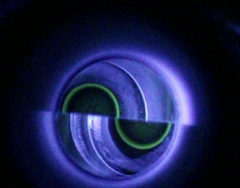
What's wrong here?
Mires too wide- gives a falsely high IOP
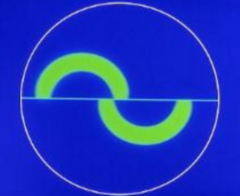
What's wrong here?
Left: Too much fluorescein
Right: Probe too low and you need to move joystick up to make semicircles equal in size
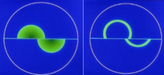
What's wrong here?
Contact with the probe is too high
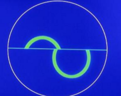
gtt
How many drops in charting
Goldmann procedure
1. Keep probe on cornea for no more than 5 sec
2. If you see 2 semi-circles on the cornea and need to make fine adjustments, do so without taking the probe off the cornea
Tono procedure
After slit lamp and before gonio (NO NEED TO INSTILL ANOTHER ANESTHETIC DROP)
1. Disinfect probe/prisms by rinsing probe for 30 sec and then putting the probe in hydrogen peroxide for 10 min then rinse for 30 sec with water followed by a 60 sec immersive soak. Pat and dry. Explain procedure to pt.
2. Instil Altafluor or tono agent
3. Check pre-tono corneal staining
4. Set pressure on probe to appropriate level (10 mmHg for lab)
5. Give pt instructions, including a target
6. Put tono in place w/ cobalt blue light on probe
7. Applanate cornea
8. Adjust biomicroscope so that the fluorescein semicircular patterns are symmetrical and centered
9. Align mires
10. Retract probe and assess cornea w/ blue filter
Ensure probe is in correct side of prism hodler
If there's a glob of fluorescein...
Pull slit lamp back and put saline on tissue while holding prism holder
Dab prism holder gentle
If the mires are shaky...
Keep control of the joy stick and pressure
Mires interlocking/too high
Dial reading set too high = circles overlap a lot
Turn dial counterclockwise (toward you) until only the inner borders touch
Mires vertically misaligned
Tono probe too high or low on cornea
Use joystick to move vertical height until circles are equal in size
Mire bands too thick
Too much fluroescein, making thick rings that overestimate pressure
Ask pt to gently dab closed eye w/ tissue and clean probe tip and restart