Autonomic Nervous System & Motor Control: Key Concepts for Neuroscience
 Call Kai
 Learn
Learn Practice Test
 Spaced Repetition
 Match
Match Flashcards
 Knowt Play
1/204

There's no tags or description
Looks like no tags are added yet.
Name | Mastery | Learn | Test | Matching | Spaced | Call with Kai | Chat |
|---|
No analytics yet
Send a link to your students to track their progress
205 Terms
Somatic Nervous System (SNS)
Voluntary, conscious control of skeletal muscles.
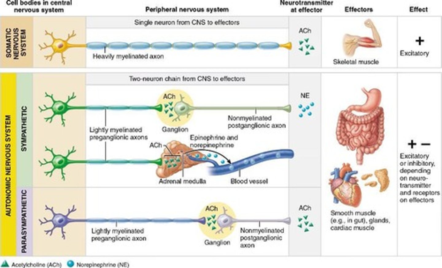
Autonomic Nervous System (ANS)
Involuntary, unconscious regulation of smooth muscle, cardiac muscle, and glands.
Effectors
Both systems can stimulate or inhibit its effectors.
Motor Fibers
Both have motor fibers but differ in effectors, efferent pathways and ganglia, and the target organ responses to different neurotransmitters.
ACh at the neuromuscular junction
All somatic motor neurons release ACh at the neuromuscular junction.
Effect on skeletal muscle
Always excitatory (induces contraction).
Postganglionic sympathetic fibers
Typically release norepinephrine (NE).
Postganglionic parasympathetic fibers
Release acetylcholine (ACh).
Target Organ Effects
The effect (excitatory or inhibitory) depends on the specific receptors on the target tissue.
Adrenergic receptors
Respond to NE; effect depends on subtype (α or β).
Cholinergic receptors
Respond to ACh; nicotinic or muscarinic subtypes, each with different responses.
Rest-and-Digest System
Supports relaxation, energy conservation, and normal body maintenance.
Key responses of the Parasympathetic Division
↓ Heart rate, constricts pupils, stimulates digestion, promotes nutrient absorption, stimulates urination and defecation, conserves energy.
Fight-or-Flight System
Prepares the body for stress, emergencies, and increased activity.
Key responses of the Sympathetic Division
↑ Heart rate and blood pressure, dilates bronchioles, dilates pupils, mobilizes energy, diverts blood flow to skeletal muscles, inhibits digestion and urinary functions.
Homeostasis
These divisions usually have opposite effects on the same organs to maintain homeostasis.
Parasympathetic Division (Craniosacral)
Parasympathetic fibers originate in the brain (cranial nerves III, VII, IX, X) and the sacral spinal cord (S2-S4).
Origin of Parasympathetic fibers
Parasympathetic fibers arise from the brainstem and sacral spinal cord (S2-S4).
Fiber Lengths
Long preganglionic fibers and short postganglionic fibers.
Cranial Portion
Preganglionic fibers travel with cranial nerves III, VII, IX, and X, controlling functions like pupil constriction, tear/salivary secretions, and major thoracic/abdominal organ regulation.
Sacral Portion
Supplies the pelvic organs and the distal half of the large intestine, helping manage urination, defecation, and reproductive organ function.
Sympathetic Division (Thoracolumbar)
Sympathetic fibers originate in the thoracic and lumbar regions of the spinal cord (T1-L2).
Origin of Sympathetic Fibers
Sympathetic fibers arise from the thoracic and lumbar spinal cord segments T1-L2.
Innervation Scope of Sympathetic Division
More complex than the parasympathetic division because it innervates more organs and all superficial structures of the body.
Unique Targets of Sympathetic Division
The sympathetic division exclusively supplies sweat glands (eccrine & apocrine), arrector pili muscles (hair-raising muscles), smooth muscle in the walls of all arteries and veins (deep and superficial), and the kidneys.
Effectors Supplied by Sympathetic Division
It innervates smooth muscle, cardiac muscle, and glands in internal body cavities—but also all superficial smooth muscle and glands.
Segment-Specific Output T1-T4
Preganglionic fibers serving the head.
Segment-Specific Output T1-T6
Preganglionic fibers serving thoracic organs.
Segment-Specific Output T5-L2
Preganglionic fibers serving the abdominal organs.
Segment-Specific Output T10-L2
Preganglionic fibers serving pelvic organs.
Parasympathetic Division Fiber Lengths
Long preganglionic fibers and short postganglionic fibers.
Sympathetic Division Fiber Lengths
Short preganglionic fibers and long postganglionic fibers.
Location of Parasympathetic Ganglia
Ganglia are located in or very close to the visceral effector organs (intramural ganglia).
Location of Sympathetic Ganglia
Ganglia lie close to the spinal cord, either in the sympathetic chain (paravertebral ganglia) or prevertebral ganglia near the abdominal aorta.
Visceral Reflex Arc
Has two consecutive motor neurons (preganglionic and postganglionic).
Somatic Reflex Arc
Has one motor neuron connecting the CNS directly to the skeletal muscle.
Afferent Pathway of Visceral Reflex Arc
Afferent fibers are visceral sensory neurons, detecting internal conditions (stretch, chemicals, etc.).
Afferent Pathway of Somatic Reflex Arc
Afferent fibers are somatic sensory neurons, detecting external stimuli (pain, temperature, touch).
Effectors of Visceral Reflex Arc
Effectors are smooth muscle, cardiac muscle, or glands.
Effectors of Somatic Reflex Arc
Effectors are skeletal muscles.
Acetylcholine (ACh)
Released by all preganglionic neurons in both sympathetic and parasympathetic divisions, released by parasympathetic postganglionic neurons, and released by sympathetic postganglionic neurons to sweat glands (an exception).
ACh
Acts on cholinergic receptors (nicotinic and muscarinic).
Effects of ACh
Can be excitatory or inhibitory, depending on the receptor type.
Norepinephrine (NE)
Released by most sympathetic postganglionic neurons.
Effects of NE
Can also be excitatory or inhibitory, depending on which adrenergic receptor subtype is present.
Epinephrine (EPI)
Acts as a hormone rather than a neurotransmitter.
Source of EPI
Secreted by adrenal medulla into the bloodstream.
Release mechanism of EPI
Sympathetic preganglionic fibers release ACh → stimulates nicotinic receptors on adrenal medulla → epinephrine secretion.
Receptors activated by EPI
α1, α2, β1, β2, β3.
Effects of EPI
Widespread sympathetic 'fight-or-flight' responses: ↑ Heart rate & contractility (β1), Bronchodilation (β2), Glycogen breakdown ↑ glucose (β2, β3), Vasoconstriction (α1) & vasodilation in skeletal muscles (β2).
Somatic motor neurons
Always release ACh, and the effect on skeletal muscle is always excitatory.
Autonomic neurons
Can release ACh or NE, and the effect on target organs varies depending on receptor type.
Autonomic Tone
Both divisions are partially active at all times, even at rest.
Purpose of Autonomic Tone
Allows precise, continuous regulation of organ function (e.g., blood vessel constriction by sympathetic tone).
Dynamic Antagonism
The two divisions often have opposing effects (e.g., sympathetic ↑ heart rate, parasympathetic ↓ heart rate).
Predominance of divisions
One division usually predominates depending on the situation: Parasympathetic: rest-and-digest, Sympathetic: fight-or-flight.
Cooperative Effects
In some cases, both divisions work together to achieve a single, coordinated outcome.
Example of Cooperative Effects
Male reproductive organs — parasympathetic → erection, sympathetic → ejaculation.
Key Concept of ANS
The ANS maintains homeostasis through tone, antagonism, and cooperation.
Thermoregulatory Responses
Sympathetic nerves dilate skin blood vessels → heat loss; constrict skin blood vessels → conserve heat.
Renin Release from Kidneys
Sympathetic impulses stimulate kidneys to release renin, triggering formation of hormones that increase blood pressure.
Metabolic Effects of Sympathetic Division
Increases metabolic rate of body cells, raises blood glucose levels (glycogen breakdown), mobilizes fats for energy (lipolysis).
Key Concept of Sympathetic Division
Helps maintain homeostasis under stress and temperature changes, regulates blood pressure, and supports energy mobilization.
Parasympathetic Division
One preganglionic neuron → one (or few) postganglionic neurons.
Acetylcholinesterase
All fibers release ACh, which is rapidly broken down by acetylcholinesterase.
Effect of Parasympathetic Division
Short-lived, highly localized control over target organs.
Sympathetic Division
Preganglionic axons branch extensively in the sympathetic trunk.
Synapse in Sympathetic Division
Synapse with postganglionic neurons at multiple levels.
Effect of Sympathetic Division
Diffuse, widespread, and highly interconnected responses throughout the body.
Key Concept of Parasympathetic Division
Parasympathetic = precise, localized actions.
Hypothalamus
Master Control Center that integrates emotional, endocrine, and autonomic responses.
Homeostasis Functions of Hypothalamus
Maintains body temperature, hunger, thirst, fluid balance.
Brainstem Functions
Contains cardiovascular, respiratory, digestive, and urinary centers.
Medulla Oblongata
Controls heart rate, blood pressure, and breathing.
Spinal Cord Functions
Mediates simple autonomic reflexes.
Example of Spinal Cord Reflex
Defecation and urination reflexes.
Visceral Sensory Neurons
Monitor internal conditions (stretch, chemicals, pressure).
Hypertension
Chronically elevated blood pressure.
Cause of Hypertension
Often due to overactive sympathetic vasoconstriction, which increases peripheral resistance.
Raynaud's Disease
Episodic spasms of small arteries, usually in fingers and toes.
Cause of Raynaud's Disease
Excessive sympathetic stimulation in response to cold or stress → vasoconstriction.
Autonomic Dysreflexia
Sudden, massive sympathetic reflex in spinal cord injury patients (usually above T6).
Cause of Autonomic Dysreflexia
Uncontrolled sympathetic response to stimuli (e.g., bladder distension, skin irritation).
Beta Blockers
Block β-adrenergic receptors, mainly β1 (heart) and β2 (lungs and blood vessels).
Effects of Beta Blockers
Lower heart rate, reduce blood pressure, decrease tremors.
Beta Blockers in Sports
Reduce physiological tremor and anxiety, improving steadiness and focus.
ANS Connection of Beta Blockers
Interfere with sympathetic nervous system signaling.
Cerebellar cortex
At the highest level of our conscious motor pathways, but not the ultimate planner and coordinator of complex motor behaviors.
Basal nuclei
Plays a role in the ultimate planning and coordination of complex motor behaviors.
Segmental level
Consists of reflexes and spinal cord circuits that control automatic movements as well as central pattern generators.
Projection level
Consists of neurons acting through direct and indirect motor pathways.
Precommand level
Consists of the cerebellum and basal nuclei, and controls the outputs of the cortex and brain stem motor centers.
Inborn (intrinsic) reflex
A rapid, predictable motor response to a stimulus that is unlearned, unpremeditated, and involuntary.
Learned (acquired) reflexes
Results from practice or repetition.
Somatic reflexes
Activate skeletal muscle.
Autonomic (visceral) reflexes
Activate visceral effectors (smooth or cardiac muscle or glands).
Flexor (withdrawal) reflex
Initiated by a painful stimulus, causes automatic withdrawal of the threatened body part.
Crossed extensor reflex
Often accompanies the flexor reflex in weight-bearing limbs and is important for maintaining balance.
Interneurons
Act as a link or bridge between sensory neurons and motor neurons within the central nervous system (CNS).
Function of interneurons
Process and interpret information received from sensory neurons, then decide how the body should respond by sending signals to motor neurons.