Pericardial Diseases
 Call Kai
 Learn
Learn Practice Test
 Spaced Repetition
 Match
Match Flashcards
 Knowt Play
1/49

There's no tags or description
Looks like no tags are added yet.
Name | Mastery | Learn | Test | Matching | Spaced | Call with Kai | Chat |
|---|
No analytics yet
Send a link to your students to track their progress
50 Terms
Pericardium =
2 layers that surround the heart and proximal great vessels
visceral pericardium =
epicardium; inner layer where coronaries sit underneath
parietal pericardium
outer layer, fibrous structure providing mechanical support
between fibrous pericardium and pericardial cavity
reflections of pericardium =
the 2 layers form a closed space with finger-like projections. this “pocket” creates sinuses
Oblique sinus
around IVC and Pulmonary veins
Transverse sinus
around Aorta and Pulmonary Artery
pericardial fluid
between 2 layers - lubricates heart so it can beat in a friction-free space
epicardial fat pad
present on anterior aspect, speckled/granular appearance
elderly, obese, diabetic, female pts
What are the 2 functions of the pericardium?
Mechanical - allows heart to beat with minimal friction
Passive restraint - controls how much heart may expand in diastole
How does the pericardium work as a passive restraint?
The fibrous parietal pericardium contributes to resting diastolic pressures and limits acute dilation
How does the mechanical restraint of the pericardium influence ventricular interdependence?
LV and RV share the IVS and is surrounded by the noncompliant pericardium → volume in one ventricle can influence pressure & filling in the other
acute pericarditis clinical presentation
dyspnea
tachypnea
palpitations
fever
In acute pericarditis, how is the positional chest pain increased/decreased?
increases with inspiration
decreases with sitting up and leaning foward
acute pericarditis EKG
reduced QRS voltage
concave ST elevation
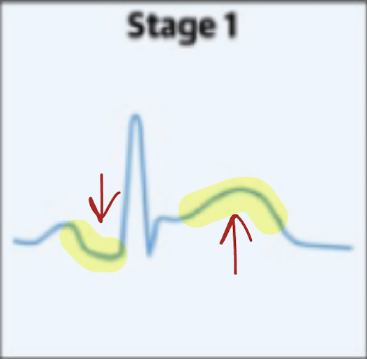
acute pericarditis EKG stage 1
ST elevation and PR depression
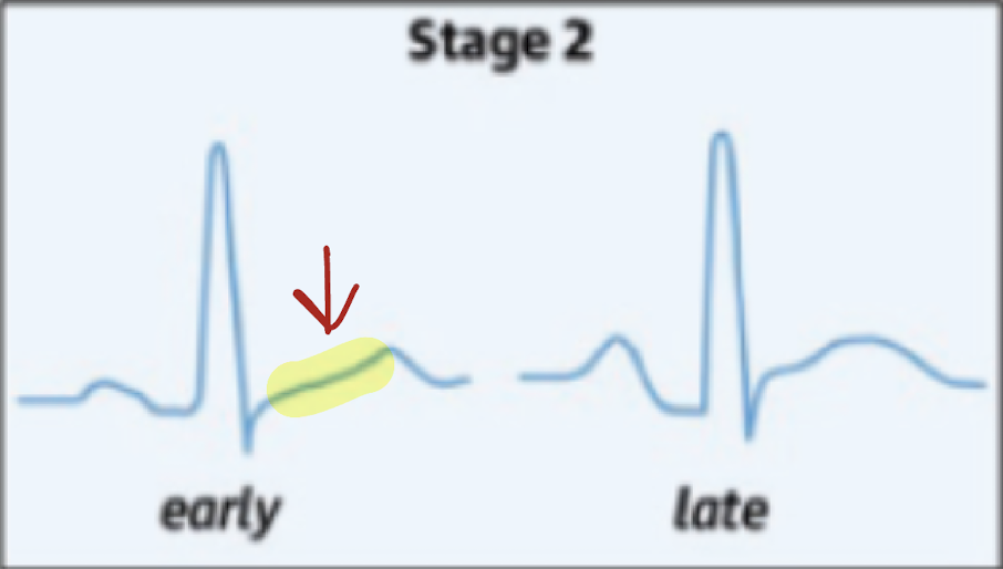
acute pericarditis EKG stage 2
J points flatten
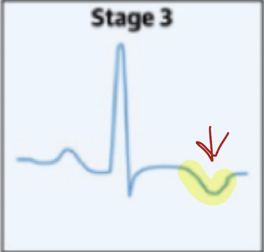
acute pericarditis EKG stage 3
T-wave inversion
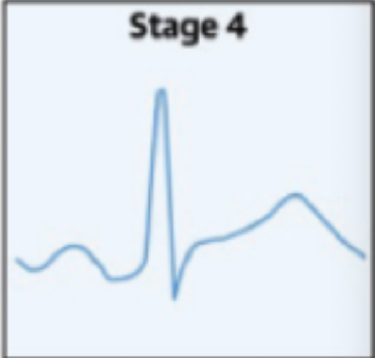
acute pericarditis EKG stage 4
Normal!
Acute pericarditis auscultation
pericardial friction rub
Ewart’s sign
Ewart’s sign
dullness on left lung
Acute pericarditis 2D echo findings
swinging heart
IVC plethora = ↑RAp
effusion
loculated effusion
increased echogenicity (blood)
Constrictive Pericarditis pathophysiology
impaired LATE diastolic filling due to noncompliant case = ↑diastolic pressures
Constrictive Pericarditis causes cessation of mid-late ventricular filling due to physical limitation of expansion. This causes what sign on the atrial and ventricular pressure maps?
rapid “y” descent and “dip and plateau”
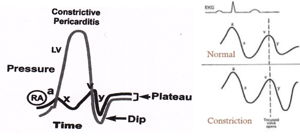
Constrictive Pericarditis clinical presentation
jugular venous distension
Kussmaul’s sign
weakness, fatigue
dyspnea, PND, orthopnea
peripheral edema
hepatomegaly + ascites
Kussmaul’s sign
rise in jugular pressures in inspiration
Constrictive Pericarditis ausultation
pericardial knock (low diastolic sound)
Constrictive Pericarditis EKG signs
diffuse T-wave flattening
low QRS voltage
afib
tachycardia
Constrictive Pericarditis 2D echo findings
thickened pericardium >3 mm
septal bounce
IVC plethora
Septal bounce
IVS bulges to left during inspiration due to abrupt cessation of diastolic filling
IVC plethora
dilated IVC with lack of variation (↑RAp)
Constrictive Pericarditis M-MODE echo findings
Railroad track sign
Posterior wall flattening
Spanish notch
Railroad track sign
parallel but separated epicardial & pericardial echoes
Posterior wall “flattening”
outward movement during mid-late diastole
Spanish notch
sudden anterior displacement and bisk posterior rebound of IVS
Constrictive Pericarditis Doppler (PW) echo findings
Trans mitral flow - increases 25% with expiration
Trans tricuspid flow - increases 40% with inspiration
hepatic venous flow - ↑diastolic reversal with expiration
TDI - annulus reversus
Annulus Reversus
septal E’ > lateral E’ due to tethering of lateral wall by pericardium
Cardiac Tamponade pathophysiology
injury to pericardium → pericardial effusion → increased intrapericardial pressures → cardiac chamber compression
Cardiac Tamponade: The increase in intrapericardial pressures causes the chamber pressures to equalize, and impairment of venous return, causing a _____ on the atrial tracing
blunted “y”
3 stages of tamponade
intrapericardial pressure rises < RVp and PCWP
intrapericardial pressure = RV but < PCWP
intrapericardial pressure = RV + PCWP
Cardiac Tamponade clinical presentation
Beck’s triad
dyspnea
shock
Pulsus Paradoxus
Total Paradoxus
peripheral cyanosis (↓CO)
Kussmaul’s sign
Ewart’s sign
Beck’s triad
Hypotension
Distended jugular veins (↑ venous pressure)
muffled heart sounds
Pulsus Paradoxus
>10 mmHg fall in systolic BP in inspiration
Total Paradoxus
complete absence of pulse with inspiration
Cardiac Tamponade EKG signs
reduced QRS voltage
ST elevation with upright T wave
electrical alternans
total alternans
tachy/bradycardia
Electrical alternans
alternating QRS amplitude
Cardiac Tamponade 2D echo findings
pericardial effusion
presystolic collapse of RA
RV + LV early diastolic collapse
LA late diastolic collapse
swinging heart
IVC plethora
dilated HpV + SVC
inspiratory bounce of IVS toward LV
What are the most significant echo findings of Cardiac Tamponade?
chamber collapse & swinging heart
Cardiac Tamponade Doppler echo findings
abnormal diastolic biventricular flow with respiration
MV ↑ in expiation
TV ↑ in inspiration
↑diastolic HpV reversal
Mitral Flow _____ with expiration and ______ with inspiration
increases (expiration)
decreases (RV fills more and IVS compresses LV)
Tricuspid Flow _____ with expiration and ______ with inspiration
decrease
INcrease with INspiration (more blood to RA → ↑volume in RV → IVS moves to left → less filling in LV)