CMA ch. 7 - 8
 Call Kai
 Learn
Learn Practice Test
 Spaced Repetition
 Match
Match Flashcards
 Knowt Play
1/94

There's no tags or description
Looks like no tags are added yet.
Name | Mastery | Learn | Test | Matching | Spaced | Call with Kai | Chat |
|---|
No analytics yet
Send a link to your students to track their progress
95 Terms
Inner most layer of the heart
endocardium
P wave

PR segment

Q segment

R wave

S wave

ST segment

T Wave

Middle layer of the heart
myocardium
Outermost layer of the heart
epicardium
Upper chambers of the heart
atria
Right atrium receives blood from
the superior and inferior vena cava
Only vein that receives oxygenated blood
pulmonary vein
Only artery that receives deoxygenated blood
pulmonary artery
Right AV valve
tricuspid valve
Left AV valve
bicuspid/ mitral valve
Lubb sound happens when
AV valve closes at the start of systole
Dubb sound happens when
semilunar valve closes at the end of systole
Mediastinum
thoracic compartment where the heart resides
Largest artery in the body
aorta
Blood exits __ side of the body out of the aorta
left
The heart account for ___% of the body’s metabolic demand
25
The heart will beat approximately __ times through a pt’s life
4.8 billion
A healthy heart is regulated by the
sinoarterial node (AV node)
Depolarization
releasing a pulse of electricity
Depolarization will occur in the SA node at a rate of
60 to 100 bpm
If the AV node is acting as the pace maker, it will depolarize at a rate of
40 to 60 bpm
His purkinje system
conducts electricity through the myocardium, allows the heart to beat in the most efficient way
Order of his purkinje system
AV node : bundle of his : bundle branches : purkinje fibers : leads to heart contraction
P waves
Atrial depolarization
PR segment
flatline, no electricity is moving and is caused by the AV delay
QRS complex
sharp deflections
Q wave
depolarization of the septum
R wave
relates to the depolarization of the apex
S wave
depolarization of the left ventricle
ST segment
isoelectric line
T wave
repolarization of the ventricle myocardium
CO
cardiac output, amount of blood being pumped out by the heart multiplied by the heart rate
Apex
the thickest region of myocardial tissue
Valve
A section of fibrous tissue to prevent the backflow of blood in the heart
AV node
resides at the junction of ventricles and atria
Bundle of his
from the av node into the interventricular septum
Bundle branches
carry the action potential through the septum to the purkinje system
Purkinje fibers
penetrate into the myocardium from the sub endo cardial space
How do the electrical signals of the heart relate to the pumping of the heart
depolarization allows for the influx of sodium ions which releases calcium ion stores allowing muscle contraction
When is 12 lead EKG used
outpatient, routine monitoring, at rest
When is telemetry used
inpatient monitoring, produces continuous monitoring
When is the holter monitor used
1 to 3 days outpatient during the commencement of normal everyday living. Used for identifying events that may not be visible in a limited window
Sinus rhythm HR
60 to 100
Sinus rhythm PR
0.12 to 0.20 sec
Sinus rhythm QRS
0.04 to 0.1 sec
Bradycardia
HR less than 60 bpm
Tachycardia
HR greater than 100 bpm
Arrhythmia
an irregular heart rhythm
Atrial rhythms
atrial flutter and atrial fibrillation
How many electrodes in a 12 lead EKG
10 electrodes
12 lead EKG gives __ perspectives of the heart
12
Right oriented EKG must be performed when
pts under the age of 2 and pts with dextrocardia (opposite oriented cardiac axis)
Telemetry monitoring can be done in __ leads
3 and 5 leads
3 lead telemetry uses
white, black and red leads
5 lead telemetry uses
white, black, red, brown, green leads
12 lead EKGs monitor (how much time frame)
10 seconds
Holter monitors monitor (how much time frame)
24 to 72 hours of continuous monitoring
When monitoring pts with the holter monitor
ask them to keep a diary of activities
2 types of Holter monitors
5 lead (2 channel) and 7 lead (3 channel)
Pt instructions for holter monitoring
keep the monitor dry, maintain a log of activities, use event button to note when they experiene symptoms, avoid strong EMF fields
EKG is a graph of
voltage over time
Standard paper feed speed
25mm/seconds
Standard gain (voltage) setting
10mm/mV
1500 method
1500/number of small boxes in the R to R interval
Size of one small box
1 mm
Size of one large box
5 mm
Sequence method
300/ number of large boxes
6 second method
count the number of QRS sequences in two of the section (3 second marks are denoted by tick marks at the top of the page) and then multiply that by 10
Method best for tachycardia or when needing precise measurements
1500 method
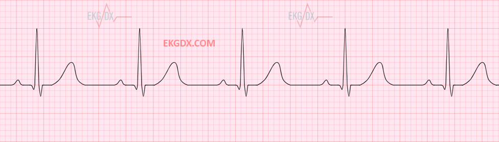
Normal sinus rhythm
(picture)
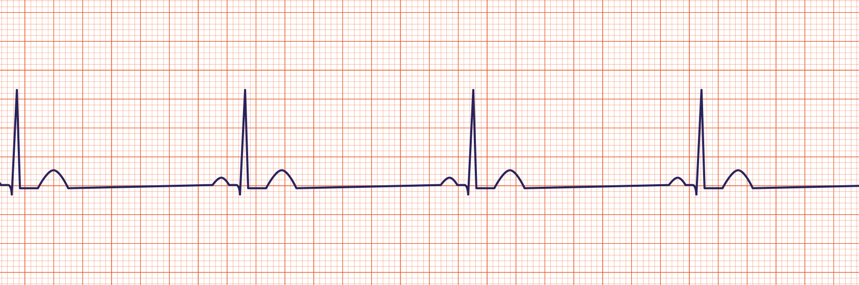
Sinus bradycardia
(picture)
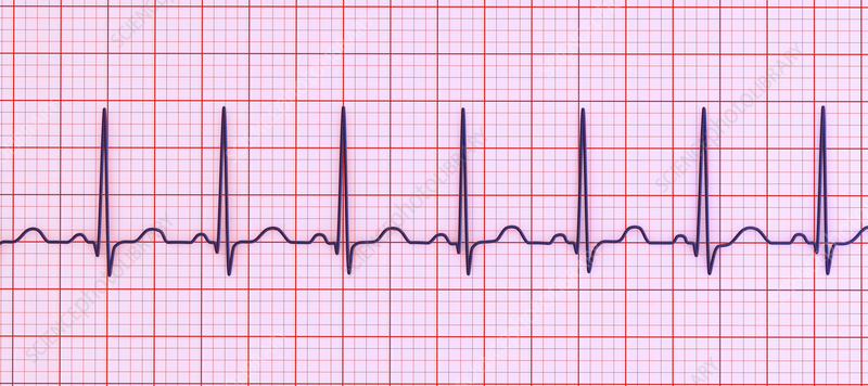
Sinus tachycardia
(picture)
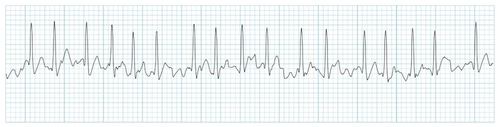
Atrial fibrillation
(picture)
Atrial flutter
(picture)
Depression of J point and ST segment indicate
myocardial infarction (MI)
Pt related artifacts on EKG
seizure, trembling, fast breathing, dry or wet skin, lotion on skin
Non patient related artifacts on EKG
expired or damaged electrodes, damaged lead cables, cell phone interference, EMF
Seziore on EKG
jagged tracing, uneven peaks
Trembling on EKG
jagged interruptions in regular tracing
Dry or wet skin/ gel problems on EKG
loose lead warning on machine, unexplained flatline
Cell phone or other electrical interference on EKG
flutter or repeating P waves
ICS
intercostal space
WTC
wilson’s central terminal
NSR
normal sinus rhythm
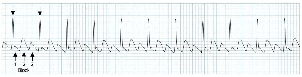
Heart block
a result of damage to the his purkinje system, represents a dealy or failure between the pacers of the atrium and the ventricles and shows up as abnormalities in the PR interval and a possibly wider QRS complex
Morphology
the study of the form or shape of an EKG tracing
Sinus rhythm
a rhythm set by the SA node
Telemetry
the process of recording and transmitting the readings of an instrument
Precordium
the portion of the body that is the anterior chest wall over the heart