Exam 1 Nerve Irritation vs Nerve Compression, Sensory LEs
 Call Kai
 Learn
Learn Practice Test
 Spaced Repetition
 Match
Match Flashcards
 Knowt Play
1/114

There's no tags or description
Looks like no tags are added yet.
Name | Mastery | Learn | Test | Matching | Spaced | Call with Kai | Chat |
|---|
No analytics yet
Send a link to your students to track their progress
115 Terms
5 components of Vertebral Subluxation Complex (VSC)
1. spinal kinesio-pathology
2. myopathology
3. histopathology
4. neuropathology
5. biomechanical changes
subluxation has ____________ as a component that contributes to S/S
nerve IRRITATION
what percent of the time is a subluxation a nerve irritation?
97% of the time
if a patient is experiencing nerve irritation what might they say? what would the sensory exam show?
"feels numb", but the sensory exam is WNL
what is the primary factor of nerve irritation?
EDEMA
-bathes the NR, irritating the tissue: prostaglandins, bradykinins, substance P, WBCs surround joint capsule
**edema does NOT compress the NR
a disc herniation at C4/C5 affects which NR?
C5 nerve root
hypertrophic changes of the uncinate processes of C5 will affect what NR?
C5 NR
hypertrophic changes of the T1 VB will affect which NR?
C8 NR
a patient with a documented anterior disc herniation that is not directly compressing the nerve root. could they still have neck pain? arm pain?
neck pain: even without direct, severe compression, a herniated disc causes localized neck pain due to tension in the annulus fibrosus, disc inflammation, or surrounding tissue irritation
arm pain: while classic radiculopathy arises from direct NR compression, arm pain can still occur via referred pain from the disc itself (chemical radiculitis) or irritation of nearby structures
*the pain is frequently a nociceptive response, where inflammation in the disc material, rather than just mechanical pressure, creates localized/radiating pain
if patient has unilateral dermatomal radiating pain, what should you suspect?
nerve root
if patient has unilateral multi-dermatomal radiating pain, what should you suspect?
plexus, peripheral nerve, or vascular
if patient has diffuse thigh pain that does NOT go beyond the knee, what should you suspect?
lumbar facet
if patient has radiating pain that goes beyond the knee and follows a dermatomal pattern, what should you suspect?
lumbar disc
if patient has pain 1" lateral to the L5 spinous process, what should you suspect?
L5 facet syndrome/irriation
myelopathy
dysfunction of the spinal cord
radiculopathy
dysfunction of a spinal nerve root
neuropathy
dysfunction of a peripheral nerve distal to the nerve root
myopathy
muscle abnormalities
dermatome
the area of skin innervated by cutaneous axons in the distribution of a single spinal nerve
myotome
refers to the muscle fibers innervated by the motor axons of a single spinal nerve
describe type A nerve fibers and characteristics of pain they carry
fast, lightly myelinated
acute, sharp, stinging sensation to nociceptive
usually localized
alerts to the presence of pain but does NOT contribute to the intensity of pain
describe type C nerve fibers and characteristics of pain they carry
slow, unmyelinated
aching, burning, lingering type of pain
usually diffuse
conduct information related to intensity of pain AND temperature
what are the three mechanistic types of pain?
1. nociceptive pain
2. neuropathic pain
3. nociplastic pain
nociceptive pain
pain arising from actual or threatened damage to non-neural tissue caused by activation of nociceptors
*pain from tissue injury and inflammation
key mediators of nociceptors
PGs, bradykinin, substance P, CGRP (calcitonin gene-related peptide), cytokines (IL-1, TNF-a)
what do the mediators of nociceptors do?
these chemicals lower nociceptor activation thresholds, producing peripheral sensitization
clinical examples of nociceptive pain
somatic pain: acute muscle sprain, ligament sprain, post-surgical pain, OA, inflammatory joint disease
radicular pain
visceral pain
neuropathic pain
pain caused by a lesion or disease of the somatosensory nervous system
*pain from nerve damage or disease
damage to peripheral or central nerves leads to
ectopic spontaneous firing
ion channel dysregulation
loss of inhibitory interneurons
central sensitization
clinical features of neuropathic pain
1. pain not necessarily proportional to injury
2. burning, electric shock-like, tingling
3. dermatomal or nerve distribution
4. may persist after healing
clinical examples of neuropathic pain
radiculopathy from disc herniation
diabetic neuropathy
post-herpetic neuralgia
carpal tunnel syndrome
trigeminal neuralgia
key pathophysiology of neuropathic pain
upregulation of voltage-gated sodium channels
microglial activation
NMDA receptor activation
increased glutatmate signaling
*these produce pathological neural firing independent of tissue injury
nociplastic pain
pain arising from altered nociception despite no clear evidence of tissue damage or nerve injury
*pain from central processing dysfunction
*CNS amplification of pain signals
-central sensitization
-reduced descending inhibition
-neuroimmune activation (microglia, astrocytes)
clinical examples of nociplastic pain
1. pain often disproportionate to injury
2. widespread, diffuse pain
3. fatigue, sleep, and mood problems
4. symptoms often persistent
-ex: persistent neck pain months after minor issues, central pain amplification despite healed tissues
-ex: fibromyalgia, chronic tension-type headache, chronic migraine, IBS, chronic non-specific low back pain
neurochemical changes in nociplastic pain
NMDA receptor activation
increased glutamate
reduced GABA inhibition
what is nociplastic pain often strongly influenced by?
stress, trauma, emotional factors, autonomic dysregulation
*the nervous system becomes hyper-responsive to normal input
pain vs nociception: which is subjective and which is objective?
pain = subjective
nociception = objective
define pain
the SUBJECTIVE, unpleasant, and often emotional experience that arises from processed neurological signals
-subjective, experiential, and highly influenced by biology, psychology, and social
**pain is biopsychosocial
pain example
Hurt back while playing basketball, now you can't work. You worry about social implications of not being able to work = increase intensity of pain
define nociception
the sensory detection and transmission of noxious (potentially damaging) stimuli
-objective, physiological, and measurable
-nociceptors detect a threat and send electrical signals up nerve fibers to the spinal cord and brain
nociception example
a pin pricks your finger, nociceptors fire and send signals to your CNS and you pull your hand away
*nociception is the transmission and modulation of noxious stimuli
which joint tissues are void of nociceptors?
articular cartilage
inner annulus of IVD
nucleus of IVD
synovial membranes
in a normal joint, the nociceptive system is?
INACTIVE
-to active the nociceptive fibers, you must first have a noxious stimuli (mechanical, thermal, or chemical)
nociceptive nerve endings are found within the annulus fibrosis. what does this mean?
annular tears can cause pain in the lower back, buttocks, SI region, and lower extremity
*this pain can occur WITHOUT nerve compression by a disc protrusion
disc protrusion that does not compress the nerve root directly can cause an inflammatory response and secondary radiculitis by?
chemically induced inflammatory neural pain leading to nerve irritation
what is the neuropathway for the patient to experience pain from a cervical disc bulge/herniation?
the sinuvertebral nerve (recurrent nerve of Luschka) conveys sensory fibers to the PLL, the dura, and the outer border of the annulus. sinuvertebral nerve re-enters the IVF, and innvervates the facet joints, the annulus fibrosus, the ligaments, and periosteum of the spinal canal, carrying pain and proprioception sensation
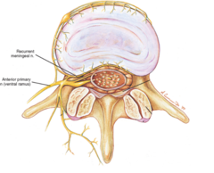
sensory deficit order of loss
lost 1st: light tough
sharp pain and temperature
lost last: vibration and position sense
when compression is removed, sensory is restored in
the OPPOSITE order
*light touch is first to go and last to return
always compare sensory ________ with the ________ side first
bilaterally, asymptomatic
with nerve irritation, the patient will say
they have sensory changes (ex: numbness) but the exam is WNL showing no sensory deficits
with nerve compression, the patient will say
they have sensory changes and the exam shows + paresthesia
paresthesia
abnormal sensations along dermatomes and described by the patient as: numbness, tingling, pins and needles, pain, prickling, aching, warmth (heat), cold, and burning
hyperesthesia
increased sensation to pain or other stimuli across dermatomal patterns
*typically seen with nerve IRRITATION, partial lesions, or secondary to healing
*typical in a chiro patient
hypoesthesia
diminished sensitivity to pain or other stimuli across dermatomal patterns
*typically seen with nerve root COMPRESSION
anesthesia
absence of sensation across dermatomal patterns
*typically seen with NR COMPRESSION
what is the MC cause of NR compression?
DISC
examples of space occupying lesions
1. disc
2. bone
3. tumor
4. ligament
5. muscle
6. hemorrhage
7. infection
if the patient does not feel the vibratory sensation equal from side to side, assume there is a deficit in the
posterior columns
if the patient does not feel the sensation at all, as in some neuropathies, you should
move up the leg to evaluate the extent of paresthesia and map it
if you find an area of hypo/hyperesthesia, then you need to determine ifit follows:
1. dermatome pattern
2. peripheral nerve pattern
3. lumbar/sacral plexus pattern
4. vascular insufficiency pattern
5. spinal stenosis
6. intermittent claudication
7. thrombophlebitis
8. visceral pain referral pattern
what is carried in the posterior columns?
position sense, vibration, pressure, texture, localization of touch, 2-pt discrimination, weight, and ability to decipher #s written on the skin
**lesions that affect these sensations while sparing light touch, pain, and temp are likely SPINAL CORD origin
for the most part, nociceptive (pain and temp) fibers travel along the lightly myelinated A delta fibers and unmyelinated C fibers of the __________
lateral spinothalamic tract
light touch is associated with which tract?
anterior spinothalamic
dermatomes with light touch deficits would indicate possible?
NR compression (along dermatomes) or possible peripheral nerve lesions (cross dermatomes)
if the pain is sharp with motion and constant, think
joint
if the pain is burning, sharp/lightning, tingling and/or numbness, think
nerve
if the pain is cramping/spasm or a dull ache with AROM and resisted PROM
muscle
if the pain is radiating dull or deep, heavy, boring, think
scleratogenous (facet, SI, disc)
if the pain is a dull ache/pain with PROM or a deep burning, think
bone/ligament
if the pain is pinpointed over paraspinal and has a crawling sensation, think
myofascial
if the pain is throbbing, think
vascular
if the pain is well localized, think
peripheral
if the pain is diffuse, think
central
if the pain is bilateral, think
CORD first
most common causes for radiculopathy include
disc herniation
synovial cyst
spinal stenosis
frequency of lumbar radiculopathy in men and women
men: 2-5%
women: 1-3%
*frequency peaks at age 44 for both sexes
why is venous congestion more common in the IVF?
because the radicular veins may be immediately compressed by reduction in foraminal diameter
-possibility of reflux from the segmental veins through pressure damaged valves
-venous congestion --> swelling within IVF --> contributes to compression of the other intra-foraminal structures, including the NR
where is circulation especially vulnerable to venous congestion?
dorsal root ganglia
why are nerve roots more vulnerable to mechanical effects than peripheral nerves?
location within the IVF makes NR vulnerable
NRs lack protection of epineurium and perineurium
NRS are dependent on a single radicular artery entering via the foramen, the margin of safety provided by collateral pathways is minimal
your patient has numbness and tingling down the right side of the neck and into the right shoulder traveling distally down into the middle finger. instrumentation break to the right at C6 with static and motion palpation fixation at C6. the sensory exam revealed a hypoesthesia to light touch and sharp pinprick along the right anterior lateral aspect of the forearm and the middle finger. what's the diagnosis?
SOL at C6 causing nerve root compression C7 on the right
thoracic spine landmarks
T4, T7, T10, T12
when the upper limb is by the side, the SP of T3 is level with the _____, and the T7 SP is level with the _____
spine of the scapula, inferior angle
T1 nerve root motor, sensory, and reflexes
motor: interosseous
sensory: hand, medial arm
reflexes: none
T2, T3, T4 motor, sensory, and reflexes
motor: intercostal
sensory: same as rib (nipple)
reflexes: none
T5, T6, T7 motor, sensory, and reflexes
motor: rectus abdominus superior
T5-T6 sensory: same as rib (epigastric)
T5-T6 reflexes: none
T7 sensory: same as rib (xiphoid)
T7 reflexes: superficial
T8-T10 motor, sensory, and reflexes
T8-T9 motor: rectus abdominus superior
T8-T9 sensory: same as rib (umbilicus)
T8-T10 reflexes: superficial (on abdomen)
T10 motor: rectus abdominus inferior
T10 sensory: umbilicus (follow around to back)
T11-T12 motor, sensory, and reflexes
T11-T12 motor: rectus abdominus inferior
T11 sensory: inferior abdomen (groin)
T12 sensory: pubic region (above pubic symphysis) (groin)
T11-T12 reflexes: superficial (abdomen)
what is a positive light touch thoracic sensory exam associated with?
anterior spinothalamic tract
what does a positive sharp touch thoracic sensory exam indicate?
hypo or hyperesthesia's nociceptive (pain and temp) fibers travel along the lightly myelinated A delta fibers and unmyelinated C fibers of the lateral spinothalamic tract
name two additional tests for a sensory exam of the thoracic spine
motor test of the same NR
DTR test of same NR
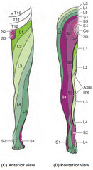
describe the LE dermatomes L1-S2
L1: inguinal
L2: anterior thigh (high)
L3: anterior thigh (low)
L4: anterior knee, medial lower leg, medial foot
L5: top of foot, anterior lateral lower leg
S1: lateral foot, lateral calf, posterior thigh
S2: posterior medial thigh, medial calf
what does a positive LE vibration sensory test indicate?
patient does NOT feel the vibratory sensation equal from side to side = assume there is a deficit of the posterior columns
2 additional tests for LE vibration sensory exam
sensory exam of LE, temperature and sharp/light touch
L4 herniated nucleus pulposus with compression
A. affects the medial aspect of the foot
B. affects the top of the foot
C. affects the lateral aspect of the foot
D. affects the anterior aspect of the thigh
B. affects the top of the foot
describe L5 posterior facet referral pain (scleratogenous referred pain)
dully achy unilateral pain found on active or passive extension
lateral to the SP of L5, radiates into posterior glute and thigh -- but NOT below the knee
what direction do lumbar discs most commonly herniate in?
posteriorly
known as: subarticular disc herniation or paracentral/lateral recess herniation
why is the exiting NR in the lumbars usually not involved in any herniations of the disc within its space?
exiting NR takes a 45 degree angle turn around the pedicle of its vertebra
-exiting NR almost never crosses the disc space below it and therefore is usually not involved
a lumbar NR is most commonly involved in herniations of the disc located ______ its point of exit
ABOVE
**traversing NR is most commonly affected in lumbars
3 locations of disc herniations in order of MC to least common
1. subarticular (MC)
2. foraminal/extraforaminal
3. central (rare)
which articulations have the greatest motion in the lumbar spine?
L4-L5 and L5-S1
**therefore, the incidence of herniated discs is greater at these areas than any other lumbar space