Wk 5: Urinary tract and water balance
 Call Kai
 Learn
Learn Practice Test
 Spaced Repetition
 Match
Match Flashcards
 Knowt Play
1/55

There's no tags or description
Looks like no tags are added yet.
Name | Mastery | Learn | Test | Matching | Spaced | Call with Kai | Chat |
|---|
No analytics yet
Send a link to your students to track their progress
56 Terms
Portal of entry: Hematogenous bacteremia
Descending bacteremia leads to embolization in the glomerulus, or within interstitial capillaries
▪ neoplastic metastases (tumor emboli)
Portal of entry: ascending from ureter
Ascending bacterial infections from external urethral skin, orifices of the intestinal or genital tracts to the bladder, ureters, and kidneys
Ascending pyelonephritis results in systemic clinical signs(fever, inflammatory leukogram, anemia)
Portal of entry: glomerular filtrate
Toxic substances secreted into the filtrate concentrate and cause injury or ischemia of renal tubular epithelium
Portal of entry: direct penetration
penetrating trauma (cystocentesis)
neoplastic invasion
Renal cysts: incidental finding(one or few)
Spherical, thin-walled distended tubules or Bowman’s space filled with clear watery fluid
tubular dysplasia
pigs and cattle
Polycystic Kidneys
may have impaired function
many cysts affecting numerous nephrons (Swiss cheese appearance)
Who gets polycystic kidneys?
Cairn & West Highland Terriers – often have cystic biliary disease
Autosomal dominant in Persians and bull terriers
Mutations in PKD-1 and/or PKD-2 genes that encode polycystin proteins essential for normal tubulogenesis
embolic glomerulonephritis
Descending hematogenous spread of bacteria (bacteremia) causes bacterial emboli within glomerular capillaries
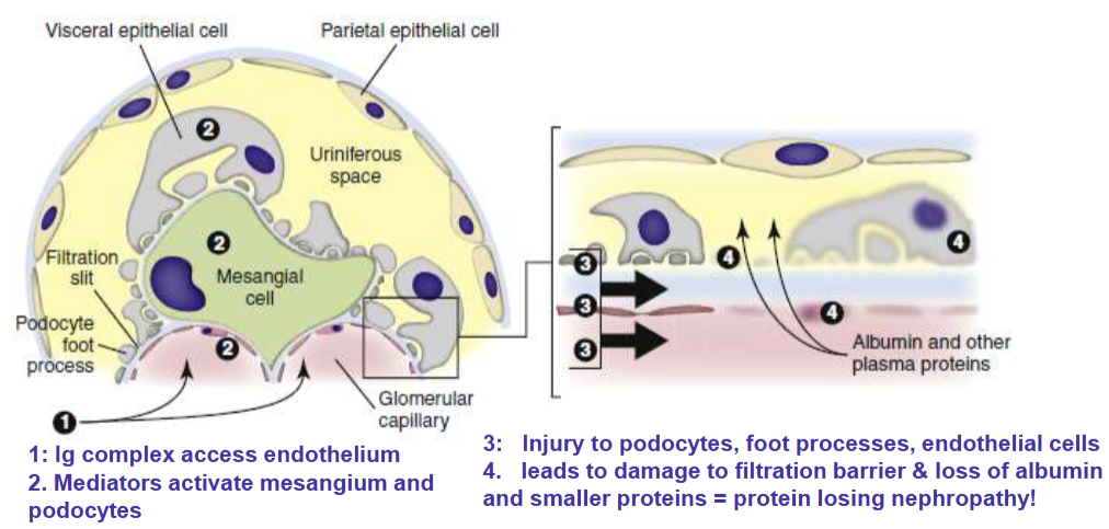
steps in pathogenesis of GN

Pathogenesis of immune complex glomerulonephritis
Membranous glomerulonephritis

Membranoproliferative glomerulonephritis

Proliferative(endocapillary) glomerulonephritis

Cyst vs pseudocyst
a true cyst has a distinct cellular lining (epithelium), whereas a pseudocyst is surrounded only by scar or granulation tissue without a true cellular wall
Immune complex glomerulonephritis Membranous glomerulonephritis
characterized by diffuse glomerular capillary basement membrane thickening due to
subepithelial deposition of immune complexes and fibrosis
Signs of Immune complex glomerulonephritis Membranous glomerulonephritis
most common form in cats & dogs
Diagnosis: proteinuria (UPC >1.0), histopathology, immunofluorescence for immune complex detection
Glomerulosclerosis
condition of aging, chronic injury, unrestricted high protein diets and high
blood pressure
What happens in glomerulosclerosis?
Glomeruli are shrunken with increased extracellular mesangial matrix leading to obliteration of capillaries and collapse of the glomerular tuft (scarring or sclerosis)
reduces the blood flow to the tubules secondary tubular degeneration and atrophy
Glomerular amyloidosis
Amyloid, insoluble fibrillar protein with β-pleated sheet
amyloidosis commonly causes proteinuria
Who gets glomerular amyloidosis
In domestic animals due to chronic inflammatory diseases reactive systemic amyloidosis (AA amyloidosis) → glomeruli are the most common site
In Abyssinian cats and Chinese Shar-Pei dogs the Medullary interstitium is the common site
Signs of glomerular amyloidosis
Gross lesions: kidneys - enlarged, pale, and have a waxy, smooth, to finely granular capsular surface
Microscopic lesion: deposition of amyloid protein within glomerular tufts
How is the perfusion of the medulla vs. the cortex?
Cortex: blood flow and O2 higher
Outer medulla: blood flow and O2 lower
Physiological and pathophysiological implications of higher perfusion of the cortex?
Physiological: maintain the hyperosmolarity in the medulla → supports the urine concentration capacity of the kidneys.
Inner medulla adapted to low oxygen due to anaerobic glycolysis
nephron injury: toxins can reach the cortex more → higher perfusion and hypoxia
Risks of medullary toxicity
Medullary hypoxic injury leads to tubular necrosis of the corticomedullary and inner medullary nephrons
Too much blood flow or too little disrupts the countercurrent exchange
What can be used during medullary toxicity?
Furosemide is used to reduce GFR and attenuate medullary hypoxia
NSAIDS, antibiotics, Hgb, Mgb, enhances tubular transport and vasoconstriction leading to outer medullary tubular hypoxia and nephrosis
Acute tubular necrosis
Most important cause of AKI
signs include oliguria or anuria with azotemia
Associated with nephrotoxicity or ischemia
Causes of acute tubular necrosis
nephrotoxic pigments - hemoglobin, myoglobin
heavy metals - arsenic, lead
pharmaceutical agents - i.e., NSAIDS
Toxins - bacterial, fungal aflatoxins, plants containing oxalates
Chemicals – ethylene glycol (anti-freeze)
Hemoglobinuric nephrosis (kidneys dark red)
Intravascular hemolysis due to oxidants leads to anemia and excess free Hgb in blood (red plasma)
Hgb passes through glomerular filtrate and exacerbates ischemic injury of the renal tubules
Free Hgb in urine (red urine – UA Heme+)
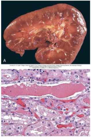
Hemoglobin casts form (Figure B) and cause renal hypoxia and tubular necrosis

Muscle necrosis releases free myoglobin in
Horses (tying-up syndrome), Capture myopathy in wild animals
Pathogenesis of Nephrotoxic pigments (Myoglobin)
Renal cortices red-brown to blue-black(Figure A) with intratubular myoglobin casts and tubular necrosis (Figure B) → Myoglobin nephrosis
Urine reddish brown (UA – heme+), Muscle enzymes elevated(CK) indicate muscle damage
Differential Diagnosis of Pigmenturia

Pathogenesis of nephrosis due to Heavy metals
Direct injury to tubular epithelial cell membranes and mitochondria
nephrosis due to Heavy metals is important in wildlife diseases and cattle:
Herbicides: arsenic
Lead: used oil, grease, old batteries, machine parts, lead paint, lead shot, environmental contamination by smelting plants
toxicological testing to confirm
Pathogenesis of bacterial pyelonephritis
Inflammation of renal pelvis (pyelitis) with concurrent tubulointerstitial nephritis
Bacterial pyelonephritis: Ascending infection from lower urinary tract more common due to
deranged vesicoureteral junction secondary to obstruction (bacterial toxins, uroliths or stones increase pressure)
recurrent cystitis compromises mucosa and ureteral peristalsis
E. coli with alpha-hemolysin most common toxin
Pyelonephritis: Gross lesions
expansion of the renal pelvis by purulent exudate (pus) and radiating steaks red/pale (see arrows in A: cat and B: cow)

Pyelonephritis: microscopic lesions
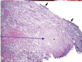
suppurative (neutrophilic) inflammation of pelvis with necrotic transitional
epithelium (short arrows) and renal medullary necrosis
Nonsteroidal anti-inflammatory drugs(NSAIDS) decrease?
synthesis of renal prostaglandins responsible for maintaining renal blood flow leads to ischemia and tubular and pelvic(medullary crest) necrosis
NSAIDs in different species
most frequent in horses phenylbutazone, flunixin meglumine (Banamine)
important in dogs/cats due to ingestion(ibuprofen, aspirin, acetaminophen)
When is antifreeze intoxication most common?
Most common in dogs and cats, occasionally pigs, cattle and horse
What does antifreeze ingestion result in?
Ingestion results in nephrotoxic metabolites (oxalates) causes calcium oxalate crystals to form in tubules → Tubular degeneration and obstruction
Infectious causes of interstitial inflammation and renal injury
Leptospirosis, Lyme dis. (common in dogs)
Lymphoplasmacytic infiltrates In interstitium causes tubular necrosis and glomerulosclerosis
Non-infectious causes of interstitial inflammation and renal injury
Chronic kidney disease
Fibrosis leads to tubular necrosis and glomerulosclerosis
Chronic kidney disease (CKD)
Geriatric cats 2X more prevalent than in geriatric dogs
Chronic diseases (i.e., infections, amyloidosis)
Environmental stress
Majority of cats with CKD have
Tubulointerstitial (mononuclear) inflammation and fibrosis with tubular atrophy
Secondary glomerulosclerosis
Atrophy of the kidneys

Renal neoplasia
x < 1% of total neoplasms
unilateral, epithelial, mesenchymal, or metastatic(lymphocytes) in origin
Epithelial tumors
Renal adenoma -rare, clinically asymptomatic
Renal carcinoma: most common primary renal neoplasm
What do epithelial tumors cause?
Typically occurs in older dogs
Paraneoplastic condition (inappropriate erythrocytosis, produce EPO)
Mesenchymal tumors
rare in the kidney
fibromas, fibrosarcomas, or hemangiosarcomas may originate in the kidneys
Hemangiosarcomas have blood-filled cavernous cavities

Metastatic tumors
any metastatic neoplasm (carcinomas/sarcomas) can occur in the kidney
renal lymphoma is a common neoplasm that may be primary or metastatic (most common in cattle and cats)
Nodular/multifocal metastatic tumor

diffuse metastatic tumor

Neoplastic lymphocyte infiltration
