Advanced Small Animal Surgery Midterm
 Call Kai
 Learn
Learn Practice Test
 Spaced Repetition
 Match
Match Flashcards
 Knowt Play
1/770

There's no tags or description
Looks like no tags are added yet.
Name | Mastery | Learn | Test | Matching | Spaced | Call with Kai | Chat |
|---|
No analytics yet
Send a link to your students to track their progress
771 Terms
What are the functions of skin?
Maintain hydration of tissues
Defense against environment
Vitamin D production
Sensory
Storage of water, fat, electrolytes, carbs, and proteins
Insulation
What are the three layers of the skin?
epidermis
dermis
hypodermis
layer of the skin composed of avascular tissue with a cuboidal/stratified epithelium and variable thickness depending on location
epidermis
layer of skin composed of hair follicles, glands, vascular plexus, lymphatics, nerves, and collagen that gives skin its viscoelasticity
dermis
layer of skin associated with the dermis mostly composed of fat and connective tissue that contains the panniculus
hypodermis
critically important landmark when performing reconstructive cutaneous surgery in dogs and cats as failure to identify and preserve it will lead to loss of blood supply and wound dehiscence
panniculus m. (aka platysma, cutanoeus trunci, supramammarius, preputialis)
panniculus fibers penetrate the ______ allowing voluntary movement of the skin
dermis
where is the panniculus muscle NOT present?
lower limbs
when undermining skin, always dissect ? the panniculus m.
under
vessels that run parallel to the skin and that serve as the skins blood supply which is unique to dogs and cats relative to humans
direct cutaneous arteries and veins
direct cutaneous arteries and veins terminate as the
deep (subdermal), middle, or superficial plexus
plexus that provides the major blood supply to the skin and is therefore of most importance with regards to cutaneous surgery
deep (subdermal) plexus
should you close bite wounds acutely
no
treatment of contaminated wounds (e.g. bite wound) prior to closure may include
lavage
abx
frequent bandage changes
topicals (e.g. silver)
negative pressure
areas of the body with extensive loose skin available for mobilization?
trunk
upper extremities
cervical region
areas of the body with limited skin available for mobilization?
eyes
ears
anogenital region
lower extremities
guide on how skin defects should be reconstructed/closed that result from gravitational and muscular forces
tension lines
if possible, incisions aare planned and made ? to tension lines to allow cosmetic closure with minimal tension
parallel
what instruments are appropriate when manipulating skin?
skin hooks
fine stay sutures
towel clamps
general suture size for reconstructive/cosmetic surgery?
2-0 to 4-0
suture material used in buried closure patterns?
absorbable monofilament (e.g. monocryl, biosyn (glycoemr))
suture material used for external skin closure?
nonabsorbable monofilament (e.g. nylon, polypropylene, and staples)
cosmetic yet time consuming suture pattern used to obtain accurate apposition
simple interrupted
efficient suture pattern with some tension relieving properties used to obtain good apposition
cruciate
cosmetic suture pattern that is primarily appositional, provides some tension relief, may be less irritating to the patient, and can be time consuming at first
intradermal/subcuticular
tension relieving suture pattern that can be combined with stents if necessary and may compromise blood supply
horizontal mattress
tension relieving suture pattern that may cause some everting of skin edges
vertical mattress
three main functions of walking sutures?
advance and appose skin edges
decrease tension
close dead space
disadvantages of walking sutures?
possible disruption of blood supply
possible pockets for seroma and abscess formation
dimply appearance
techniques for local skin mobilization and tension relief?
Undermining
Relaxing incisions
Various Plasty incisions (V to Y)
Local (subdermal plexus) flaps
easiest flaps to perform that rely on the subdermal plexus
local (subdermal plexus) flaps
the length to width ratio for local (subdermal) flaps must be equal to or less than
1.5
examples of local (subdermal) flaps ?
single pedicle advancement flap
bipedicle advancement flap
rotation flap
what local (subdermal) advancement flap provides improved blood supply and greater tissue coverage?
bipedicle advancement flap
flap using a region with a named direct cutaneous artery and vein
axial pattern flap
Axial pattern flap examples?
Caudal superficial epigastric
Caudal auricular
Deep circumflex iliac
Thoracodorsal
Tibia "crus"
Lateral Genicular
advantages of the caudal superficial epigastric (CSE) axial pattern flap?
Immediate and robust blood supply
Infection resistanc
Durable
Cosmetic
Can close large wounds
disadvantages of the caudal superficial epigastric (CSE) axial pattern flap?
Can't reach distal extremities
Large donor site
Time consuming
what is the maximum number of caudal mammary glands that can be used for a caudal superficial epigastric (CSE) axial pattern flap in dogs?
4 glands
what is the maximum number of caudal mammary glands that can be used for a caudal superficial epigastric (CSE) axial pattern flap in cats?
3 glands
Principles of axial pattern flap reconstruction?
Make a definitive incision
Dissect beneath panniculus
Use landmarks and transillumination
Dissect distal to proximal
Bridging incisions
Position with towel clamps
Use multiple people/teams for suturing
axial pattern flaps to know for exam
Caudal superficial epigastric
Caudal auricular
Antitragus
what is structure A?
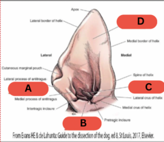
Tragus
what is structure B?

Anthelix
what is structure C?
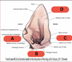
Scapha
what is structure D?
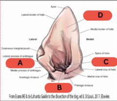
structures of the external ear?
vertical canal
horizontal canal
cartilages
glands (e.g. seruminous)
hair
the middle ear is also known as the
tympanic cavity
structure of the middle ear that connects to the nasopharynx
eustachian tube
species with a middle ear that is divided into two compartments by a septum
cat
structures of the inner ear?
labyrinth
semicircular canals
pinna laceration tx options?
Manage as wound
Primary wound closure
Pinnectomy
when can pinna lacerations be treated with primary wound closure?
If adequate and healthy tissue is present
Injury occurred within 6 hours
Suture patterns to close skin of pinnae lacerations?
Simple interrupted
Ford interlocking
Suture patterns to close skin and cartilage of pinna laceration together?
Vertical mattress
Figure of eight
criteria for partial or complete pinnectomy?
malignant tumor
ulcerated tumor
painful tumor
a ? thickness partial pinnectomy is indicated for malignant tumors
full
painful hemorrhage within layers of the pinna causing the layers of cartilage to separate due to trauma or an autoimmune response
aural hematoma
aural hematoma tx?
Address underlying cause
Treat quickly
Continuous drainage
Steroids/PRP possibly
TF needle aspiration of aural hematoma is an effective treatment
F
Methods for continuous drainage of aural hematoma?
teat cannula
penrose drain
closed suction drain
S shaped incision
circular fenestration
goals of aural hematoma tx?
remove clot
remove fibrin
close dead space
provide cosmesis and function
prevent recurrence
Indications for external ear surgery?
Stenosis
Poor/no response to medical management
Ear masses
Dystrophic mineralization
Trauma
TF most external ear surgeries are salvage procedures
T
Best diagnostic imaging method for evaluation of ear canal, extent of disease, and secondary changes from otitis externa/media?
CT
three most common surgical procedures employed for otitis externa?
Lateral wall resection
Vertical canal ablation
Total ear canal ablation and lateral bulla ostectomy (TECALBO)
rarely indicated procedure through which the lateral wall of the vertical canal is opened
lateral wall resection
indications for lateral wall resection?
Small tumor of lateral wall
Mild otitis externa affecting vertical canal
Congenital stenosis
rarely indicated procedure through which the vertical canal is removed and an opening is created for the horizontal canal
vertical canal ablation
vertical canal ablation indications?
Small tumor of vertical canal
Mild otitis externa affecting vertical canal
Congenital stenosis
TF lateral wall resection and vertical canal ablations are useful and effective procedures to treat otitis
F
commonly fails if treating otitis and becomes stenotic
procedure defined by complete removal of the vertical and horizontal canal as well as the lateral wall of the bulla that requires special equipment
total ear canal ablation and lateral bulla ostectomy (TECALBO)
indications for TECALBO?
severe chronic otitis
neoplasia
Steps of TECALBO?
Make incision around ear canal opening
Incision along vertical canal and make a flap for erect ears
Elevate auricular muscles from canal
Elevate canal from opening of bulla
Remove lateral wall of bulla
Remove epithelial lining from bulla cavity
Lavage
Culture and submit for histopath
Complications of TECALBO?
Facial nerve injury (temporary vs. permanent)
Deep infection (draining tract/abscess)
Hemorrhage
Damage to inner ear
Pinna necrosis
Airway swelling
Incisional complications
Loss of hearing
more common and temporary facial nerve injury that results from stretching of the nerve after ear surgery - resolves in 6-8 weeks
neuropraxia
failure to ? in a TECALBO can and often will result in infection/abscesses and draining tract formation within weeks to a year of surgery
remove all of the canal cartilage and epithelial lining of bulla
TF facial nerve injury is a very serious and life-threatening complication following ear surgery
F
mainly cosmetic, need to lubricate eyes with artificial tears to prevent corneal ulcers
important vessels to avoid when performing ear surgery?
retroarticular vein
external carotid
performing a bilateral TECABO raises the risk of what complication?
airway swelling
TF inner ear clinical signs that appear AFTER ear surgery are typically permanent
F
typically resolve in time
TECABO post op care?
Pain management
Abx and change based on culture
Incision care
benign pedunculated growth of fibrovascular tissue that arise due to congenital predisposition or chronic inflammation
inflammatory polyp
inflammatory polpys may arise from three locations?
Lining of auditory (eustachian tube)
Nasopharynx
Middle Ear
What are the consequences of polyps arising from the middle and/or inner ear?
Mass effect of infection
Horner's syndrome
Vestibular signs
Bony infection
what is the most common signalment for inflammatory polyps?
Cats
Average 2yo
Hx of URI
Hx of OE
external ear inflammatory polyp clinical signs?
Chronic OE that is unresponsive to topicals
Head shaking
Otorrhea
Visible or palpable painful mass
nasopharyngeal inflammatory polyp clinical signs?
Nasal d/c
Stertor
Sneezing
Dysphagia
Dyspnea
Megaesophagus
Middle ear inflammatory polyp clinical signs?
Head tilt
Horners
Ataxia
Nystagmus
Circling
Facial n. paralysis
Always do a ? if you suspect an inflammatory polyp to rule out other differentials
sedated otic exam
inflammatory polyp of nasal turbinates
hamartoma
TF facial palsy is a typical clinical sign of an inflammatory polyp in the middle ear
F
most likely neoplasia/large tumor
inflammatory polyp diagnostics?
PE
Radiographs to assess bulla and nasopharynx
CT
MRI if neurologic
Otoscopy
Anesthesized oral/nasopharyngeal exam
external ear inflammatory polyp tx?
traction avulsion
steps for external ear polyp traction avulsion?
Place patient under GA
Place hemostats, allis tissue forceps or alligators across stalk
Apply constant traction and rotate
Control hemorrhage with gentle pressure for 2-5 minutes
Clean residual debris from ear
unique steps for nasopharynx polyp traction avulsion?
Place patient under GA
Ensure adequate ET tube cuff inflation
Retract soft palate rostrally with spay hook or stay sutures
Pack laryngeal region/suction afterwards to control hemorrhage
What are your options for an inflammatory polyp after traction avulsion?
Medical management
Surgery (e.g. myringotomy, VBO, TECALBO)
when is medical management indicated for a polyp after traction avulsion ?
Clean bulla on imaging
No imaging will be performed
No signs of middle ear disease
what does medical management of a polyp consist of?
Prednisolone 1mg/kg 4-6 week taper
Veraflox for 2 weeks
Topical ear medication for 2 weeks
when is surgical management indicated for a polyp after traction avulsion ?
Recurrent polyp
Clinical signs of middle ear disease
Middle ear disease detected on imaging