Cerebral Cortex from Cells to Function
1/93
There's no tags or description
Looks like no tags are added yet.
Name | Mastery | Learn | Test | Matching | Spaced |
|---|
No study sessions yet.
94 Terms
What makes up a neuron
cell body, dendrites, axon
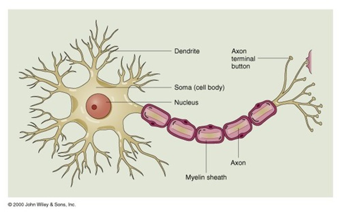
What is the axon hillock?
This is a part of the neuron, between the cell body and the axon, that controls traffic down the axon through summation of excitatory and inhibitory postsynaptic potentials.
Are all neurons equally branched
No
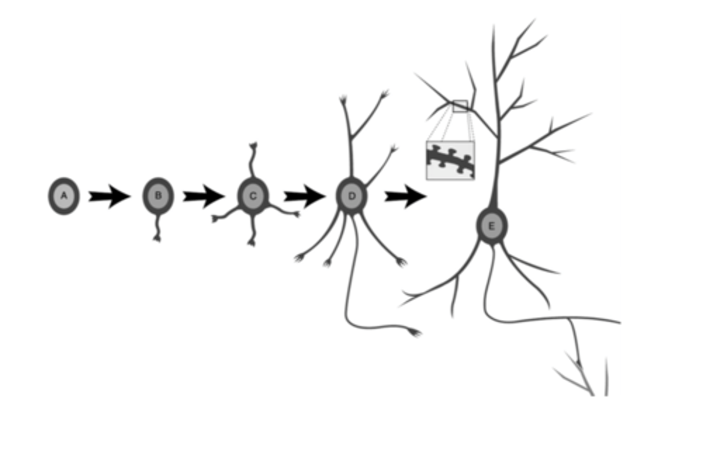
Are largely branched neurons unipolar or multipolar neurons
Multipolar neurons
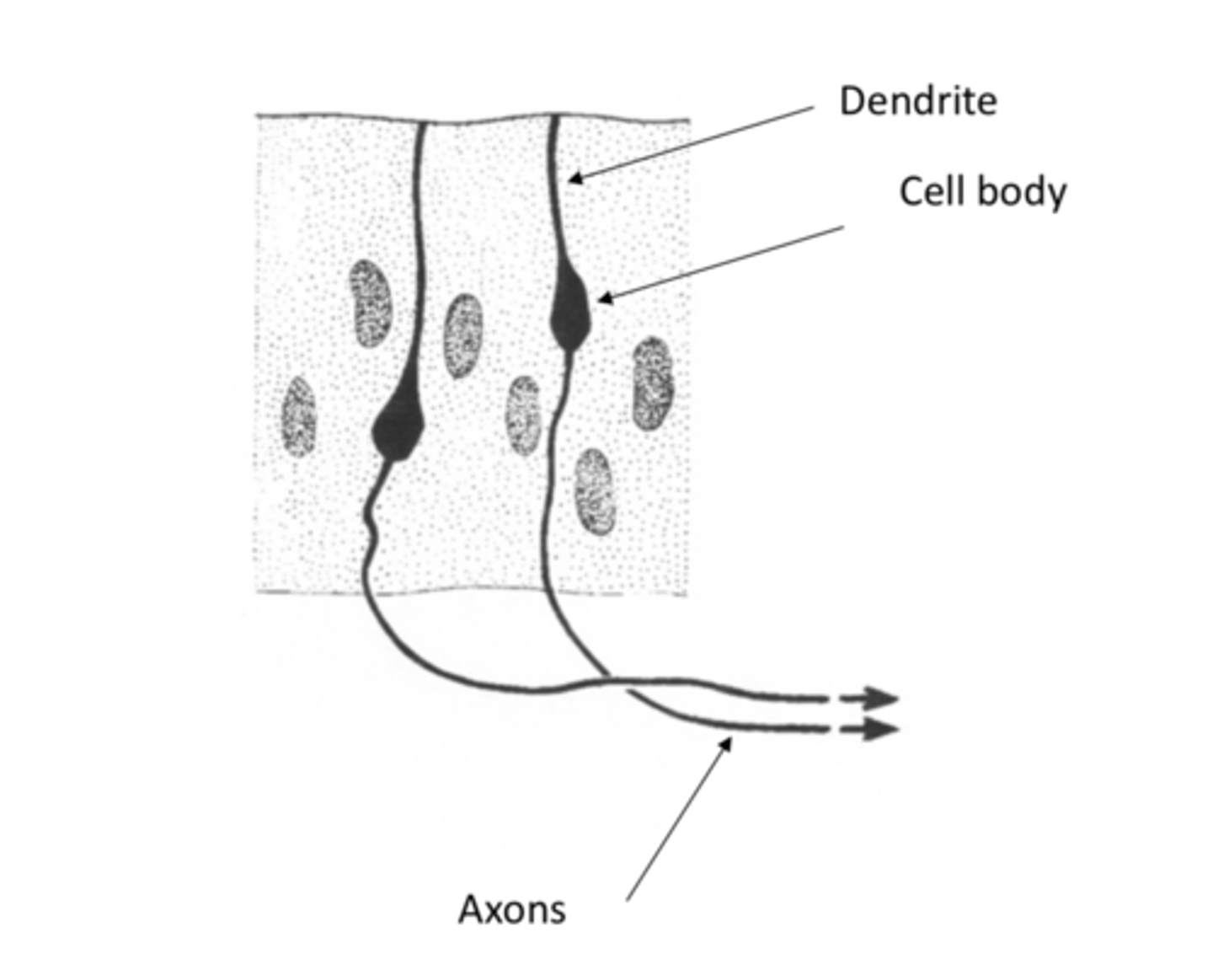
What are unipolar neurons?
A unipolar neuron is a neuron in which only one process, called a neurite, extends from the cell body. The neurite then branches to form dendritic and axonal processes.
Why are unipolar neurons also known as "modified" bipolar neurons or pseudo unipolar cell?
It appears to have a single process but through development it was actually a bipolar cell that has fused and now there is only a single pole to the cell which branches. It has a central and peripheral sprout. The peripheral sprout collects sensory information via this process which is functionally an axon.

Where are pseudounipolar neurons found?
dorsal root ganglia
Where are true bipolar nerve cells found?
Special sense organs (ear, eye, nose)
Describe the function of bipolar neurons
They have a single dendrite that senses stuff and sends info towards the cell body that then goes into the CNS
What cell type are the vast majority of cells in the CNS
Multipolar neurons
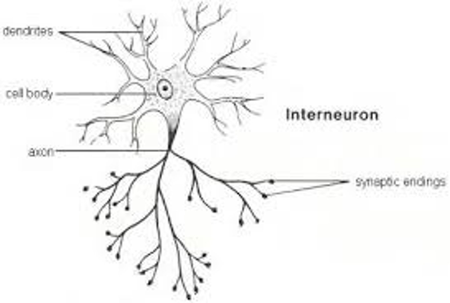
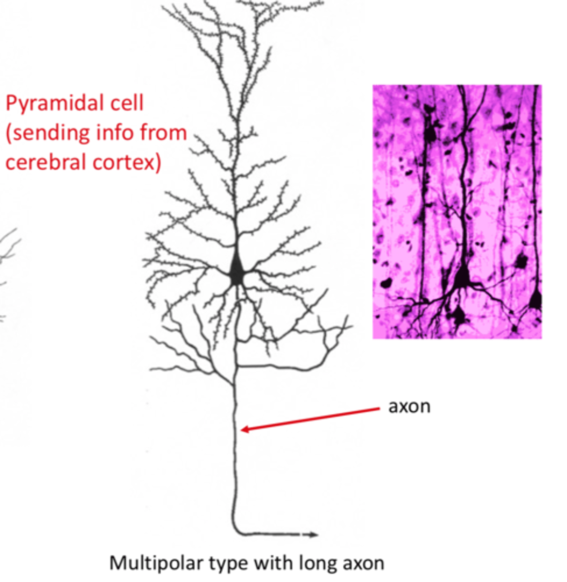
What are multipolar neurons?
Neurons with lots of dendrites emerging from the cell body with only one axon.
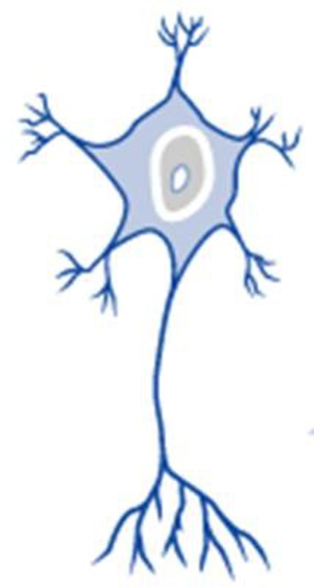
Give examples of some types of multipolar neurons
Interneurons
Pyramidal cells
What is the function of interneurons
They communicate with cells close by and are important in processing local information. Carry nerve impulses from sensory to motor neurons
What is the function of pyramidal cells
Pyramidal cells, or pyramidal neurons, are a type of multipolar neuron found in areas of the brain including the cerebral cortex, the hippocampus, and the amygdala. Pyramidal neurons are the primary excitation units of the mammalian prefrontal cortex and the corticospinal tract. They have very long axons, large amount of nuclei to accommodate for the large amount of ribosomes.
What is the function of myelination
To insulate the neuron and prevent excessive loss of electrolytes into the interstitial fluid and it helps the transmission of the action potential be propagated faster down it's length.
What cell is responsible for myelination in the PNS
Schwann cell
What cell is responsible for myelination in the CNS
Oligodendrocyte
Are all axons myelinated?
No
What happens to unmyelinated axons in the PNS
Schwann cells 'envelope' unmyelinated axons contacting 1 or more axons
What happens to unmyelinated axons in the CNS
Unmyelinated axons are not associated with glial cells like in the PNS
Describe conduction in an unmyelinated axon
Unmyelinated axons have 'continuous conduction' of action potentials due to passive current flow (low conduction)
Give examples of unmyelinated axons
Examples of unmyelinated axons are sensory (C) fibres carrying pain, temperature, itch
What is multiple sclerosis?
Phasic disease where axons become demyelinated due to inflammation as a result of T cell/macrophage immune attack. This causes conduction blocks, crosstalk (paraesthesia)
What is white matter?
Collections of nerve fibres many of which are coated with insulating fatty myelin. Called white as it is fatty
What is grey matter?
Cell bodies, synapses and processes of neurons
What are glial cells?
cells in the nervous system that support, nourish, and protect neurons
Name the glial cells of the CNS
• Oligodendrocytes (myelination)
• Astrocytes
• Microglia
• Ependyma (lining cells of the CNS cavities)
Name the glial cells of the PNS
Schwann cells (myelination)
Satellite cells (support cells in ganglia)
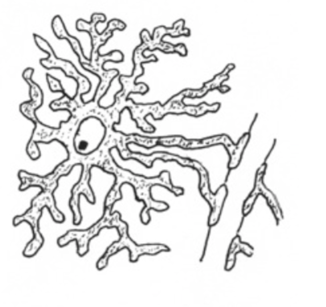
What are astrocytes?
These are abundant, star-shaped cells that account for nearly half of the neural tissue. Important in metabolic and mechanical support (in CNS scar tissue also)
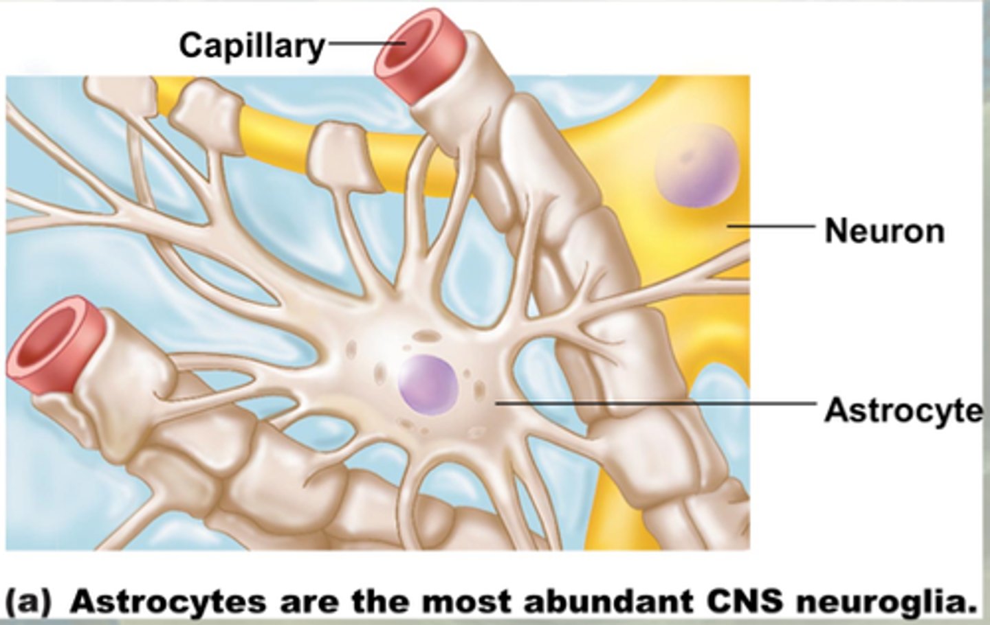
Name the functions of astrocytes
Control water distribution
Potassium buffering - prevents accumulation of K+ in the extracellular space
ROS scavenging (ROS stands for reactive oxygen species)
Define architecture
Regulate migration/pruning/synaptogenesis
Help maintain but do not make up the BBB
What astrocytes are present in white matter
Fibrous astrocyte
What astrocytes are present in grey matter
Protoplasmic astrocyte
What is the BBB composed of?
A barrier composed of endothelial cells AND their tight junctions
How do astrocytes help maintain the BBB
Integrity highly dependent on astrocyte 'end feet
So the barrier consists of endothelial cells and their tight junctions and the astrocytes help to maintain it
What are microglial cells?
Resident macrophages of the central nervous system
What is the function of microglial cells?
Phagocytosis and antigen presentation (immune response)
Synaptic pruning - if there are unused or defective synapses they can be pruned or degraded by phagocytosis
What are ependymal cells?
Ciliated cuboidal epithelial cells, line ventricle as part of plexus and secrete (also reabsorb) CSF
What is CSF
Clear, cell-free fluid produced in specialised ependyma on choroid plexus
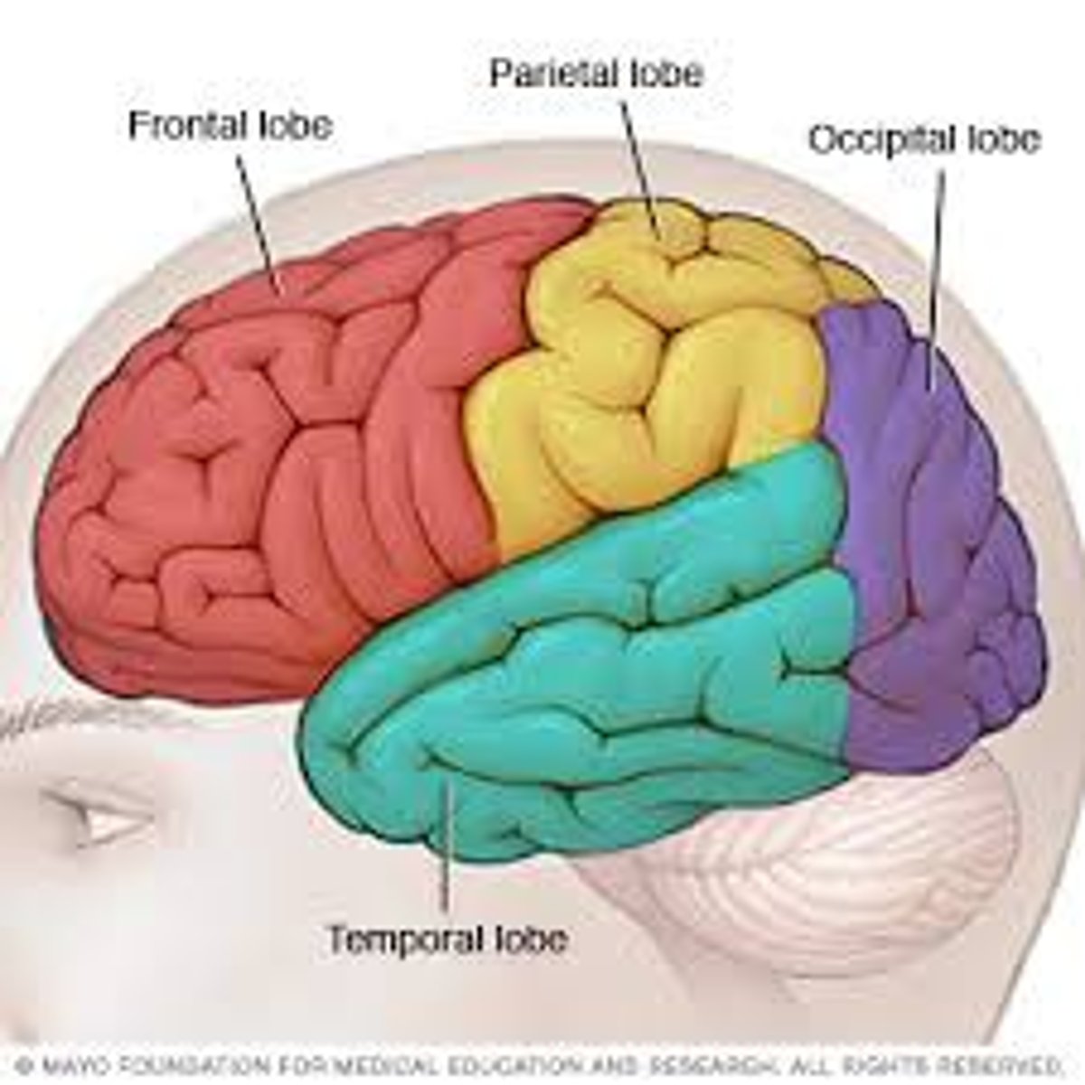
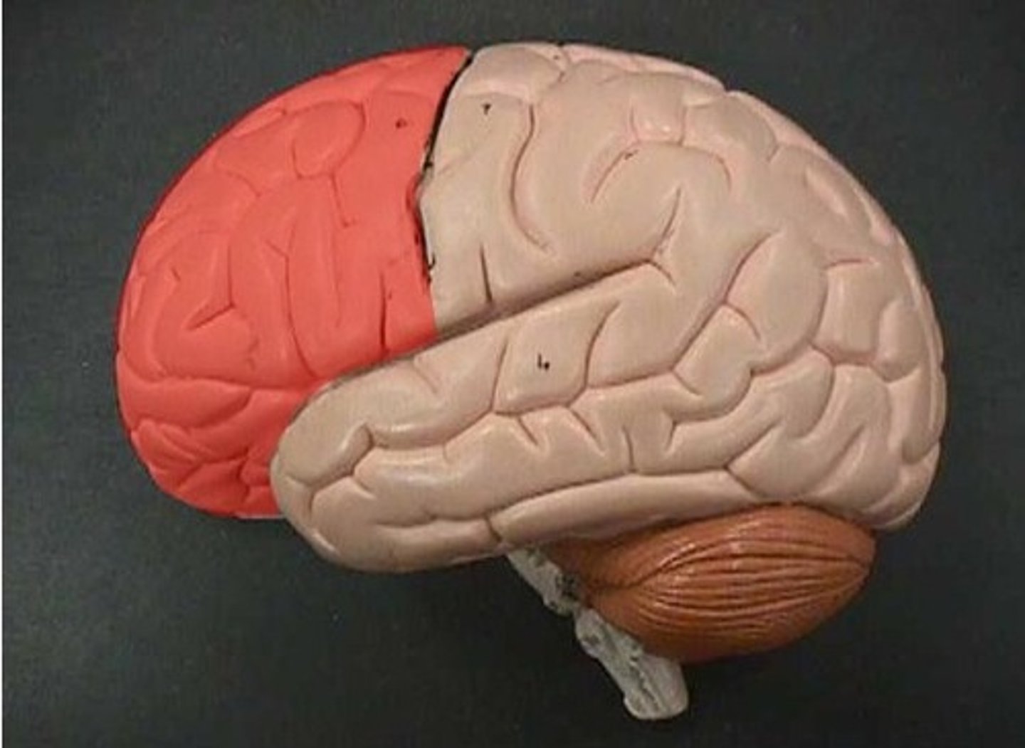
What are the 4 lobes of the brain?
frontal, parietal, temporal, occipital
What is lamination?
Lamination is the biological process by which cells are arranged in layers within a tissue during development. Laminated structures in the nervous system include the cerebral cortex, retina and hippocampus
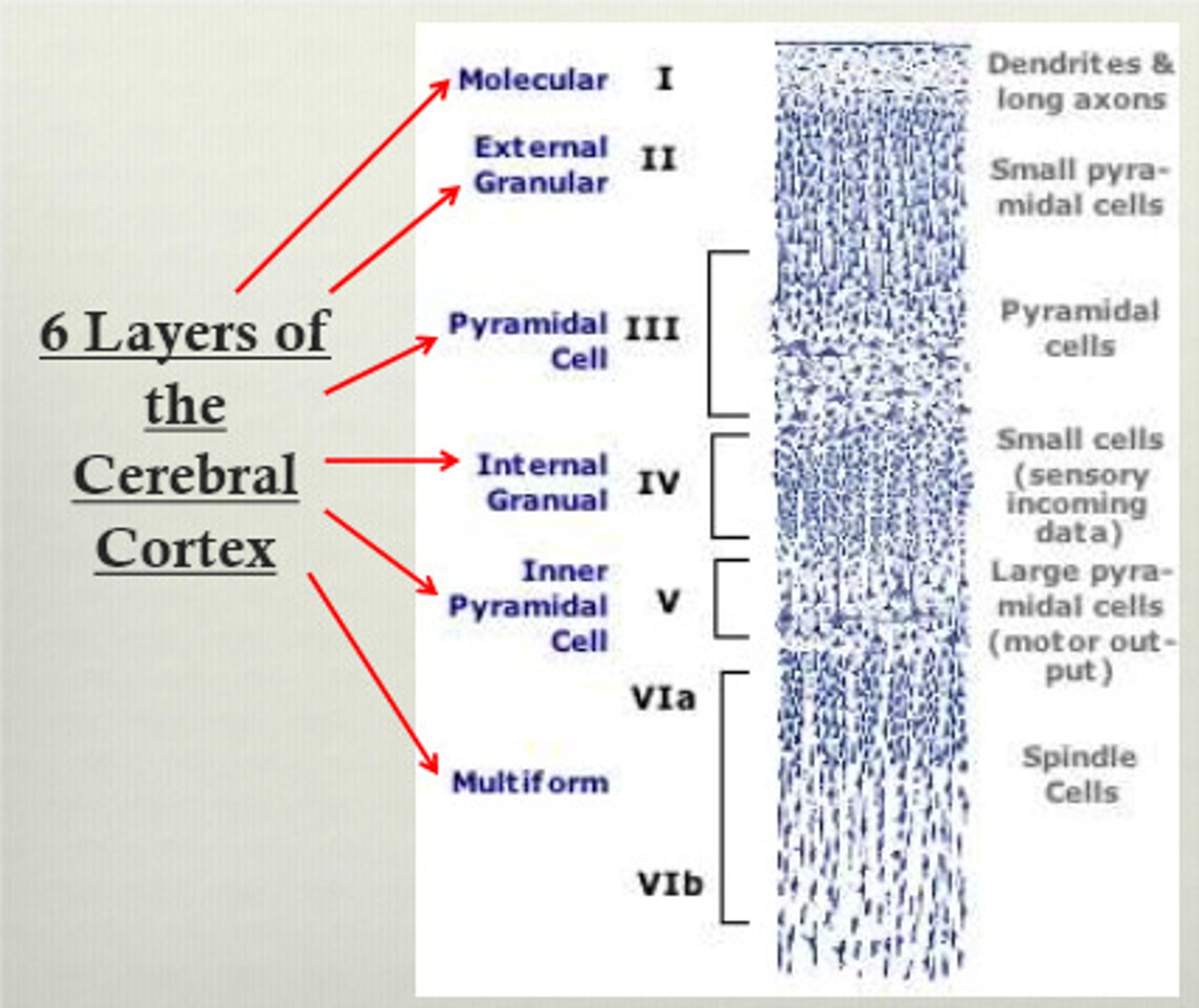
How many layers are in the cerebral cortex
6
How can layering in the neocortex be seen
Can be seen histologically and sometimes by eye
What are the layers of the cerebral cortex
Molecular layer
External granular layer
External pyramidal layer
Internal granular layer
Internal pyramidal layer
Multiform layer
What 2 neuron classes define the main layers of the cerebral cortex
Stellate interneuron - Granular layers (input and processing)
Pyramidal neurons - Pyramidal layers (output)
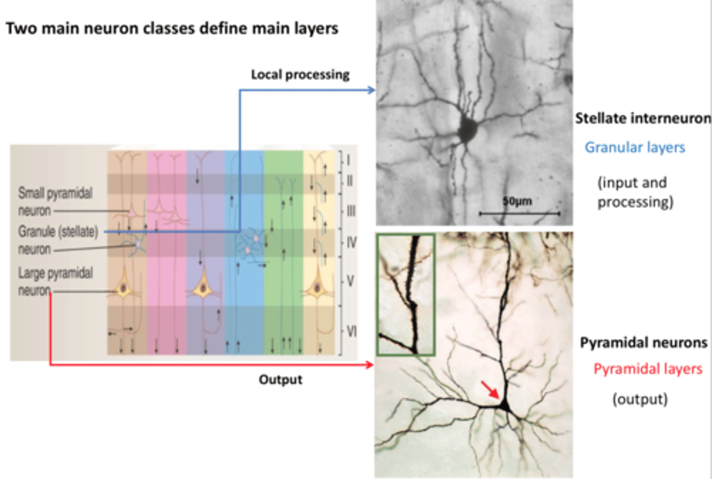
The more peripheral you are in the brain...
The more talking to other neurons and processing information there is
The deeper you go into the brain...
The more output and long distance of information to other structures there is
Where does the external granular layer receive information from
Local areas from the other cortex
Where does the external pyramidal layer send information to
Local areas in the cortex
Where does the internal granular layer receive information from
Deeper structures. It receives sensory input from the thalamus
Where does the internal pyramidal layer send information to
Very distant structures such as the brain stem and spinal cord
Are the pyramidal cells bigger in the internal or external layer
Internal as they have to send information much further away
The layers of the brain vary in
Thickness with function from region to region. The variation in thickness between these layers maps into their function. Layer IV well developed prominent makes a striped appearance
Where are interneurons more numerous
Sensory cortex
Where is pyramidal output more marked
Motor cortex
What are Brodmann's areas?
Brodmann's areas of the cortex refer to 52 regions of the cerebral cortex that were identified by German Neurologist, Korbinian Brodmann, based on cytoarchitectonic (cell size, spacing or packing density, and lamination) differences.
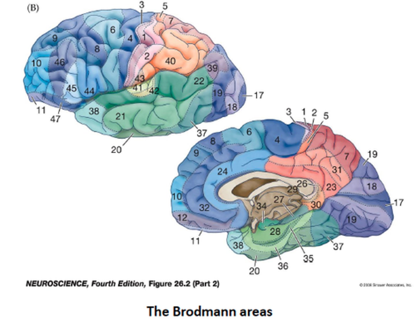
What area is motor function located in?
4 i.e. if area 4 was stimulated your leg would move
The occipital lobe does nothing but...
Visual processing
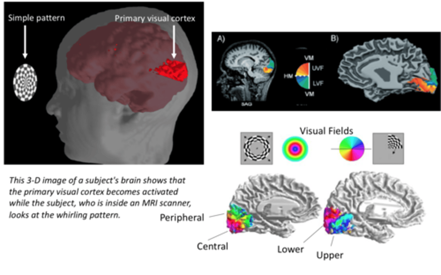
What is the primary visual cortex?
Seen on the extreme posterior tip of the occipital lobe. Receives visual information from the retinas.
What are association cortices?
Association cortexes are areas of integration, which are responsible for more complicated sensory information. For example in the occipital lobe you are not just processing visual information from the primary visual cortex but integrating it with information coming from other areas in the occipital lobe to do with motion, object recognition to create a full picture. Most of the occipital lobe, apart from the primary visual cortex is the association visual cortex
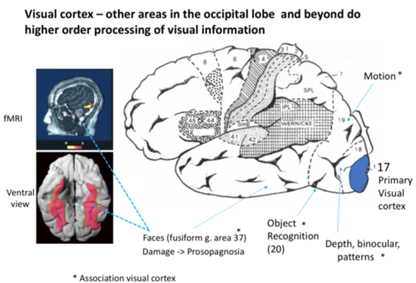
What is the fusiform face area?
The FFA is the location that is responsible for interpreting familiar faces.
What happens if damage occurs to the fusiform face area
It results in prosopagnosia - face blindness - which is the inability to recognise someone's face
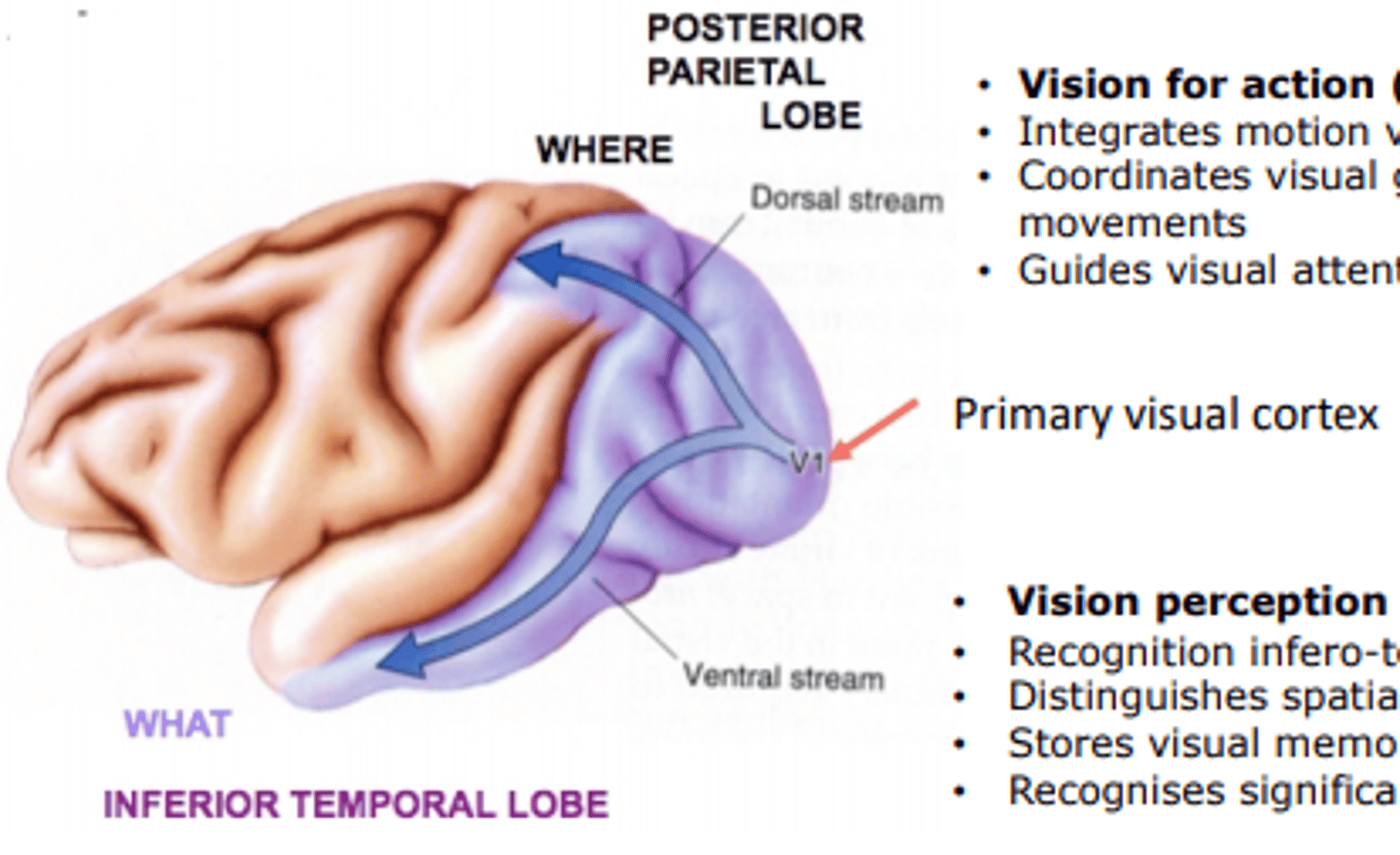
What is the visual cortex dorsal and ventral dual stream model
The idea that there is a "where" and a "what" pathway in the brain. As visual information exits the occipital lobe, and as sound leaves the phonological network, it follows two main pathways, or "streams". The ventral stream (also known as the "what pathway") leads to the temporal lobe, which is involved with object and visual identification and recognition. The dorsal stream (or, "where pathway") leads to the parietal lobe, which is involved with processing the object's spatial location relative to the viewer and with speech repetition
What is a primary area (within the brain)
An area that is either the first one to receive the input or the final path out
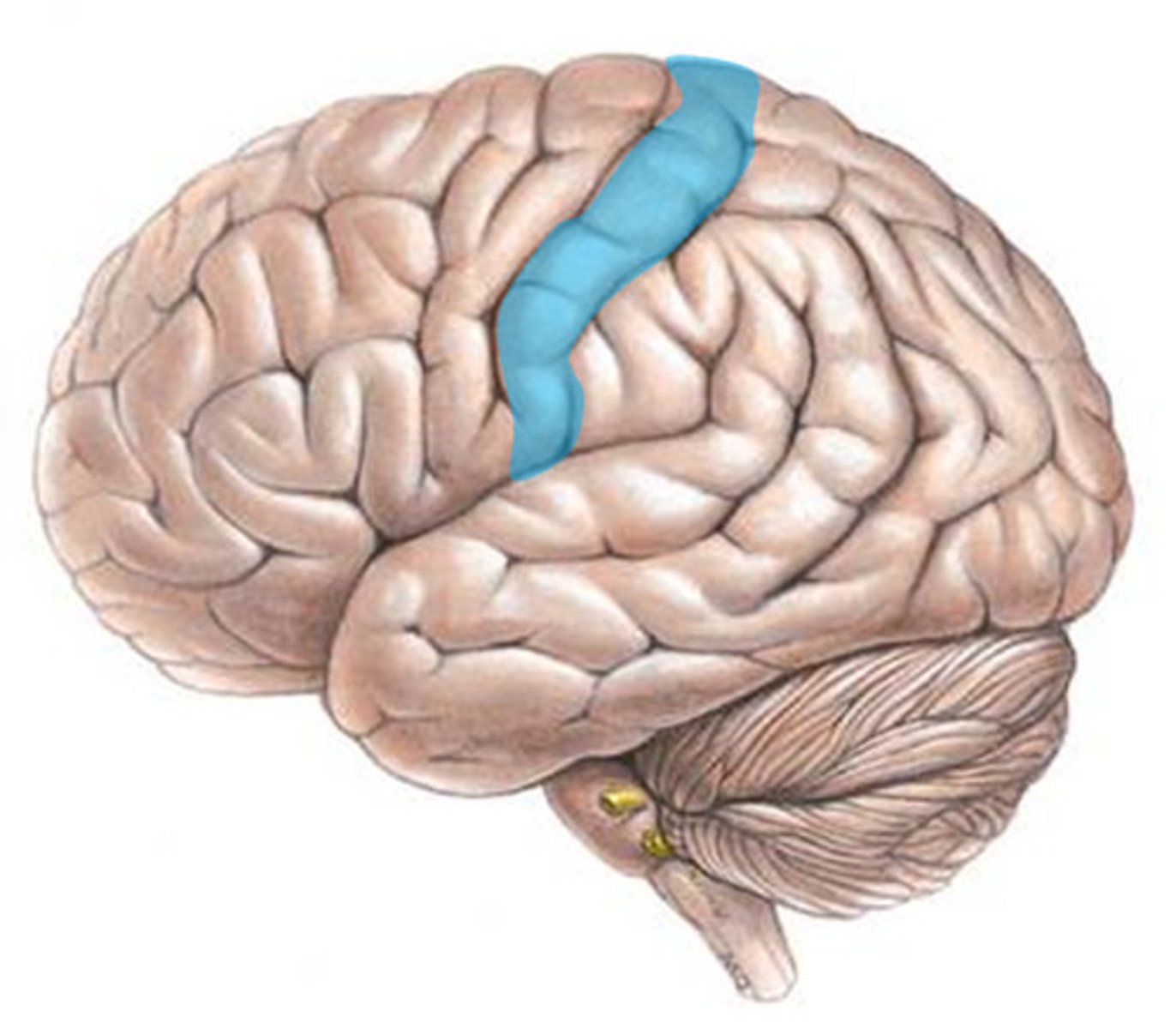
Where is the primary motor cortex located?
precentral gyrus of frontal lobe
What is the function of the primary motor cortex
allows conscious control of precise, skilled, voluntary movements
What other areas does motor output come from
Motor association cortex and some areas in the sensory cortex
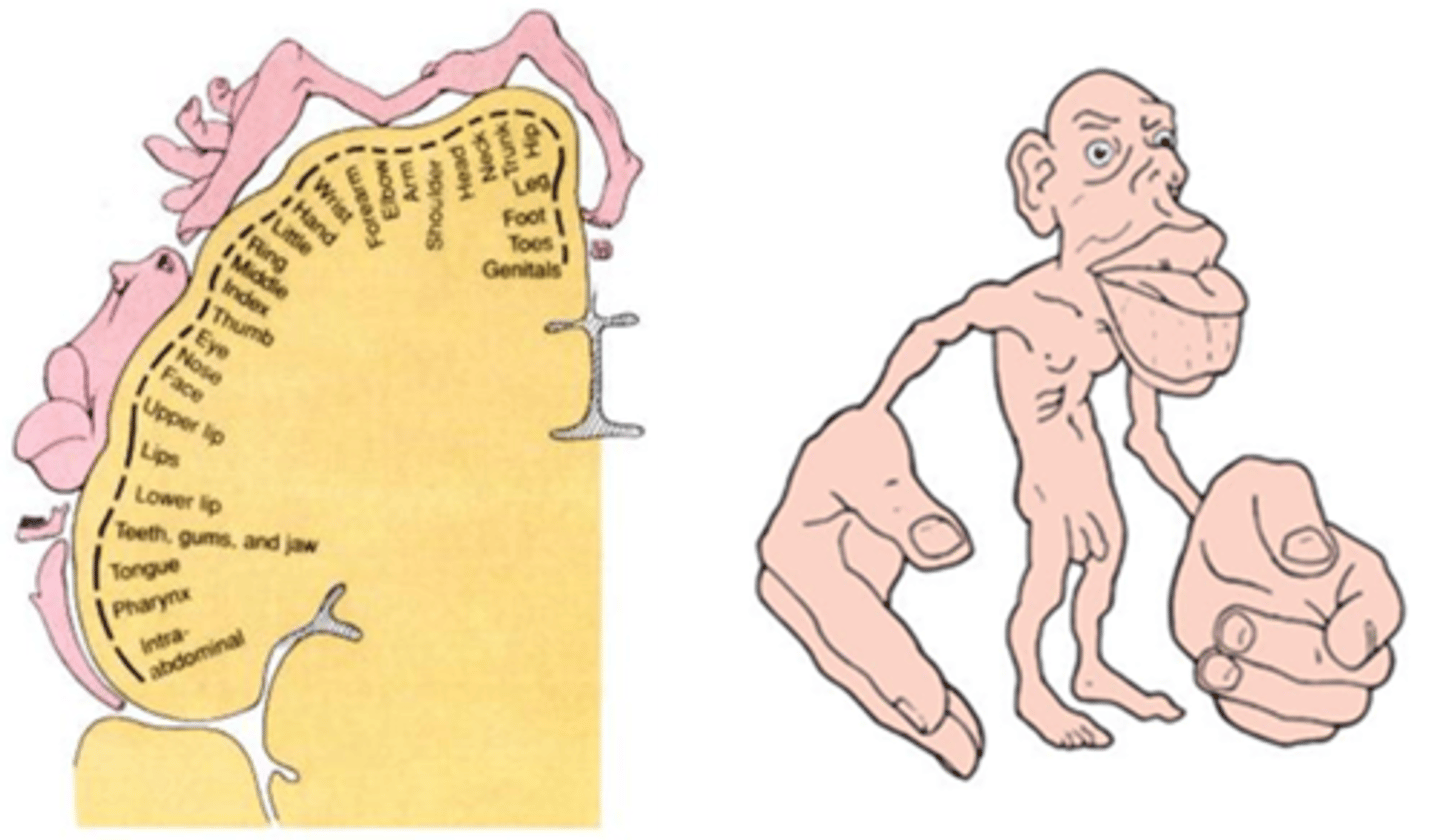
What is somatotopy?
point-to-point correspondence between an area of the body and an area of the CNS
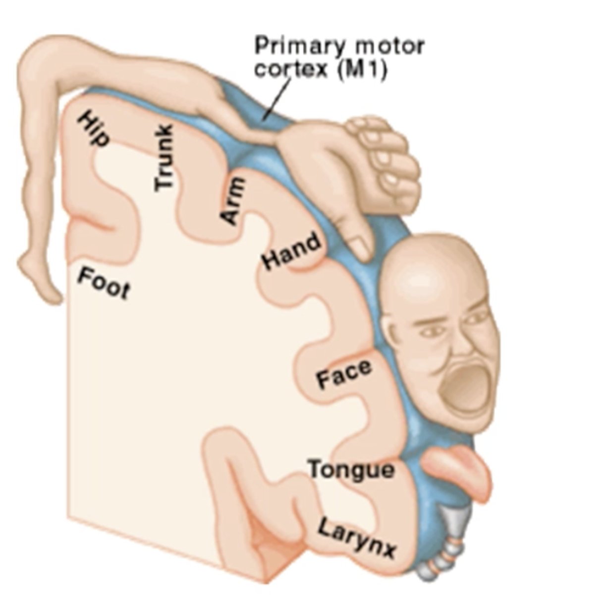
Which parts of the body have enlarged representation
Area that require fine motor movement e.g. hands and face
What is the homunculus?
Topographical representation of sensory and motor areas in the cerebral cortex. Used to localize lesions (in blood supply) leading to specific deficits.
What are the motor association cortices
Supplementary motor area
Premotor cortex
Posterior parietal cortex
What is the function of the supplementary motor area
Important for complex movements and planning them
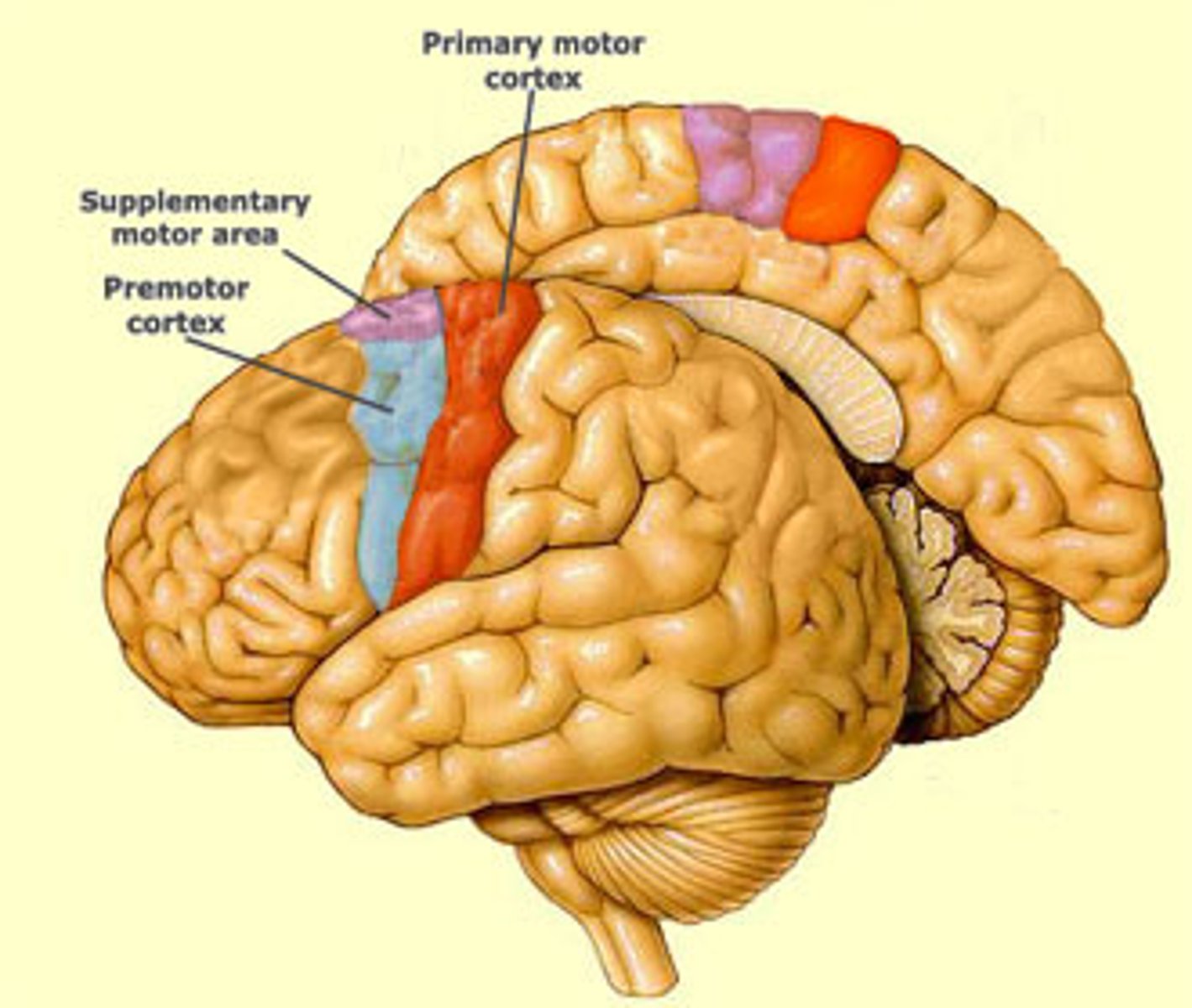
What is the function of the premotor cortex
Preparation for action - posture and gait (integration of spatial information and planned movement, grasping)
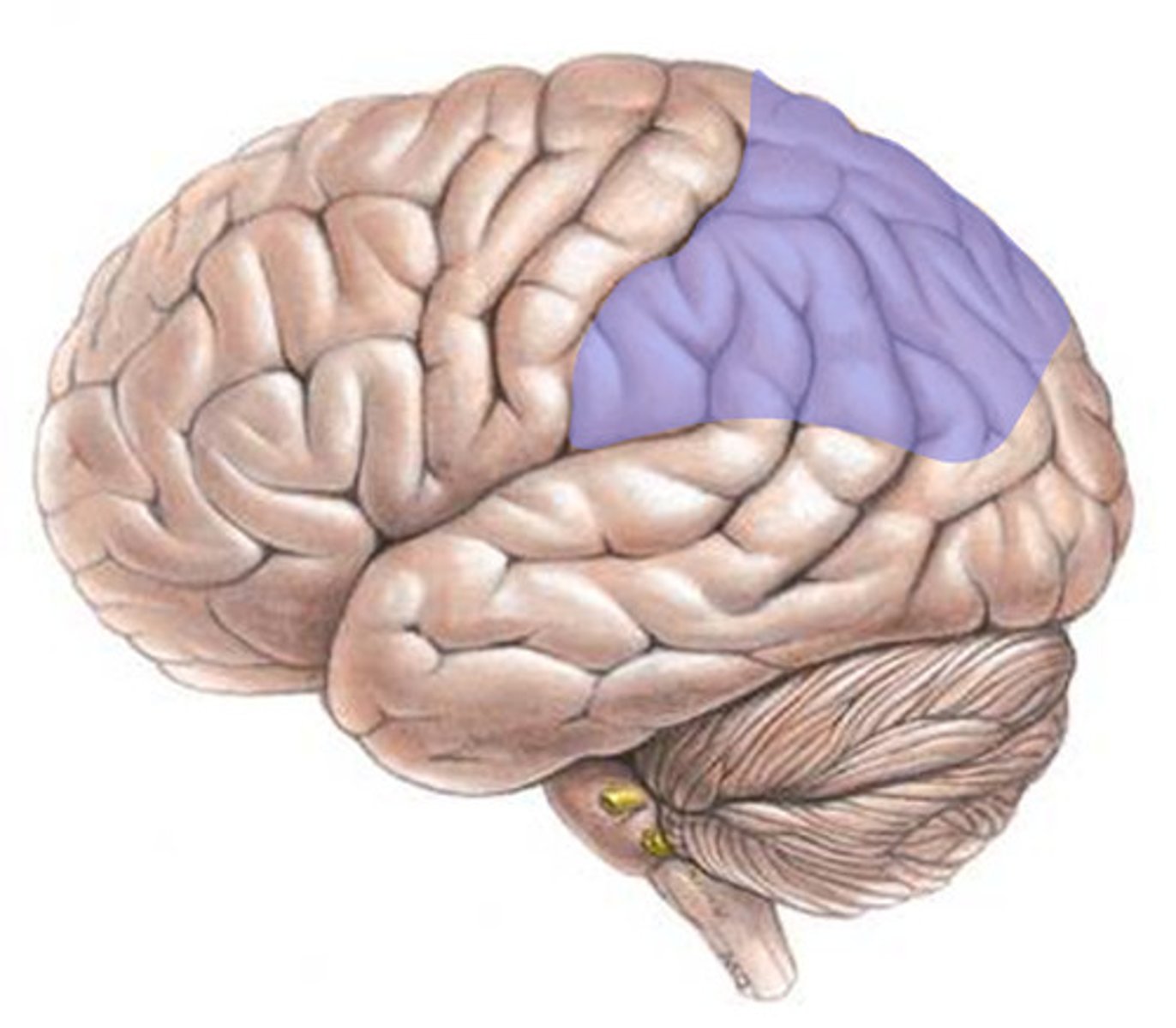
What is the posterior parietal cortex
Integrating visual information to formulate motor commands
What are the 2 classes of cerebral cortex specialisation?
Primary and association
What are the 2 types of association cortex
Unimodal and polymodal
What is a unimodal association cortex?
One type of modality input/output
What is a polymodal association cortex
Multiple modal input (e.g. vision and somatic sensation in the dorsal stream)
What is the primary somatosensory cortex
- located in the postcentral gyrus
- receives information from the skin and skeletal muscles
- exhibits spatial discrimination
- located next to primary motor cortex to prevent buildup of white matter bundles
What is the function of the association somatosensory cortex
Awareness/perception in space (orientation), rearrangement of memories, organising grasping reaching movement, number processing, mirror neurons here!
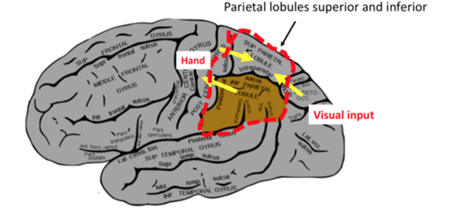
What happens if damage occurs to the inferior parietal lobule
Contralateral neglect (especially right lesions) where they ignore the left side of the body
Either hemisphere - astereognosis (inability to recognize objects by touch)
What happens if damage occurs to the superior parietal lobule
Problems with visuomotor integration (optic ataxia)
What is the function of the pre-frontal cortex?
Contributes to attention
Morality
Planning
Working memory
Conscious decision making
Social behavior regulation
Features of frontal lobe damage
(1) Personality changes
(2) Deficits in planning
(3) Perseveration
(4) Primitive reflexes (suckling reflex)
(5) Abulia (slowness of intellect)
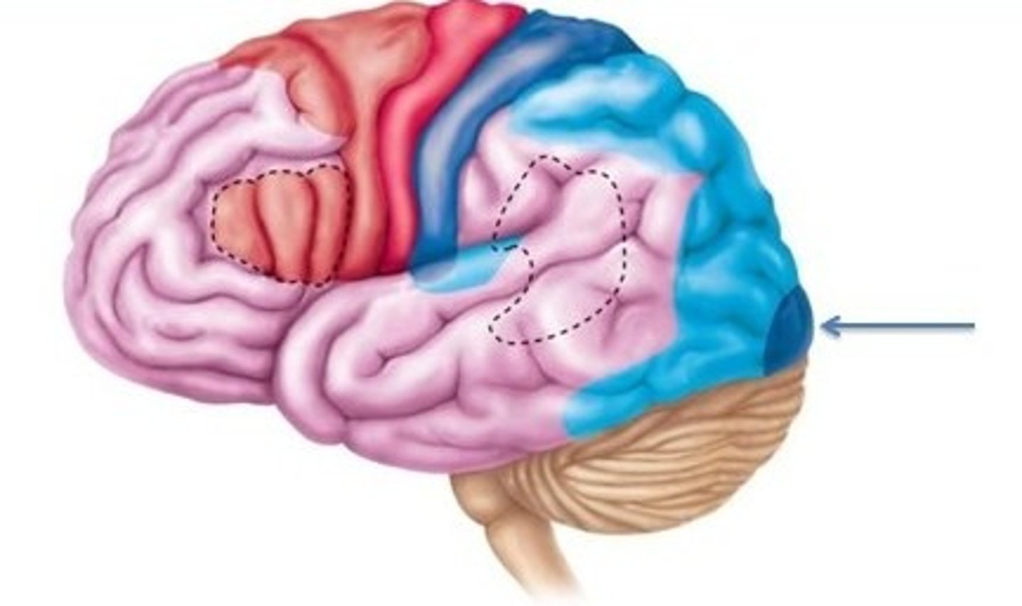
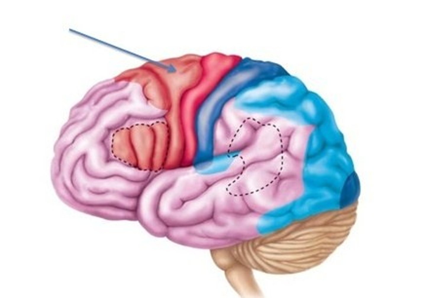
What is Broca's area?
A motor speech area that directs muscles of the tongue
Is active as one prepares to speak
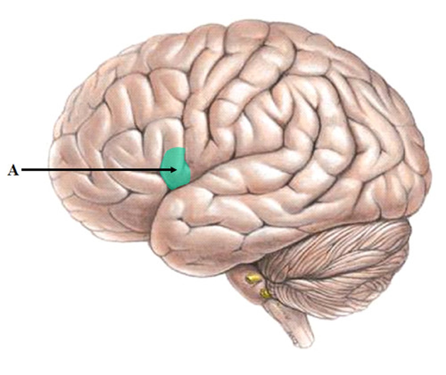
What happens if damage occurs to Broca's area
Expressive aphasia
Language is reduced to disjointed words and sentence construction is poor
Comprehension OK
What is Wernicke's area?
speech comprehension
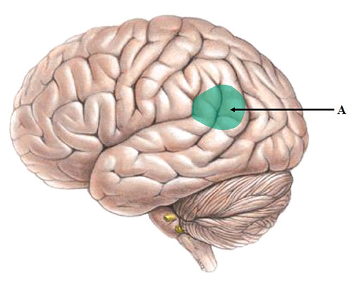
What happens if damage occurs to Wernicke's area
Receptive aphasia (unable to understand/generate meaningful language)
How do Broca's area and Wernicke's area communicate
Via the arcuate fasciculus (association bundle)
What happens if damage occurs to arcuate fasciculus
Conduction aphasia (difficulty repeating words/phrases)
What is the most common dominant hemisphere for language processing
Left (96%) - Many of the 4% with right sided language control have damaged left hemisphere
Therefore hemispheric specialisation can switch sides
Where is short term memory located?
Parietal and frontal areas but there are widespread cerebral cortex regions involved
When would you surgically separate the corpus callosum
In epilepsy
What is the feel the keys experiment
Done in split brain patients. Ask someone with their left hand to feel an object in their left hand pocket and tell you what it is but they can't. Left hand feels, right cortex recognises, but cannot activate the language centres in the left hemisphere to speak the word keys.
How do brain cells and brain regions talk to each other
Commissural - cortex to cortex cross over the midline
Association - cortex to cortex "stay" on the same side
Projection - communicate with other structures inside and outside the brain